All published articles of this journal are available on ScienceDirect.

Photogrammetric and Cephalometric Analyses of Ricketts' Esthetic Line in Malaysian Malay Adults: A Cross-sectional Study
Authors Info & Affiliations
Abstract
Objective:
To compare and correlate the Ricketts Esthetic Line measurement between photographs and lateral cephalogram. The norm for evaluating Ricketts' E-Line in the Malaysian Malay population is to be established.
Materials and Methods:
This was a cross-sectional study involving 126 pre-existing photographs and lateral cephalometric radiographs of skeletal Class I Malaysian Malay aged 19 to 40 recruited from the Orthodontic Clinic of Universiti Teknologi MARA (UiTM). Photogrammetric and cephalometric analyses of the upper and lower lips to Ricketts' E-line were performed. A paired t-test was performed to determine any statistically significant difference between the photographic and cephalometric variables. In addition, Pearson correlation was conducted to estimate the correlations between the photographic and cephalometric variables.
Results:
There was no significant difference between photographs and lateral cephalogram methods in the upper and lower lip to E-line measurement (p>0.05). For the photogrammetric analysis, the distance from the upper lip to the E-line was -0.30mm ±2.46, whereas the cephalometric analysis was -0.43mm ±2.63. The lower lip distance to the E-line for the photogrammetric analysis was 1.44mm ±2.8, whereas the cephalometric analysis was 1.24mm ±3.08. The Pearson correlation coefficient (r) for photographs and lateral cephalometric variables was close to 1 (0.914 and 0.898, respectively), indicating a robust positive correlation between the two variables.
Conclusions:
Photographic and cephalometric analysis can be used synonymously in measuring the Ricketts E line. Malaysian Malay has a more protrusive upper and lower lip to the E-line, compared to Caucasians, and the norm for the investigated population was established.
1. INTRODUCTION
Cephalometry and facial photography are essential diagnostic tools for diagnosis and orthodontic treatment planning. Clinicians obtain orthodontic diagnostic records alongside dental casts to execute an accurate plan for treatment course and a predictable treatment outcome [1]. Over the years, multiple analyses were introduced to facilitate orthodontic treatment decisions. When making decisions, it is imperative for clinicians to apply specific analysis and to take into account the ethnic distinctions in skeletal and facial features. However, normative standardised values were used as the reference values, which can be inaccurate as the orthodontic patient's morphology varies in anthropology and ethnicity [2-4].
Radiographs and cephalometric analyses are performed to determine the association between the soft tissues, dentition, and maxillofacial relationship. Orthodontists have an obligation to ensure the right indication before radiation prescription. The dosage must always be justified and kept “as low as reasonably practicable.” The usefulness of lateral cephalometric images may vary with the severity of cases [5]. The routine radiographs may also expose radiosensitive organs to radiation; the threshold dose below which the patients are not at risk is yet determined [6]. There is disagreement over the necessity of lateral cephalometric imaging as a routine procedure in orthodontic diagnosis and treatment planning. A group of researchers found that the lateral cephalometric image had no influence on the extraction pattern [7], with a similar trend of results found by Durao et al. in 2015 [1]. Clinical examination and photographs were often able to contribute details for diagnosis. Most recently, Dinesh et al. 2020 concluded that lateral cephalometric radiographs have no significant impact on diagnosis and treatment planning [8].
High-quality clinical images are indispensable in the field of orthodontics and aesthetic dentistry as a diagnostic tool to track treatment progress and for educational and medical-legal reasons. Photographs have the advantages of being relatively cost-efficient, without radiation risks, and could quickly provide sufficient information on soft tissue structures. Photograph documentation is routine in the field; however, they are analysed subjectively due to a lack of objective measurement for quantitative analysis [9]. Wayne Rasband, at the National Institutes of Health (NIH), created the robust, frequently cited image-processing platform known as ImageJ. ImageJ has excelled in numerous scientific initiatives and research works since its commencement in 1997 [10]. By manipulating the scale in Image J, life-size measurements can be obtained from the image. The software can then be utilised to calculate the linear dimension of the real object. Adaptation of Image J software for clinical soft tissue measurements can act as an alternate diagnostic tool that is affordable, reproducible, logistically feasible, safe, and accurate is especially needed in underdeveloped nations [11, 12].
Ricketts Esthetic line (E-line) is a soft tissue analysis to describe the relationship between the nose, lips, and chin in view of the aesthetic approach of a person's profile perspective. Robert Ricketts initiated this concept in 1957 to help better understand the protrusiveness of the lips to the E line, which is drawn from the greatest contour of the tip of the nose (pronasale) to the soft tissue chin (pogonion) [13]. The ideal soft tissue morphology described by Ricketts is that the upper lip distance is 4 mm posterior to the E line and the lower lip 2 mm posterior to the line, with consideration and proper judgment from the orthodontist of the normal variation of the person's ethnicity. According to Ricketts, lips that fall beyond the line were described as retrusive and unattractive [14]. This ideal measurement was based on the average Caucasian population that differs anthropologically from other craniofacial morphology. For Mongoloids racial groups, particularly among Asians, the dental protrusion is much greater than in Caucasians. The Thais, Malays, Chinese, and Filipinos are among the Southeast Asians [15-18] who opprobriously exhibit bimaxillary dental and hence soft tissue protrusion. Neglecting the significance of taking ethnic differences into account when evaluating individual patients could jeopardise treatment outcomes, particularly enhancing aesthetics exclusive to ethnicity and its traits.
Thus, this study aimed to compare Ricketts' Esthetic Line measurement between photographs and lateral cephalograms and to determine their correlation. The null hypothesis was that there is no difference between Ricketts' Esthetic Line measurement using photographs and lateral cephalograms. In addition, the differences in Ricketts' Esthetic Line measurement using photographs and lateral cephalograms between males and females were compared in order to generalise the Esthetic Line result to both genders. The norm for evaluating Ricketts E-Line in Malaysian Malays with Class 1 dentoskeletal patterns and aesthetically pleasing profiles will be established.
2. MATERIALS AND METHODS
This was a cross-sectional study involving one hundred and twenty-six (126) pre-existing lateral cephalometric radiographs of the non-growing Malaysian Malay population aged 19 to 40 years, which were randomly retrieved and recruited from the clinical records of the Orthodontic Clinic of Universiti Teknologi MARA (UiTM) from February 2022 to December 2022. Ethical approval was obtained from the UiTM Research Ethics Committee, REC/11/2021 (MR/884). Written consent was obtained when the patient was called for the review to confirm Malay ethnicity. The sample size was determined using G*power software to find correlations between bivariate normal models using a two-tailed test by taking α=0.05, power of 0.95; the sample size needed was one hundred and fifteen (115). The sample recruitment was based on the following criteria:
2.1. Inclusion Criteria
- Class 1 dentoskeletal pattern.
- Permanent dentition with all teeth present from upper lower 7 to 7.
- Good facial symmetry and balanced proportions (absence of gross facial asymmetry and craniofacial deformity that affects facial symmetry).
- Malay race with no interracial marriages in two generations predecessor [19-22].
2.2. Exclusion Criteria
- Previous orthodontic treatment.
- Previous maxillofacial/plastic surgery.
- Lateral photos are not in natural head position (natural head position is the patient's head in an upright posture, and the eyes focus on a point in the distance, indicating the visual axis is horizontal).
- Radiographs with inadequate quality for analyses.
2.3. Lateral Photographs
One hundred and twenty-six (126) recruited subjects, one hundred and one (101) female subjects, and twenty-five (25) male subjects were reviewed clinically in the Orthodontic Clinic of UiTM. Sample with Class I dentoskeletal pattern were identified through profile photos using a visual method looking at the distance between the soft tissue point A and B. During the review, patients consented to participate in the study and confirmed their Malay ethnicities by filling out a form. It consists of information about the patients and inquiries about their ancestors' Malay heritage going back at least two generations [21, 22]. The widest length of the auricle was recorded with a digital calliper by 2 authors, and the mean of two readings was recorded. This measurement was recorded to calibrate the pre-existing records of their extraoral photographs. It acts as known distance/measurements during photo analysis with Image J software version 1.54d National Institutes of Health, Bethesda, Maryland, USA.
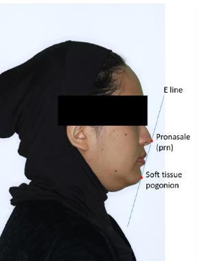
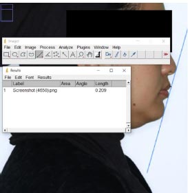
Two calibrated operators digitalised landmarks to minimise errors (Fig. 1). Using a known distance of the widest length of the right auricle, life-size measurements were obtained from the image by setting the measurement scale using Image J software. The real object linear measurement was calculated using the software by setting the scale based on the known size. The linear distance between the upper lip and lower lip to the Esthetic Line was computed for all records (Fig. 2).
2.4. Lateral Cephalograms
Each subject's information was recorded and analysed using Dolphin Imaging software version 11.95, USA (patient's name, age, gender, and lateral cephalometric radiographs). Landmarks were digitised directly from the radiograph using Dolphin Imaging software, and the linear distance between the upper lip and lower lip to the Esthetic Line was measured using Dolphin Imaging software by entering the cephalometric landmarks, and the value was automatically generated by the software (Table 1).
| No. | Landmarks | Definition |
|---|---|---|
| 1 | Pronasale (prn) | The most prominent midline point on the nose tip is identified on the lateral view. |
| 2 | Soft tissue pogonion (pog') | The most prominent point on the soft tissue chin. |
| 3 | Ricketts E Line (Ricketts 1979) | The line joining the nasal tip (prn) with the soft tissue pogonion (pog'). |
2.5. Statistical Data Analysis
Data analysis was performed using IBM Statistical Package for the Social Sciences (SPSS) software version 24.0 (Chicago, IL, United States of America). The significance level was set at p < 0.05. The operator was trained, calibrated, and analysed for any errors by an expert orthodontist. Twenty photographs and radiographs assessed by the two operators were subjected to inter-operator and intra-operator class correlation (ICC) to determine reliability for consistency.
Descriptive statistics were calculated for all photographic and cephalometric variables, including mean and standard deviations. Then, a paired t-test was performed to determine whether there was any statistically significant difference between the photographic and cephalometric variables. In addition, Pearson correlation was conducted to estimate the correlations between the photographic and cephalometric variables.
3. RESULTS
A total of 126 participants were included in this study. There were 101 female subjects with a mean age of 33.1 and 25 male subjects with a mean age of 32.3.
Twenty radiographs and photographs were randomly selected for the measurement to be repeated by the same operator and a second operator. Measurements were repeated within two-week intervals to assess measurement error. The reliability assessment for consistency of photogrammetric and cephalometric among two operators using intra-class correlation coefficient (ICC) shows excellent and good correlation for both intra-operator (0.990) and inter-operator (0.831) assessments, respectively. The ICC gives a measure of the reliability of the measurement in terms of consistency, with values between 0.75 to 0.9 considered good and more than 0.9 as an excellent correlation [24-26].
| Variable |
Male Mean (S.D.) |
Female Mean (S.D.) |
Mean Difference (95% CI) |
t-statistics (df) |
P-value |
|---|---|---|---|---|---|
| Photographs | - | - | - | - | - |
| Upper Lip to E-line | -0.96 (2.63) | -0.14 (2.40) | -0.82 (-1.90, 0.26) | -1.498 (124) | 0.137 |
| Lower Lip to E-line | 1.28 (3.04) | 1.49 (2.76) | -0.21 (-1.46, 1.03) | -0.338 (124) | 0.736 |
| Lateral Cephalogram | - | - | - | - | - |
| Upper Lip to E-line | -0.98 (2.65) | -0.29 (2.62) | -0.68 (-1.85, 0.48) | -1.166 (124) | 0.246 |
| Lower Lip to E-line | 1.11 (3.26) | 1.27 (3.05) | -0.16 (-1.53, 1.21) | -0.233 (124) | 0.816 |
| Variable | Photograph Mean (S.D.) |
Lateral Cephalogram Mean (S.D.) |
Mean Difference (95% CI) |
t-statistics (df) |
P-value |
|---|---|---|---|---|---|
| Upper Lip to E-line | -0.30 (2.46) | -0.43 (2.63) | 0.13 (-0.06, 0.32) | 1.361 (125) | 0.176 |
| Lower Lip to E-line | 1.44 (2.80) | 1.24 (3.08) | 0.21 (-0.03, 0.45) | 1.701 (125) | 0.092 |
| - | Correlation Coefficient, r | Strength of Correlation | P-value |
|---|---|---|---|
| Upper Lip to E-line | 0.914 | Strong Positive Correlation | <0.001 |
| Lower Lip to E-line | 0.898 | Strong Positive Correlation | <0.001 |
Table 2 analyses the presence of sexual dimorphism. No significant difference was observed between male and female subjects in relation to the upper line to E and lower line to E in both photographs and lateral cephalograms (p>0.05). The cephalometric measurements for both genders were pooled for further analyses.
Table 3 compares Ricketts's E-Line between the photograph and lateral cephalogram. There was no significant difference in the upper and lower lip to E-line measurement between photographs and lateral cephalogram methods (p>0.05).
Table 4 tabulated the Pearson's correlation coefficient and significance values of Ricketts' E-Line between the photograph and lateral cephalogram. The correlation coefficient (r) for both variables is close to 1 (0.914 and 0.898, respectively), indicating a robust positive correlation between the two variables. The correlation strength is described as a “Very Perfect Correlation” for both variables. The p-values for both variables are less than 0.001, indicating that the observed correlations are statistically significant at a significance level of 0.1%. This suggests a strong relationship exists between the two variables, and changes in one variable are likely to be associated with changes in the other variable. Overall, the results suggest a robust positive correlation between Upper Lip to E-line and Lower Lip to E-line measurements using photograph and lateral cephalogram) and this correlation is statistically significant.
4. DISCUSSION
Gender influence on perceptions of health care and orthodontic therapy is indisputable, and there's obviously an increased proportion of females seeking orthodontic therapy than males. Hence, a higher number of female subjects was observed in this study. Girls have significantly more aesthetic concerns [27, 28] and Holmes et al. in 1992 discovered that despite a lack of objective data supporting this belief, more females believed they had less beautiful dentitions and needed more treatment [29]. However, no significant difference was observed between male and female subjects in relation to the upper lip to E-line and lower lip to E-line in both photographs and lateral cephalograms analysis (p>0.05), as presented in Table 2. This agrees with previous cephalometric studies among Chinese with little gender disparity [27]. Hence, we can generalise the result to both genders.
The results in Table 3 show that the differences between Ricketts' E-Line between the photograph and lateral cephalogram were not statistically significant. Therefore, the null hypothesis was accepted. Photographic analysis can better assess the harmonic relationships between the exterior craniofacial structures because they are economical, do not expose the patient to potentially hazardous radiation, and are less time-consuming [9]. It makes no significant difference concerning the Ricketts E line. In addition, Pearson correlation analysis of the final values taken from lateral cephalograms, and lateral photographs showed a highly significant correlation between all the tested variables. There is no difference between the Ricketts E line to the upper and lower lips from photographs and lateral cephalometric. Through reliability tests, previous studies confirmed that the linear and angular measurements relevant for characterising facial morphology can be measured with photogrammetric analysis [30-34]. Therefore, photogrammetric analysis can be considered an acceptable technique for evaluating the Ricketts E line.
Due to changes in magnification, the photogrammetric analysis possesses some limitations, such as distortion of the photographic images compared to lateral cephalograms [34]. The posture of the head is another source of inaccuracy. It is imperative that the head position is consistent between cephalograms and photographs. Value discrepancies may be accounted for, partly through minor deviations from the natural head position, which can profoundly impact the analysis [35]. Furthermore, straining the lips or stretching the jaw, mainly when the mentalis muscle is hyperactive, can influence the measurement outcomes [31, 36].
Malay being the majority race of Southeast Asia, data from this study will benefit the orthodontic practice in the local vicinity and be applicable to Southeast Asian patients. The Malaysian Malay ethnic group was the focus of this study as Purmal in 2013 conducted work on the relationship of the lips using the E-line for the Malaysian Chinese and Malaysian Indians [16]. In line with the findings of this study, it revealed that the Malaysian Malay population's upper and lower lips were more protrusive than the Caucasians. For the photogrammetric analysis, the distance from the upper lip to the E-line is -0.30mm ±2.46, whereas the cephalometric analysis is -0.43mm ±2.63. The lower lip distance to the E-line for the photogrammetric analysis is 1.44mm ±2.8, whereas the cephalometric analysis is 1.24mm ±3.08. There is also a close agreement between the results with the literature suggesting that the Malaysian Malay population has more proclined upper and lower incisors and more protrusive maxilla and mandible [20, 21]. This strongly indicates that this is the population variation of characteristics and needs to be emphasised with soft tissue assessment.
An investigation into a bimaxillary protrusion sample revealed considerable differences from the Caucasian population, whereby the sample exhibits more protrusive features [19]. Furthermore, a Malay population norm for cephalometry has been established [20, 21]. These works emphasised how the distinctive Malaysian Malay community differs from Caucasian ideals. Malaysian Malay portrays normal to mild skeletal dental bimaxillary protrusion compared to Caucasians. There has been a report on the facial anthropometry morphology of Malay adults [22] with a comparison of the outcomes to earlier studies that were similar [23, 24]. Unique features present in the Malay face are significantly diverse compared to the Caucasians [22-24].
From the data of this study, we rejected the hypothesis as there is a significant difference between Caucasians and Malaysia Malay E-line. The Malaysian Malay Ricketts E-line measurement concerning the upper and lower lips has been established. It expedites diagnosis and treatment planning. It also helps meet patients' expectations and enhance local patient education. If the anthropological distinction is not taken into account, practitioners and patients should be made aware that what will alter is the population's normal and distinctive appearance.
Limitations of the present study include its cross-sectional design, with the results being biased by the sample size of both genders. The male subjects were lower than the females. However, the differences between the upper and lower lip to the E-line for both genders were less than 0.8mm, which clinically and statistically can be considered insignificant. This study's norm values for the Malay population provide valuable data for population studies.
CONCLUSION
Photographic and cephalometric analysis can be used synonymously in measuring the Ricketts E line. The Malaysian Malay population has more protrusive upper and lower lips, with the established value from photogrammetric analysis for the upper lip to E-line being -0.30mm ±2.46 and for the cephalometric analysis is -0.43mm ±2.63. The lower lip distance to the E-line for the photogrammetric analysis is 1.44mm ±2.8, whereas the cephalometric analysis is 1.24mm ±3.08.
AUTHOR’S CONTRIBUTION
1. Conceptualisation: JML, MM, LTW, NAH
2. Data curation: JML
3. Formal analysis: JML
4. Funding acquisition: NAH
5. Investigation: JML, MM, LTW, NAH
6. Methodology: JML, MM, LTW, NAH
7. Project administration: JML, MM, NAH
8. Resources: JML, MM, NAH
9. Supervision: MM, LTW, NAH
10. Writing – original draft: JML
11. Writing – review & editing: JML, MM, LTW, NAH
ETHICS APPROVAL AND CONSENT TO PARTICIPATE
Orthodontic Clinic of Universiti Teknologi MARA (UiTM) from February 2022 to December 2022. Ethical approval was obtained from the UiTM Research Ethics Committee, REC/11/2021 (MR/884). Written consent was obtained when the patient was called for the review to confirm Malay ethnicity.
HUMAN AND ANIMAL RIGHTS
No animals were used in this research. All procedures performed in studies involving human participants were in accordance with the ethical standards of institutional and/or research committees and with the 1975 Declaration of Helsinki, as revised in 2013.
AVAILABILITY OF DATA AND MATERIALS
The data and supportive information are available within the article. The data supporting the findings of the article is available in the Zenodo at https://zenodo.org/record/8433360, reference number https://doi.org/10.5281/zenodo.8433360.

