All published articles of this journal are available on ScienceDirect.

Facial Esthetic Analysis of Nepalese Subjects
Abstract
Background:
Facial beauty is a prime concern in facial esthetic treatments and facial plastic surgery. The cephalometric can be used in the diagnosis, treatment planning, assessing the growth pattern in the craniofacial complex and skeletal disproportion with the relationships of the teeth to their supporting bone.
Objective:
The objective of this research was to analyze the face, evaluate the variability between males and females, and determine the cephalometric norms of Nepalese subjects based on Ricketts analysis.
Materials and Methods:
Craniofacial analysis of 106 Nepalese Brahmins subjects (60 females and 46 males) was done using the lateral cephalogram. The samples were criteria selected who met the criteria; well-balanced face, class I canine relation, and class I molar relation with normal occlusion. Ten parameters of Ricketts analysis were measured and analyzed using SPSS 20. The results were compared among males and females using Independent T-test with P value = 0.05.
Results:
The mean values of the facial axis, mandibular plane angle, mandibular arc, convexity at A were higher among the Brahmin females compared to Brahmin males. While the mean values of lower facial height, lower incisor to A Pog distance, lower incisor to A Pog angle, upper molar to PTV, and distance from lower lip to E plane were higher among the Brahmin males compared to Brahmin females. Female had smaller craniofacial variables compared to males. The mean values of upper molar position are higher for both males and females compared to the standard value of 12 ±3 mm; however, the mean values are significantly higher among males compared to females (P = 0.001). It showed that the facial axis, chin position, lower facial height, and facial depth were no significant differences between Nepalese males and females.
Conclusion:
There was sexual dimorphism in craniofacial features in Nepalese Brahmins. Female had smaller craniofacial variables compared to males. This research provided an overview of craniofacial features and they can be a norm for Nepalese Brahmins in orthodontic diagnosis and treatment planning.
1. INTRODUCTION
The human perception of facial attractiveness is data-driven and largely irrespective of the perceiver. Facial beauty is a prime concern in modern esthetic facial treatments and facial plastic surgery. Facial proportion and teeth proportions aid in facial esthetics [1-3]. Beautiful faces have esthetic facial proportions [1, 2]. Various methods are used for face analysis [4]. In 1931, the introduction of cephalometric radiographs was done by Broadbent in the United States and Hofrath in Germany [5]. Cephalometrics can be used in assessing the growth pattern in the craniofacial complex diagnosis, treatment planning. In addition, it can be used to study the malocclusion and skeletal disproportion, the relationship of teeth, jaw, and the cranial base, evaluation of pre- treatment and post-treatment evaluation [6]. Most of the cephalometric analysis was performed in Caucasians and those values were used as a reference value in diagnosing and treating cases of individuals of different ethnic groups [7-9]. Since the morphologic features of different races and ethnic groups appear in a geographic cluster, the culture, climate, and geographic boundaries are bound to influence the facial morphology.
In 1961, Ricketts cephalometric analysis was a computerized analysis tool developed by Robert Ricketts [10, 11]. It is one of the most used cephalometric analyses. Ricketts proposed to use points, planes, and axes in addition to the traditional landmark for specific analysis, to analyze the chin in space, facial convexity, teeth position, and facial profile [12, 13].
Nepal is a country with a large number of racial subgroups and interracial mixtures. Chhetri is the largest group, accounting for 16.6% (4,398,053) of the total population, followed by Brahmins (12.2%: 3,226,903) [14]. Patients most commonly undergo orthodontic treatment at around 14-27 years of age, and priority should be given to obtaining solid norms for this age group. At present, there are limited studies for the Brahmin of Nepal and no published Nepalese Brahmins cephalometric norms using Rickets analysis. Knowing their cephalometric values would help to best treat these groups according to their facial characteristics. The objective of this research was to determine the cephalometric norms of Nepalese subjects based on Ricketts analysis and to evaluate the variability between males and females.
2. MATERIALS AND METHODS
A total of 106 Nepali Brahmins (60 females and 46 males) were included in this cross-sectional study. The study subjects included patients, dental students, and staff meeting the selection criteria. The ethical clearance was taken from the Institutional Review Board of Tribhuvan University and People's Dental College and Hospital, Kathmandu. In all the subjects, digital lateral cephalometric radiographs were taken. The study was done from October 2017 – December 2018. The details of the subjects are shown in Table 1. The inclusion criteria of the subjects were 18-27 years of age (Mean 22.5 years old and standard deviation ±3.4 years), Class I molar and canine relationship, Brahmins ethnic origin traced, overjet and overbite not exceeding more than 2-4 mm, no or little incisor crowding or rotations, presence of all permanent teeth except third molars, acceptable facial profile, good quality of cephalometric records [15-18]. The exclusion criteria were previous history of orthodontic treatment, orthognathic surgery, apparent skeletal or dental deformity, history of facial trauma or craniofacial disorder, such as cleft palate, missing permanent teeth except for third molars.
| Subject Details | Frequency |
|---|---|
| Total subjects • Female • Male |
106 60 (56.60%) 46 (43.40%) |
| Mean age Standard deviation Range |
22.5 years old ±3.4 years 18 – 27 years old |
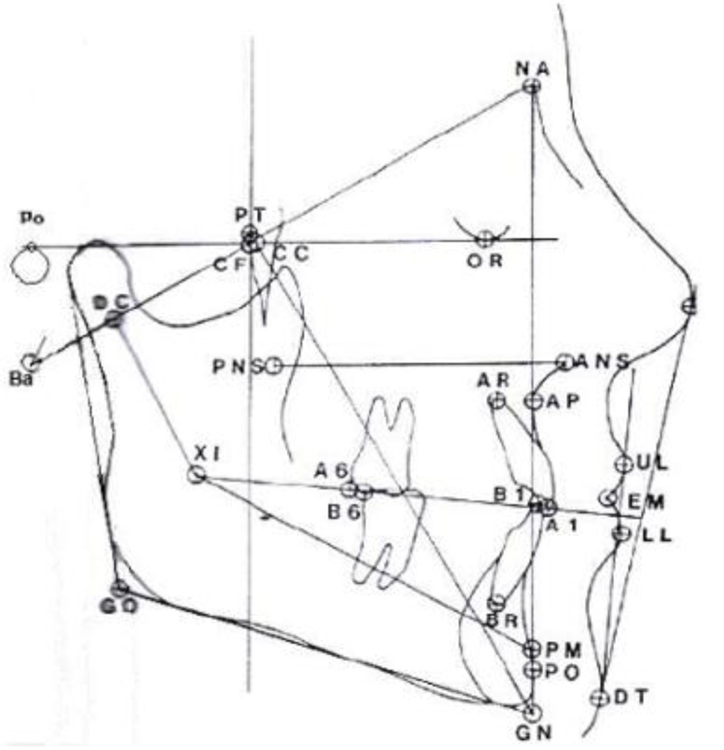
In the lateral cephalometric radiograph, various points were marked, and lines were traced (Fig. 1). Fig. (2) shows a lateral cephalometric radiograph showing various points and lines. The location and definition of various points are shown in Table 2. The description of various planes and angles used in this study is shown in Table 3. The description of various axes used in this study is shown in Table 4.

| Points | Location | Description |
|---|---|---|
| A6 | Upper molar | A point on the occlusal plane located perpendicular to the distal surface of the crown of the upper first molar |
| B6 | Lower molar | A point on the occlusal plane located perpendicular to the distal surface of the crown of the lower first molar |
| C1 | Condyle | A point on the condyle head in contact with and tangent to the ramus plane |
| DT | Soft tissue | The point on the anterior curve of the soft tissue chin tangent to the esthetic plane or E-line |
| CC | Center of cranium | The point of intersection of the basion-nasion plane and the facial axis. |
| CF | Points from planes at pterygoid | The point of intersection of the pterygoid root vertical to the FH plane |
| PT | PT point | The junction of the pterygomaxillary fissure and the foramen rotundum |
| DC | Condyle | The point in the center of the condyle neck along the Ba-N plane |
| En | Nose | A point on the soft tissue nose tangent to the esthetic plane or E- line |
| Gn | Gnathion | A point at the intersection of facial and mandibular planes (cephalometric Gn as opposed to anatomic Gn) |
| Go | Gonion | A point at the intersection of the ramus and the mandibular planes (cephalometric Go as opposed to anatomical gonion) |
| PM | Suprapogonion | The point at which the shape of the symphysis mentalis changes from convex to concave- also known as protuberant menti |
| Pog | Pogonion | The point on the bony symphysis tangent to the facial plane |
| PO | Cephalometric | The intersection of the facial planes and the corpus axis |
| TI | TI point | The point of intersection of the occlusal and the facial planes |
| Xi | Xi point | To locate the Xi point, it is explained in the following Figure and the accompanying explanation |
| Various Planes and Angles. | Description |
|---|---|
| Frankfort horizontal | Extends from porion to orbitale |
| Facial plane | Extends from nasian to pogonion |
| Mandibular plane | Extends from gonion to gnathion |
| PtV(pterygoid vertical) | A vertical line drawn through the distal radiographic outline of the pterygomaxillary fissure and perpendicular to the Frankfort horizontal |
| Basion-nasion plane | Extends from basion to nasion: divides the face and cranium. |
| Occlusal plane | The functional occlusal plane is represented by a line extending through the first premolars and premolars. |
| A-Pog line | A line from point A to pogonion is often referred to as the dental plane |
| E-line | An esthetic line or plane extending from the soft tissue tip of the nose (En) to the soft tissue chin point (DT) |
| Facial axis (Y-axis) | The angle between Ba-N and Pt-Gn lines |
| Facial angle | The angle between the FH plane and N-Pog plane |
| MD plane to FH | The angle between the mandibular plane and FH plane |
| Facial taper | The angle between the mandibular plane and N-Pog line |
| Lower Facial Height | Angle formed by intersection of the line from anterior nasal spine (ANS) to Xi point and the corpus axis (Xi-Pm) |
| Mandibular Arc | The angle formed by condylar axis (DC-Xi) and the distal extrapolation of the corpus axis |
| Convexity at point A | The convexity of the middle face is measured from point A to the facial plane (N-Pog) |
| Lower incisor to A-Pog | The A-Pog line or plane is referred to as the denture plane |
| Upper molar to PtV | Distance from the pterygoid vertical to the distal of the upper molar |
| Lower incisor to A-Pog inclination | The angle between the long axis of the lower incisor and the A-PO plane (1 to A-PO) |
| Lower lip to E plane | The distance between the lower lip and the esthetic (nose-chin) |
The data were entered in Microsoft Excel and then transferred to Statistical Package of Social Sciences (SPSS) 20 (SPSS, Chicago Inc., USA) for the analysis. Mean values of all the ten parameters were calculated using Rickett's analysis [19, 20]. Descriptive statistics were calculated. The categorical variables like sex were expressed in percentages. An Independent T-test was used to test to compare the results between males and females at P-value = 0.05.
3. RESULTS
It showed that the 106 Nepali Brahmin included students and staff with esthetic facial profiles and excluded the orthodontic patients who were undergoing orthodontic treatment. More than half (56.6%) of the study participants were female (Table 1). The results of 10 parameters studied for cephalometric evaluation through Ricketts analysis in the study are shown in Table 4. These ten parameters show the cranial relations i.e., mandible position, maxilla/mandible relations, denture relations, and esthetic relations of the Brahmin patients of Nepal.
Table 4 shows the results of various parameters of the study subjects for Rickett's analysis. This table gives the overall value for the Nepalese Brahmin subjects. The Comparison of various parameters between Nepali Brahmin males and females is shown in Table 4. The mean values of the facial axis, mandibular plane angle, mandibular arc, convexity at A were higher among the Brahmin females compared to Brahmin males. While the mean values of lower facial height, lower incisor to A Pog distance, lower incisor to A Pog angle, upper molar to PTV, and distance from lower lip to E plane were higher among the Brahmin males compared to Brahmin females.
| Various Axis | Description |
|---|---|
| Facial axis | A line extending from the foramen rotundum (PT to Gn) |
| Condylar axis | Extends from DC to Xi point; used to describe the morphologic features of the mandible |
| Corpus axis | Extends from Xi to PM; used to describe the morphology of the mandible and to evaluate dentition changes |
Table 5.
| Parameters | Mean ±SD | Std. Error | Range |
|---|---|---|---|
| Facial Axis ψ | 90.64 ±3.30 | 0.32 | 84.0 - 99.0 |
| Facial Depth ψ | 88.95 ±2.86 | 0.27 | 82.0 - 96.0 |
| Mandibular plane angle ψ | 22.42 ±5.33 | 0.51 | 10.0 - 34.0 |
| Mandibular Arc ψ | 37.57 ±7.51 | 0.72 | 21.0 - 56.0 |
| Lower Facial Height ψ | 41.85 ±5.35 | 0.52 | 27.0 - 50.0 |
| Convexity at A § | 0.63 ±2.36 | 0.22 | -05.5 - 04.0 |
| Lower Incisor to A- Pog ψ | 2.62 ±2.74 | 0.26 | -04.0 - 10.0 |
| Lower Incisor - A-Pog angle ψ | 26.59 ±5.91 | 0.57 | 10.0 - 42.0 |
| Upper Molar to PtV§ | 20.45 ±4.76 | 0.46 | 10.0 - 35.0 |
| Lower lip to E plane§ | - 01.76 ±2.34 | 0.22 | -07.0 - 04.0 |
| Parameters |
Males (Mean ±SD) |
Females (Mean ± SD) |
Mean Diff. | 95% CI | P value | |
|---|---|---|---|---|---|---|
| Lower | Upper | |||||
| Facial Axis ψ | 90.4 ±3.5 | 90.8 ±3.1 | -0.42 | -0.42 | -1.70 | 0.51 |
| Facial Depth ψ | 88.9 ±3.23 | 88.9 ±2.5 | -0.05 | -1.17 | 1.05 | 0.91 |
| Mandibular plane angle ψ | 20.7 ±4.9 | 23.7 ±6.1 | -2.96 | -4.96 | -0.96 | 0.004* |
| Mandibular Arc ψ | 37.3 ±9.0 | 37.7 ±6.1 | -0.36 | -3.46 | 2.73 | 0.81 |
| Lower Facial Height ψ | 42.1 ±6.0 | 41.6 ±4.7 | 0.53 | -1.62 | 2.70 | 0.61 |
| Convexity at A § | -0.3±2.5 | 1.3 ±1.8 | -1.74 | -2.60 | -0.88 | <0.001* |
| Lower Incisor to A- Pog ψ | 2.9 ±2.8 | 2.3 ±2.6 | 0.58 | -0.47 | 1.65 | 0.27 |
| Lower Incisor - A-Pog angle ψ | 28.1 ±5.6 | 25.4 ±5.9 | 2.66 | 0.41 | 4.91 | 0.02* |
| Upper Molar to PtV§ | 22.1 ±4.5 | 19.1 ±4.5 | 3.05 | 1.28 | 4.81 | 0.001* |
| Lower lip to E plane§ | -1.6 ±2.4 | -1.8 ±2.3 | 0.22 | -0.68 | 1.14 | 0.622 |
The average values of facial depth between both males and females are similar. Furthermore, it showed that mandibular plane angle was significantly lower among the Brahmin males compared to females (P = 0.004), indicating that males have a higher tendency for a closed bite and forward growth of the mandible. The Brahmin males have a significantly lower value (P <0.001) for measurement from point A to the facial plane, indicating a concave profile of Brahmin males compared to females. The Brahmin males have significantly lower incisor inclination i.e., higher mean value of lower incisor to A Pog angle compared to Brahmin females (P = 0.02).
The mean values of upper molar position are higher for both males and females compared to the standard value of 12 ±3 mm; however, the mean values are significantly higher among males compared to females (P=0.001) (Table 5). It shows that the average values indicate that separate cephalometric norms for Brahmin males and females of Nepal are required.
The results of the mean values of Rickett's analysis among Brahmin male and female subjects are shown in Table 5. It showed that the facial axis, chin position, and facial depth were no significant differences between Nepalese males and females. The mandibular plane angle of females was significantly higher than males (P=0.004), which shows that the Nepalese female has a more vertical growth pattern compared to Nepalese males. But the mean mandibular arc between Nepalese Brahmin males and females was not statistically significant. The growth pattern, when compared between Nepalese males and females, is the same. The lower facial height between males and females was not statistically significant; that is, they have the same growth pattern (Table 6).
The convexity at point A between Nepalese Brahmins male/female was statistically significant (P<0.001), showing that Nepalese Brahmin male individuals (-0.3 degree) have skeletal Class III pattern as compared to the female (1.3 degrees) who had skeletal Class II pattern. The lower incisor to A-Pog between Nepalese Brahmin male and female was not significant that is they have nearly same the location of lower incisor in relation to A-Pog line. The lower incisor was placed 2.9 mm and 2.3 mm ahead in males and females, respectively to A-pog line.
4. DISCUSSION
Nepal is a country with many racial subgroups and several religious and interracial mixtures. The present study is conducted to establish cephalometric norms for Brahmins using Ricketts analysis. The inclusive criteria and methodology are oriented to identify normative mean values that can assist in the diagnosis and treatment planning of the Nepalese Brahmins seeking orthodontic treatment. The mean and the standard deviation of ten variable of Rickets analysis has been determined. The study is performed to analyze the craniofacial structure of male and female individuals within the Brahmin ethnicity. It showed that the craniofacial structures of Nepalese Brahmin males are larger than Nepalese Brahmin females.
The cephalometric norms of Caucasians for many decades were being applied to the population groups all over the world [7-9]. But with time, many investigators concluded that there was a variation of the craniofacial morphology between different racial/ethnic groups. It was realized that cephalometric norms were inadequate for application to different racial or ethnic groups and proven that the 'norms' should be based on ethnic, sex, and age differences for better clinical evaluation [21-26].
It showed that the lower incisor to A-Pog angle of Nepalese Brahmin males and females is statistically significant (P=0.02). The Nepalese Brahmins male has proclined lower incisor as compared to Nepalese female Brahmins. The Nepalese Brahmins male has anteriorly placed upper molar than Nepalese Brahmins female (P=0.001). The Lower lip to E-plane between Nepalese males and females is statistically not significant. The lower lip is behind the E-plane and there is a soft tissue balance between lip and profile in Nepalese males and females.
Janson et al. [27] compared the skeletal, dental, and soft-tissue characteristics of Caucasian and Afro-Caucasian Brazilian subjects with normal occlusion and evaluated sexual dimorphism within the groups. They found that the upper posterior facial height was larger in the Caucasian subjects than in the Afro-Caucasian subjects, which is similar to the African subjects. The Afro-Caucasian subjects had more proclined and protruded maxillary and mandibular incisors, more protruded upper and lower lips, and a smaller nasolabial angle than the Caucasian subjects. These characteristics show that Afro-Caucasian subjects have dental and soft tissue components similar to African ancestors. The Afro-Caucasian females had lesser mandibular protrusion and smaller total posterior facial height and upper posterior facial height than males. It is common among African ancestors for males to have a greater mandibular protrusion than females. The literature also shows that African female subjects have smaller posterior vertical dimensions in the face than males. In our study, we compared the craniofacial structure of male and female individuals within Brahmin ethnicity and we found that the craniofacial structures of Nepalese Brahmin males are larger than Nepalese Brahmin females. A study by Rokaya et al. [28] did a study on the mentolabial sulcus in Nepalese to compare the sulcus between males and females. They found that the mean mentolabial sulcus angle in Nepalese was 118.19° ±12.28° (male: 119.43° ± 9.99° and female: 117.61° ± 13.23°). There was no statistically significant difference in sulcus angle between males and females (P=0.098). In total students, the average was more predominant followed by deep and shallow.
This degree of difference justified separate cephalometric standards for Nepalese Brahmins. Orthodontic treatment, including orthognathic surgeries, is performed based on clinical examination and other clinical records, knowledge of normative cephalometric values provided by this study can be an additional help. Various factors affect craniofacial growth and features [29]. In this present era, where the paradigm has shifted towards soft tissue, besides skeletal analysis, soft tissue should be focused, and treatment planning should always be centered on the patient's needs and desires. It would be preferable to use specific Nepalese Brahmins norms and separate for gender since a comparison has revealed a statistically significant difference in the most variable between males and females. This study can be extended to compare the craniofacial structures among the Caucasian subjects and Asian/ Nepalese subjects.
CONCLUSION
This study determined the cephalometric norms of Brahmins based on Rickets analysis and evaluated the variability between males and females. One hundred and six Brahmin individuals meeting our inclusion criteria were selected. There was sexual dimorphism in craniofacial features in Nepalese Brahmins. This research provided an overview of craniofacial features, and they can be a norm for Nepalese Brahmins in orthodontic diagnosis and treatment planning.
ETHICS APPROVAL AND CONSENT TO PARTICIPATE
The ethical clearance was taken from the Institutional Review Board of Tribhuvan University and People's Dental College and Hospital, Kathmandu.
HUMAN AND ANIMAL RIGHTS
No animals were used in this study. The reported experiments on humans are in accordance with the ethical standards of the committee responsible for human experimentation (institutional and national), and with the Helsinki Declaration of 1975, as revised in 2013.
CONSENT FOR PUBLICATION
Not applicable.
STANDARD OF REPORTING
STROBE guidelines and methodologies were followed in this study.
AVAILABILITY OF DATA AND MATERIALS
Not applicable.
FUNDING
None.
CONFLICT OF INTEREST
The authors declare no conflict of interest, financial or otherwise.
ACKNOWLEDGEMENTS
Declared none.

