All published articles of this journal are available on ScienceDirect.

Chronological Age Assessment Based on the Root Pulp Visibility in Lower Third Molars in a Group of Thai Population: Panoramic Radiographic Study
Authors Info & Affiliations
Abstract
Background:
Radiographic visibility of the root pulp in lower third molars is one of the reliable methodologies for chronological age estimation.
Objective:
To evaluate the radiographic visibility of the lower third molar root pulp using digital panoramic radiographs of the Thai population, and determine the appropriation of this technique in age assessment, especially at the age of 18 years.
Methods:
A total of 746 digital panoramic radiographs of the Thai population aged from 16 to 26 years were recruited for the present study. The visibility of root pulp of lower third molars with completed root formation was recorded in the four stages. Descriptive statistics were performed for each stage in both males and females.
Results:
The first appearance of stage 0 is 16.71 years in males and 17.00 years in females. The minimum age of stage 1 was found at 17.08 years in males and 17.18 years in females. The beginning of stage 2 was 18.33 years in males and 18.17 years in females. For stage 3, the earliest presence was started at 18.58 years in males and 18.83 years in females.
Conclusion:
The occurrence of stages 2 or 3 in both males and females indicated that the subject is over 18 years of age. Further study on the Thai population should be conducted to confirm the results of the present study.
1. INTRODUCTION
In recent years, many undocumented immigrants have entered Thailand, with a proportion who cannot reveal their true age. Therefore, it is necessary to find out whether those individuals have reached the age of majority to process their asylum processes correctly [1]. Dental age is an accurate, minimally invasive, and thoroughly performed method of assessing an individual’s age but is only reliable for the closure of the second molar root apices [1]. Many human biological markers were used to determine chronological age, including secondary dentin deposition, bone growth apposition, narrowing and obliteration of root canals, and periodontal ligament visibility on radiographs. However, in many cases, the periodontal ligament could not be assessed due to fused roots or narrowed furcation. It appears to be of limited value for forensic age estimation. Root pulp visibility of lower third molars seen on panoramic radiograph is the other interesting method for human age estimation. In 2010 Olze et al. [2] described the four-stage system root pulp visibility of lower third molars on panoramic radiographs in Germans. The authors concluded that this technique could help assesspatients over 18 years and over 21 years. The root canals were assessed for the proportion of root pulp obliteration. This methodology is indicated to be reliable and reproducible [3].
There are also studies of root pulp visibility of lower third molars seen on panoramic radiographs in the other races [3-8]. However, the previous studies suggested that differences between studies in different populations are evident, and specific population standards should be used when applying this technique. To our knowledge, there was no report about chronological age estimation by using root pulp visibility of lower third molars seen on radiographs in the Thai population. Therefore, the present study was performed to evaluate the radiographic visibility of the lower third molar root pulp using digital panoramic radiographs of the Thai population, to determine the appropriation of this technique in age assessment, especially at the age of 18 years.
2. MATERIALS AND METHODS
The institutional ethics committee approved the present study (IRB No. P10068/63). A total of 800 digital panoramic images from Thai patients who came for oral health evaluation at oral diagnosis and medicine clinic, dental hospital, Naresuan University were assessed. Digital panoramic images were taken at the oral and maxillofacial radiology clinic in our dental hospital between 2018 and 2020.
The visibility of root pulp of lower third molars with completed root formation, as defined by Olze et al. [2], was recorded in the four stages, (Fig. 1).
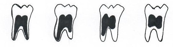
Stage 0: The root pulp is visible along the full length of all roots;
Stage 1: The root pulp is invisible in one root from apex to more than half root;
Stage 2: The root pulp is invisible along almost the full length of one root or along part of the root in two roots or both;
Stage 3: The root pulp is invisible along almost the full length of two roots.
A descriptive analysis of the stages of visualization of the root pulp according to age was done. To assess reliability, 100 randomly selected panoramic images were evaluated by two radiologists (WT and ST), and the first author re-examined the same 100 images after a 1-month interval.
3. RESULTS
A total of 746 digital panoramic images (299 males and 447 females) were recruited for the present study (Table 1). Fifty-four panoramic radiographs (7.24%) could not be assessed because of single, malformed roots or narrowed furcation.
| Age (Years) | Sex | Total | |
|---|---|---|---|
| Male | Female | ||
| 16 | 3 | 0 | 3 |
| 17 | 32 | 22 | 54 |
| 18 | 28 | 29 | 57 |
| 19 | 54 | 100 | 154 |
| 20 | 29 | 96 | 125 |
| 21 | 43 | 60 | 103 |
| 22 | 20 | 49 | 69 |
| 23 | 20 | 23 | 43 |
| 24 | 25 | 25 | 50 |
| 25 | 25 | 26 | 51 |
| 26 | 20 | 17 | 37 |
| Total | 299 | 447 | 746 |
Table 2 demonstrated the stages of root pulp visibility on panoramic radiographs of teeth 38 and 48 in males and females. It can be seen that the first appearance of stage 0 is 16.71 years in males and 17.00 years in females. The minimum age of stage 1 was found at 17.08 years in males and 17.18 years in females. The beginning of stage 2 was 18.33 years in males and 18.17 years in females. For stage 3, the earliest presence was 18.58 years in males and 18.83 years in females. The minimum age increase is related to the increasing stage of root pulp visibility with the older in females, except in stage 2. Also, our investigation showed that from stage 2, all individuals were over 18 years of age.
| - | Tooth | Stage | n | Min | Max | Median | Mean | SD |
|---|---|---|---|---|---|---|---|---|
| Males | 38 | 0 | 110 | 16.17 | 25.00 | 19.33 | 19.63 | 1.70 |
| 1 | 83 | 17.08 | 26.92 | 22.00 | 22.25 | 2.29 | ||
| 2 | 57 | 18.58 | 26.92 | 23.08 | 23.01 | 2.44 | ||
| 3 | 9 | 18.92 | 26.92 | 24.83 | 23.49 | 2.90 | ||
| 48 | 0 | 103 | 16.17 | 25.17 | 19.33 | 19.56 | 1.76 | |
| 1 | 87 | 17.75 | 26.92 | 21.67 | 21.98 | 2.21 | ||
| 2 | 66 | 18.33 | 26.92 | 24.00 | 23.42 | 2.26 | ||
| 3 | 13 | 18.58 | 26.92 | 25.58 | 23.78 | 3.37 | ||
| Females | 38 | 0 | 153 | 17.00 | 24.75 | 19.50 | 19.82 | 1.55 |
| 1 | 117 | 17.18 | 26.17 | 20.50 | 21.13 | 1.88 | ||
| 2 | 110 | 18.17 | 26.92 | 22.46 | 22.76 | 2.20 | ||
| 3 | 8 | 19.75 | 26.50 | 21.71 | 22.54 | 2.62 | ||
| 48 | 0 | 149 | 17.00 | 24.75 | 19.75 | 20.01 | 1.69 | |
| 1 | 105 | 17.75 | 26.92 | 20.58 | 21.07 | 1.91 | ||
| 2 | 116 | 18.58 | 26.92 | 22.50 | 22.80 | 2.38 | ||
| 3 | 12 | 18.83 | 25.08 | 22.55 | 22.25 | 2.33 |
The medians of stage 0 range between 19.33 years and 19.75 years, stage 1 between 20.50 years and 22.00 years, stage 2 between 22.46 years and 24.00 years, and stage 3 between 21.71 years and 25.58 years, respectively. The mean chronological age of the subjects varies between males and females, namely, between 19.56 years and 20.01 years in stage 0, between 21.13 years and 22.25 years in stage 1, between 22.76 years and 23.42 years in stage 2, and between 22.25 years and 23.78 years in stage 3, respectively. Regarding the mean age of the stages of root pulp visibility on panoramic radiographs of teeth 38 and 48, it was not statistically significant different both in males and females (Table 3).
| - | Mean age | p Value | ||
| Tooth 38 | Tooth 48 | |||
| Males Stage |
0 | 19.63 | 19.56 | 0.75 |
| 1 | 22.25 | 21.98 | 0.44 | |
| 2 | 23.01 | 23.42 | 0.33 | |
| 3 | 23.49 | 23.78 | 0.84 | |
| Females Stage |
0 | 19.82 | 20.01 | 0.32 |
| 1 | 21.13 | 21.07 | 0.82 | |
| 2 | 22.76 | 22.80 | 0.88 | |
| 3 | 22.54 | 22.25 | 0.80 | |
The Kappa statistics revealed a good agreement between both intra- and inter-observer agreement (K = 0.85 and 0.82, respectively). These values demonstrated high reproducibility and repeatability.
4. DISCUSSION
Chronological age anticipation is significant, especially at the age of 18, when legal procedures come into effect in Thailand. In Thailand, the Criminal Code states that “For any person over 15 years of age but not yet 18 years of age, it shall reduce the scale of punishment as provided for such offense by one – half (section 75)” [9]. Therefore, the accuracy of age estimation methods for suspects with unknown chronological age is needed in the interest of justice [10]. We decided to estimate the probability of an adolescent being older than 18 years from a medico-legal aspect, which indicates adulthood status in our country. We performed the present study to evaluate the radiographic visibility of the lower third molar root pulp using digital panoramic radiographs of the Thai population to determine the appropriation of this age assessment technique, especially at the age of 18 years. Fifty-four radiographs (7.24%) were excluded from the study because of the abnormal morphology. However, the data from 746 radiographs (92.76%) from the present study may be used as the Thai population's reference.
In the present study, the minimum age of stage 0 was 16.17 years in males and 17.00 years in females. Our results were the same as the previous studies' results in various countries, ranging from 16-17 years [1-7]. In contrast, the study in a group of Chinese population reported the minimum age found in stage 0 was older than ours (between 17 and 18 years) [8].
Considering stage 1, the first detected was 17.08 years in males and 17.18 years in females, similar to the study in UK Caucasians [3]. However, our findings are younger than the minimum age of the same stage in the previous reports. In the study of the Turkish population [5], the authors determined the earliest age in stage 1 was 19.1 years in males and 19.4 years in females. Timm et al. [6] found the minimum age of 21.0 years in males and 20.6 years in females in the German population. Even in a similar race, the minimum age of stage 1 was 19.25 years in males and 20.73 years in females of the Chinese population [8]. In addition, we found that the minimum age in stage 1 of our results was older than the study in the Maltese population, which reported the first occurrence of stage 1 at the age of 16.28 years in males and 16.08 years in females [1].
For stages 2 and 3, our study's first observation was not older than 18 years, both in males and females. The first evidence of stage 2 in our study are similar to that of Al Qattan et al. [1], Lucas et al. [3], Pérez-Mongiovi et al. [4], Akkaya et al. [7]. However, the minimum age in stage 3 of the present study was 1 to 5 years younger than the previous report as mentioned above. Therefore, in the Thai population, it may be confirmed that males and females presenting stages 2 or 3 were 18 years or older. Moreover, Timme et al. [6] confirmed that males and females presenting stage 1 of root pulp visibility were older than 18 years. And the presenting of stage 2 indicated the age older than 21 years. Guo et al. [8] concluded that stages 1, 2, and 3 could be used to verify that a person is over 18 years of age. They also indicated that if stages 2 or 3 are determined, it is possible to ensure that an individual has already reached 21 years of age. As mentioned above, the difference between studies may be ascribable to differences in study design, statistical analysis, the age range of the samples, the races, and the observer variations. Third molar mineralization is unique in various populations, which may influence pulp visibility [7]. Therefore, the potential factors such as eating behavior, systemic diseases should be further analyzed.
To the best of our knowledge, the present study reports the first assessment of this methodology in the Thai population. Therefore, we believe that our study’s results may be used as another applicable reference for age estimation in Thai. Our study's limitation is the variation in morphology of lower third molars, which may be the same as the previous studies. In an attempt to reduce this problem, some alternative methods have been offered. Recently, there have been new approaches in age estimations rather than third molar root pulp visibility. Balla et al. [11] analyzed the radiographic visibility of the root pulp of mandibular first molars using a classification of Olze et al. [2] as a biologic marker at the age of 18 years. They concluded that when the mandibular first molars reach stage 3 of pulp visibility in males, there is a very high probability of being above 18 years and a high probability for females above 18 years. Root pulp visibility stages in mandibular first molars are useful to judge the age threshold of 18 years. Besides, Suvarna et al. [12] evaluated the radiographic visibility of the root pulp in mandibular second molars using the stage classification of Olze et al. [2] for determining the age over 18 years and indicated that root pulp visibility in lower second molars might be useful in the identification of the age older than 18 years. These new methodologies may be recommended to use in parallelism with other age estimation methods. The other limitation in our study is the balance between the number of subjects in each age group. Further study should be performed to eliminate this issue.
Panoramic radiography is the most common radiographic modality in evaluation for many purposes. Most dental professionals are familiar with this imaging modality. It revealed the tooth structures and surrounding structures, including the temporomandibular joint, some cervical vertebrae, submandibular areas, etc. In Thailand, panoramic radiography is one of the most common modalities used for dental purposes, including chronological age estimation. Our previous study was performed by evaluation of third molar development from panoramic radiograph to estimate the chronological age. It was concluded that the determined probability of the Thai population being younger or older than 18 years for legislation might be valuable in future forensic practice [13]. Accordingly, the present study results may be an additional marker for forensic age estimation in Thai.
CONCLUSION
The root pulp visibility of lower third molars might be the appropriate technique in diagnosing the age of over 18 years in the Thai population. The occurrence of stages 2 or 3 in both males and females indicated that the subject is over 18 years of age. Further study on the Thai population should be conducted to confirm the results of the present study.
AUTHORS' CONTRIBUTIONS
WT, ST, CD are responsible for study design, concepts, clinical studies, and data acquisition. WT, RC are responsible for data analysis and manuscript preparation. WT, ST, CD, RK are responsible for literature search, manuscript editing, and manuscript review.
ETHICS APPROVAL AND CONSENT TO PARTICIPATE
This study was approved by the Institutional Review Board at Naresuan University, Phitsanulok, Thailand (IRB No. P10068/63).
HUMAN AND ANIMAL RIGHTS
No animals were used in this research. All human research procedures were followed in accordance with the ethical standards of the committee responsible for human experimentation (institutional and national), and with the Helsinki Declaration of 1975, as revised in 2013.
CONSENT FOR PUBLICATION
Written consent was obtained from all participants in the study.
AVAILABILITY OF DATA AND MATERIALS
The data that support the findings of this study are available from the corresponding author [W.T.] upon reasonable request.
FUNDING
None.
CONFLICT OF INTEREST
The authors declare no conflict of interest, financial or otherwise.
ACKNOWLEDGEMENTS
Declared none.

