All published articles of this journal are available on ScienceDirect.

Sensitivity and Specificity of Mandibular Third Molar Calcification at Chronological Age and Hand Wrist Maturation Stage to Discriminate Between Female and Male at Pubertal Growth Period
Authors Info & Affiliations
Abstract
Background:
The mandibular third molar is the last tooth that is not completely developed by the time pubertal growth has been finished. Maturation of the mandibular third molar is one of the physiological maturation indicators that can be used to determine the stage of pubertal growth.
Objective:
The aim of the study was to determine the sensitivity and specificity of mandibular third molar calcification at chronological age and hand wrist maturation stage to discriminate between female and male at pubertal growth period.
Methods:
It is a retrospective study with a cross-sectional approach using panoramic and hand-wrist digital radiographs of 279 females and 144 males, age 8-17 years, with a total of 423 panoramic radiographs and 423 hand-wrist radiographs. Statistical analysis was performed using Excel Mega Stat. ANOVA to analyze the differences between mandibular third molar calcification at chronological age and hand-wrist maturation stage, and t-test was used to analyze the differences between females and males. Spearman rank correlation was used for the analysis of the correlation between mandibular third molar calcification with chronological age and hand-wrist maturation stage; sensitivity and specificity were used to discriminate the pubertal growth period between mandibular third molar calcification and hand-wrist maturation stage.
Results:
There were significant differences found in mandibular third molar crown maturation stage B and C, but no significant difference was observed between mandibular third molar stage A, D, E, F, G and H, between females and males. The highest percentage of mandibular third molar crown formation in females was observed at stage D (6.68%) at MP3u, and in males, it was observed at stage D (8.83%) at SMI-4. The highest percentage of root formation in females was stage E (8.24%) at the SMI-10 stage, and males stage F (4.86%) at MP3u. The correlation was observed between mandibular third molar calcification with hand-wrist, females 0.22 and males 0.43, and chronological age 0.60 for females and 0.69 for males. The highest sensitivity of mandibular third molar calcification of 97.0% was observed in female at SMI-4 of hand-wrist maturation with specificity of 100%, while in male, a sensitivity of 94.5% was observed at SMI-2 stage with a specificity of 99.99%.
Conclusion:
There were significant differences found in mandibular third molar calcification between females and males except for stage B and stage C; weak correlation was observed between mandibular third molar calcification and hand-wrist, and moderate correlation was observed with chronological age. The sensitivity and specificity in females and males indicate that mandibular third molar calcification is only useful for diagnosing the pre-pubertal growth phase.
1. INTRODUCTION
The development of tooth calcification is one of the physiological maturation indicators that can be used to predict pubertal growth. The development of mandibular third molar based on chronological age varies greatly from the initial formation until the completion and can be used to estimate chronological age at pubertal growth period [1-3]. In the Orthodontic treatment, the evaluation of mandibular third molar development is important to estimate whether it needs to be extracted or not because mandibular third molar eruption can disrupt the stability of the orthodontic treatment results [4, 5]. The development of mandibular third molar as other teeth can be observed using a panoramic radiograph which is routinely used for assessing dental maturation in orthodontic treatment. The study by Thorson et al. shows that mandibular third molar is accurate and precise as an indicator of chronological age [6]. The development of the mandibular third molar is under strong genetic control and has less environmental influence compared to other physiological maturation [7].
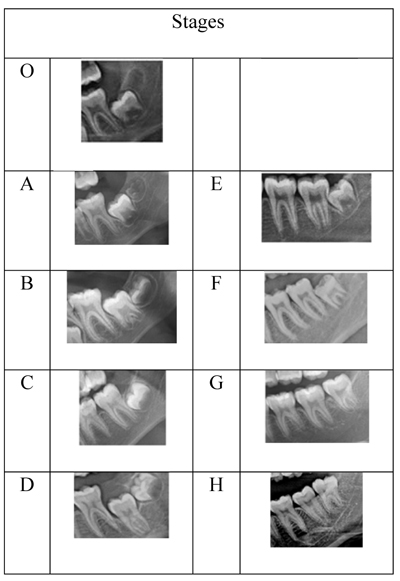
The most common method for assessing the dental calcification stage is Demirjian and Goldstein method which consists of 8 stages A to H. Stages A to D represent the crown formation starting from the appearance of the cusp until the completion of the crown, and stage E to stage H represent root formations from radicular bifurcation to apical closing. The stages of tooth development proposed by the Demirjian and Goldstein method were based on the ratio of the crown and root formation and not on the changes of linear measurements.
Previous studies have found that the mineralization of the mandibular third molar is a population-specific process and varies according to chronological age in different ethnic groups. Therefore, estimation of tooth development should ideally be carried out in the same population [8-12]. Many studies conducted in different countries have analyzed the levels of mandibular third molar calcification based on chronological age and hand-wrist maturation stage at the pubertal growth period [13-17]. Considering that the level of mandibular third molar calcification is influenced by many factors, it is possible that Indonesian subjects have different maturation stages compared to other populations. The purpose of the study was to determine the sensitivity and specificity of mandibular third molar calcification at chronological age and hand wrist maturation stage to discriminate between females and males at pubertal growth period.
2. MATERIALS AND METHODS
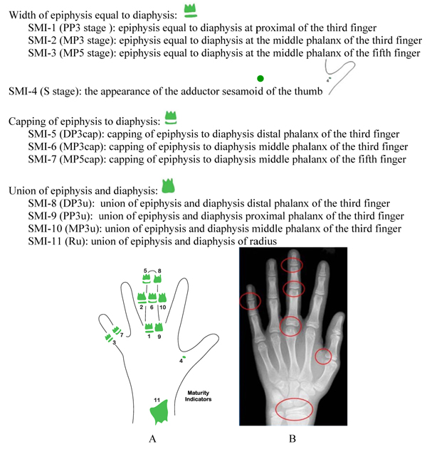
This was a retrospective study with a cross-sectional approach. The subjects consisted of 279 females and 144 males, ages 8 to17 years, with each subject providing good quality digital panoramic and hand-wrist radiographs, making a total of 423 panoramic and 423 hand-wrist radiographs. All participants were orthodontic patients in Orthodontic Clinic, Faculty of Dentistry, University of Padjadjaran. Inclusion criteria involved Indonesian, healthy patients, free from systemic or serious illness, who never had previous orthodontic treatment, or trauma on the face, hand and wrist, and those not missing mandibular third molar on both sides of the lower jaw. Hand-wrist maturation was analyzed by the Fishman method [18], and mandibular third molar calcification was done according to modification of Demirjian and Goldstein method on panoramic radiographs [8].
2.1. Statistical Analysis
ANOVA was used to analyze the differences between mandibular third molar calcification in chronological age and hand-wrist maturation stage, and post hoc using t-test was used to analyze the differences between females and males. Spearman rank correlation was used to analyze the correlation between mandibular third molar calcification with chronological age and hand-wrist maturation stage at pubertal period (P<0.05). Sensitivity and specificity tests were carried out using screening test Thornier-Remain 2x2. Statistical analysis was performed using Excel Mega Stat. Gaussian distribution was as follows: male P= 0.1231 and female P= 0.0956. The reliability test was conducted by taking 10 random panoramic and hand-wrist radiographs of female and male participants. Repeated measurements were done 3 times by three researchers, with 2-week intervals. Cohen’s Kappa interrater coefficient showed no significant difference in mandibular third molar calcification (0.852 in females and 0.873 in males) and hand-wrist maturation (females 0.910 and 0.879 in males).
Hand-wrist maturation stage of Fishman method consists of 11 stages, which can be divided into three stages of maturation: pre-pubertal growth (SM-1 to SMI-4), pubertal growth (SMI-5 to SMI-7) and post-pubertal growth (SMI-8 to SMI-11).
3. RESULTS
The distribution of subjects according to gender and chronological age is shown in Table 1. Mandibular third molar calcification stage of females and males in chronological age is shown in Table 2. The highest mandibular third molar calcification was achieved by 81 female subjects (29%) at 12.65 years while in male 40 subjects (27.77%) at 12.70 years; thus no significant difference was observed between females and males (P> 0.05). Calcification mandibular third molar stage H only occurred in 2 male subjects (1.39%) at 17.00 years old. Mandibular third molar calcifications stage B and C differed significantly between females and males (P<0.05), while mandibular third molar calcification stage A to G did not differ significantly between male and female subjects (P>0.05) (Table 2). The distribution of mandibular third molar crown calcification stage A, B, C and D in females was at 10.9 -12.65 years and males at 10.0-12.70 years. Root formation of mandibular third molar stage E, E, G, in females was at 14.5-15.57 years, and in males, the stage E, F, G was at 12.70-17.00 years. This indicates that root formation was not finished at pubertal growth, and only two male subjects reached stage H (Table 2).
| Age (Year) | 8 | 9 | 10 | 11 | 12 | 13 | 14 | 15 | 16 | 17 | Total |
| Male | – | 19 | 21 | 15 | 15 | 26 | 18 | 11 | 13 | 6 | 144 |
| Female | 5 | 32 | 28 | 46 | 33 | 43 | 32 | 22 | 25 | 13 | 279 |
| Stage | Females | Males | Comperativ Females vs Males | |||||
| n | Average | std | n | Average | std | t -test | p-value | |
| 0 | 37 | 11.59 | 2.68 | 18 | 11.00 | 2.3 | 4.26 | 0.00* |
| A | 23 | 10.09 | 1.62 | 10 | 10.00 | 0.82 | 0.7 | 0.2433 |
| B | 32 | 10.28 | 1.35 | 14 | 10.07 | 1.14 | 2.46 | 0.009* |
| C | 35 | 11.34 | 1.21 | 27 | 11.74 | 1.75 | -5.66 | 0.000* |
| D | 81 | 12.65 | 1.64 | 40 | 12.70 | 1.38 | -1.2 | 0.1155 |
| E | 40 | 14.5 | 1.24 | 18 | 14.42 | 1.69 | 0.98 | 0.1661 |
| F | 21 | 15.57 | 1.03 | 13 | 15.54 | 1.27 | 0.32 | 0.3740 |
| G | 7 | 15.57 | 2.64 | 2 | 17.00 | 1.41 | -0.71 | 0.2495 |
| H | − | − | − | 2 | 17.00 | 0 | − | − |
| *Significant | - | - | - | - | - | - | - | - |
The stage of mandibular third molar calcification at hand-wrist maturation stage is shown in Tables 3 and 4. The highest calcification of crown formation was at stage D at MP3u (6.68%) for females and males at SMI-4 (Sesamoid/S), 8.33%. The highest root formation of mandibular third molar calcification for female stage E was 8.24% at SMI-10 (MP3u), while in males, it was at stage F (4.86%) at SMI-10 (MP3u) The last stage for females at stage G was 2.15% at SMI-11(Ru) and males 0,16%; only male samples (1.39%) had reached stage H on SMI-11 (Ru) (Table 4). The mandibular third molar calcification in female subjects was at SMI-4 (S stage) of hand-wrist maturation with 97.0% sensitivity and 100% specificity; in males, sensitivity of 94.5% and specificity of 99.99% were observed at SMI-2 (stage MP3). SMI-4 (stage S) and SMI-2 (stage MP3) were prepubertal growth stages of hand-wrist maturation. This result indicates that the ability of mandibular third molar calcification for identifying pubertal growth at the hand-wrist maturation stage was only for prepubertal growth stage (Table 5). The correlation between mandibular third molar and hand-wrist maturation stage was estimated at 0.43 in males and 0.22 in females, which was found to be lower than the correlation observed between mandibular third molar calcification and chronological age, with females showing 0.60 and males 0.69 (P<0.0001) (Table 6).
4. DISCUSSION
In the field of orthodontic treatment, the determination of diagnosis, treatment plan, and appropriate time of orthodontic treatment in patients during pubertal growth is more accurate than chronological age using physiological maturation indicators. There are various physiological maturation indicators to assess pubertal growth status, such as height, weight, secondary sexual maturation, dental and skeletal maturation [19]. Dental maturation can be assessed from either the phase of tooth eruption or the stage of tooth calcification. Tooth eruption is influenced by local or systemic conditions but not tooth calcification, therefore tooth calcification is more reliable for the determination of pubertal growth stage [8, 20, 21]. Demirjian and Goldstein proposed a method for estimation of dental maturity on panoramic radiograph for the seven left permanent mandibular teeth at eight calcification stages. Stages A, B, C, D were for crown formation and stages E, F, G, H were for root formation (Fig. 1) [8]. The use of mandibular third molar calcification has an advantage because mandibular third molar calcifications still continue even though pubertal growth has been completed; therefore, it can be used to determine the stage of pubertal growth [8, 9, 21, 22].
| - | Stage Hand-Wrist | M3 Calcification Stage | Percentage |
| Male | SMI-4 (S) | D | 8.33 |
| Females | SMI-10 (MP3u) | D | 6.68 |
| - | Females | - | |
| - | Stage Hand-Wrist | M3 Calcification Stage | Percentage |
| Males | SMI-10 (MP3u) | F | 4.86 |
| Females | SMI-10 (MP3u) | E | 8.24 |
| - | Females | Males | ||||
| Stage Hand-Wrist | Sensitivity | Specificity | Stage Hand-Wrist | Sensitivity | Specificity | |
| Highest | SMI-4 (S) | 97.0 | 100.00 | SMI-2 (MP3) | 94.5 | 100.00 |
| Lowest | SMI-11 (Ru) | 2.0 | 100.00 | SMI-11 (Ru) | 7.6 | 100.00 |
| Stages | Females | Male | ||||
| r | t | p-value | r | t | p-value | |
| M3 and hand-wrist | 0.22 | 3.71 | .00013* | 0.43 | 5.61 | .0001* |
| M3 and chronological age | 0.60 | 12.55 | .0001* | 0.69 | 11.22 | .0001* |
| *Significant | - | - | - | - | - | - |
The evaluation of dental maturation using mandibular third molar on panoramic radiograph would be an adjunctive tool for pubertal growth assessment in combination with hand-wrist maturation stage, but it should be taken into consideration because there are many variations in conjunction with chronological age and hand-wrist maturation [9, 23, 24]. In this study, we used the modification of Demirjian and Goldstein system to assess left mandibular third molar maturation, know its correlation with chronological age and hand-wrist maturation stage, and to test the sensitivity and specificity with respect to hand-wrist maturation [11, 17]. In Thai subjects, it has been shown that males tend to be more advanced as compared to females in relation to skeletal maturity stages [24], while in Spanish, mandibular third molar crown formation is complete at the age of 14 years and the root formation finishes at an average age of 18.5 years [12]. In a study by Kullman et al., the crown formation of mandibular third molar was completed at 13.5 years and root formation began at about 15.1 years completed at an average age of 19.3 years [25]. In the Australian subject, crown completion of mandibular third molar at stage A to D was found at 14.77 y ±1.68 years and root formation without closure of the apex (stage G) was observed at a mean age of 19.00±1.48 years [23]. In our study, as shown in Table 2, female crown formation of mandibular third molar was observed at 10.09 years which finished at 12.65 years, and root formation of mandibular third molar was observed to start at 14,5 years and stage G at 15.57 years, while for male, the crown formation started at 10.00 years and finished at 12.70 years, and root formation started at 14,42 years and reached stage G at 17 years. Only 2 subjects were at stage H at 17 years. With respect to the hand-wrist maturation, the highest percentage of mandibular third molar stage E in female subjects was 8.24% at MP3u, while for males, stage D was 8.33% at S stage (Table 3). This difference can be attributed to different race, ethnicity, sample size and research methods [19].
Many studies have reported the sensitivity and specificity of mandibular third molar maturation as a physiological maturation index. The sensitivity and specificity of mandibular third molar maturation in this study were evaluated in the context of the association between mandibular third molar calcification stage and hand-wrist maturation divided into three categories: pre-pubertal (SMI-1 to SMI- 4)PP3-S), pubertal (SMI=5 to SMI-7), and post-pubertal periods (SMI-8 to SMI-11). The results show that the higher sensitivity in female subjects was 97.0% with specificity 100% at SMI-4 of hand-wrist maturation, while in males, sensitivity was 94.5% and specificity 99.99% at SMI-2; both females and males were at the prepubertal stage. The lowest sensitivity of females was at 2% and males at 7.6%; this indicates that at the end of pubertal growth, the stage of mandibular third molar calcification cannot be used as an indicator of pubertal growth maturation because mandibular third molar calcification was not yet finished in both females and males. This result was in concordance with a previous research indicating dental calcification as only useful for diagnosing the pre-pubertal growth phase [26, 27].
Many studies have reported strong relationships between mandibular third molar calcification and hand-wrist maturity, but in contrast, Demirjian and Buschang et al. reported a weak relationship between dental maturity and skeletal maturity. A previous research study showed a significantly weak correlation between mandibular third molar and skeletal maturity in females 0.31 and males 0.47 [19], but Ëngstrom and coworkers found a strong correlation between dental maturity and skeletal maturation (r = 0.72) [21]. In Thai subjects, mandibular third molar showed the lowest correlation with hand-wrist maturation stage at r = 0.47 in male subjects, and female subjects r = 0.31 [24]. In our study, a weak correlation was observed between mandibular third molar and hand-wrist maturation stage, such as in males, it was 0.43, while in females, it was 0.22; however, correlation of mandibular third molar with chronological age was moderate, with females 0.60 and males 0.69. This result of our study was found to be in accordance with the result of mandibular third molar calcification in the Korean population, having a strong correlation with chronological age [9]. The use of mandibular third molar calcification from other studies or other national standards is certainly not appropriate because it can cause overestimated or underestimated degrees of development [19, 28, 29]. Based on our study, mandibular third molar calcification stage at panoramic radiographs can be used as an adjunctive tool in combination with hand-wrist maturation as a diagnostic tool to estimate the timing of the pubertal growth, especially at prepubertal growth.
CONCLUSION
The purpose of the study was to determine the sensitivity and specificity of mandibular third molar calcification at chronological age and hand wrist maturation stage to discriminate between female and male at pubertal growth period from 8 years to 17 years. The following conclusions have been obtained.
(1) No significant difference in mandibular third molar calcification stage B and C between females and males has been observed, but significant differences were found in mandibular third calcification stage between females and males at stages A, D, E, F, G and H (P<0.05).
(2) The highest percentage of mandibular third molar crown formation at hand wrist maturation stage in females was at stage D (6.68%) at SMI-10, and males, it was at stage D (8.83%) at stage SMI-4. Root formation in females was at stage E (8.24%) at MP3u, while in males, it was observed at stage F (4.86%) at SMI-10.
(3) Regarding the sensitivity and specificity of mandibular third molar calcification at hand wrist maturation stage, in females, at S stage, 97% sensitivity and 100% specificity were observed, and in males, 94,5% sensitivity at SMI-2 and 99.99% specificity have been observed, indicating mandibular third molar calcification to be only useful for diagnosing the pre-pubertal growth phase.
(4) There was a moderate correlation observed between mandibular third molar stage and chronological age (females 0.60 and males 0.69), and low correlation between mandibular third molar and hand-wrist maturation (females 0.22 and males 0.43). This result indicates that mandibular third molar calcification stages might be clinically used as a maturity indicator of the pubertal growth period. However, further study is recommended in a larger sample size.
The limitation of our study is that the samples were obtained only from the Deutero-Malay racial group, which is the largest race group in Indonesia, and from the urban city of Bandung that may not represent the socio-economic demographics of the entire population of the Deutero-Malay subjects and other races in Indonesia. Another limitation is that we were unable to control the nutrition, systemic illnesses, sports hobbies or other factors that could affect pubertal growth and development unless based on anamnesis.
ETHICS APPROVAL AND CONSENT TO PARTICIPATE
Ethical approval was given by Health Research Ethical Committee of Faculty of Medicine, Universitas Padjadjaran (Protocol No. 395/UN6.CI.3.2/KEPK/PN/2016).
HUMAN AND ANIMAL RIGHTS
No animals were used in this research.All human research procedures followed were in accordance with the ethical standards of the committee responsible for human experimentation (institutional and national), and with the Helsinki Declaration of 1975, as revised in 2013.
CONSENT FOR PUBLICATION
The participants provided written informed consent to be involved in this study.
AVAILABILITY OF DATA AND MATERIALS
The data sets analyzed during the current study are available from the corresponding author [E.M] upon reasonable request.
FUNDING
None.
CONFLICT OF INTEREST
The authors declare no conflict of interest, financial or otherwise.
ACKNOWLEDGEMENTS
The researchers gratefully acknowledge the Orthodontic Department at Padjadjaran University for research facilities.

