All published articles of this journal are available on ScienceDirect.

Face Proportions, and Analysis of Maxillary Anterior Teeth and Facial Proportions in a Thai Population
Authors Info & Affiliations
Abstract
Background:
Esthetics is the primary consideration for patients looking for prosthetic treatment. Determining the facial types of Thai populations is important because most studies conducted in Caucasian populations mention their facial types, and describe the gender comparisons, and GP of the teeth.
Objectives:
This study compared facial proportions, analyzed the maxillary anterior teeth proportions and determined the relationships between teeth with facial measurements using the Golden Proportion (GP) and Golden Standard (GS) in a Thai population.
Methods:
A total of 200 students (18-35 years) with well-aligned maxillary anterior teeth participated in this study. Face heights and widths were measured from the face using digital calipers and faces were classified as narrow, average, and broad. Teeth and face proportions were compared with GP and GS. One-way ANOVA was performed to compare maxillary anterior teeth proportions and facial proportions with different face types (α= 0.05). Pearson’s correlation was used (α= 0.05) to determine the correlation between facial proportion and teeth dimension among different facial types.
Results:
The findings showed 82.50% narrow face, 13.50% average, and 4% broad face among Thai subjects. No GP or GS was found in the face and anterior teeth, but teeth and face proportions other than GP and GS were found. No significant correlation was found between the Width of the Central Incisor (WCI) with interpupillary distance and alar width.
Conclusion:
In this study, the GP was not found in the facial and teeth proportion. The data obtained from this study may help to provide guidelines for prosthetic and orthodontic management and help to select the proper size and form of the maxillary anterior teeth to achieve proper esthetic outcomes in the Thai population.
1. INTRODUCTION
Esthetics is the primary consideration for patients looking for prosthetic treatment. Facial landmarks are important to produce a pleasant facial harmony expression in estheticrestorations. The human face has a variety of shapes [1]. The facial esthetic evaluation has become an essential component for facial esthetics assessment, cosmetic dentistry, and reconstructive surgeries [2-4]. Maxillary central incisors play an important role in esthetics and are mostly seen from the front [5-8]. The importance of dental proportion with different facial types was emphasized by Lombardi et al. [9] and they found a recurring ratio noted between the face and the teeth from the central incisor to the first premolar. The facial index is a numerical expression of the ratio between the facial heights (nasion to menton), (NMH), and the bizygomatic width (BZW) of a living person [4, 10]. Faces can be classified using a facial index. Usually, faces are classified into three types: broad, average, and narrow. Many studies have investigated the maxillary anterior teeth proportion with face types in various regions of the world.
Among the esthetic principles, the proportion can be predicted with a formula that defines the ratio of the component from one constituent to the next. The GP has been used from studying beauty to design esthetic restorations [11-13] and constitutes a valuable tool to evaluate symmetry, dominance, and proportion in diagnosing tooth arrangement and applying esthetic dental treatment [5]. Dental and facial aesthetics are optimized when the proportion between widths of the maxillary anterior teeth is repeated as the patient is viewed from the front [14]. In this manner, the visible WLI is 62% (0.618) of the central incisor and the visible width of the canine is 62% (0.618) of the lateral incisor [6, 15, 16]. Leven also mentioned that the ratio of 2 WCI to the central incisor mean height (2WCI/HCI) is equal to the GP (1.618) [14]. The Golden Standard (GS) is the ratio of the actual width to height (W/H) of the maxillary central incisors; it should be 80% [17, 18]. The anterior teeth with a higher W/H ratio indicate short teeth, while a lower ratio indicates long teeth. The study conducted on GS suggested a range of 66 to 88% for GS for maxillary anterior teeth [5, 19]. Facial types play an important role in prosthetic management in selecting artificial teeth especially anterior teeth.
Regarding teeth proportion, gender variations in the dimension of the anterior teeth have been noted in most racial groups with men and women. Men exhibit wider teeth than women [20-22]. Several anatomic measurements are used to identify the correct size and shape of anterior teeth in relation to the face. Intercommissural width, BZW, alar width, and interpupillary distance are mostly discussed [23-28].
Determining the facial types in a Thai population is important because most studies conducted in Caucasian populations mention their facial types and describe gender comparisons and the GP of the teeth. However, to our knowledge, no study has investigated the maxillary anterior teeth proportion in a Thai population. This study aimed to compare the anterior teeth proportion among three facial types and compare them with the GP.
2. MATERIALS AND METHODS
A total of 200 participants (67 males and 133 females) participated in this cross-sectional study using the criteria: (a) subject is willing to participate in the study, (b) age of the male and female participants is 18-35 years, (c) no missing maxillary anterior teeth, (d) no gingival or periodontal conditions or therapies (e) mild interdental spacing between anterior teeth, not more than 0.5 mm, (f) minimal crowding as defined by Little’s Irregularity Index (score of linear displacement of five anterior contact points of each of the maxillary anterior teeth from the adjacent teeth ranging from 1 to 3), (g) no anterior restoration, (h) no intruding or extruding anterior teeth, (i) no anterior open bite and (j) no apparent loss of tooth structure due to attrition, fracture or caries. The study protocol and ethics were approved by the Institutional Review Board of the Faculty of Dentistry, Mahidol University. All participants were requested to read the informed consent form and sign it before participating. Each participant was measured for BZW, face height (NMH), alar width, and mouth width using digital calipers (Model CD 6, Mitutoyo Co., Kannagawa, Japan) (Fig. 2). The facial index was calculated using the BZW/facial height (NMH) in percentage and the facial type was classified as broad, average, or narrow [29].
Then, irreversible hydrocolloid (Jeltrate, Dentsply, PA, USA) was used to make an impression of the maxillary arch from each participant and a dental cast was made using type IV dental stone (Vel-Mix stone, Kerr Co., CA, USA). All these materials were used according to the manufacturer's instructions. The perceived width of each anterior tooth was measured according to the method mentioned by Al-Marzok et al. [30] and Rokaya et al. [29]. For perceived width measurement using a plain paper, the first line was drawn and the cast was placed on that line to measure the perceived width of the six anterior teeth from the front side. The mesial and distal borders of the teeth were noted, and lines were drawn on the paper. Then, digital calipers were used to measure the distance between these lines to calculate the perceived width of the six anterior teeth. The mean perceived ratios of the central incisor to the lateral incisor and canine to the lateral incisor and the mean ratio of the width of 2 central incisors to the height of the central incisor were calculated. The mean results of CI/LI, C/LI, and W2CI/HCI in the total studied population were compared with the three facial types and with the GP. For the actual width of the anterior teeth measurement, maximum mesiodistal distance, and maximum vertical distance from the cervical margin to the incisal edge were measured from the cast. Then, the mean actual width to the height ratio (W/H) was determined. These ratios were calculated in percentages. The mean W/H ratio was used to compare among the three facial types in the total studied population.
All the measurements were made by a single investigator. Each measurement was measured three times and mean values were calculated. Statistical Package for Social Science (SPSS 18.0) was used with the level of significance (α) = 0.05 for statistical analysis.
3. RESULTS
In all, 67 males and 133 females participated in this study. The 3 facial types classified included 8 broad faces (4%), 27 average faces (13.5%) and 165 narrow faces (82.5%). In the male group, 6 broad, 12 average, and 49 narrow facial types were identified, and in the female group, 2 broad, 15 average, and 165 narrow facial types (Table 1). The facial types comprised broad 139.50 (2.95%), average 130.40 (5.79%) and narrow 124.25 (6.56%); while the mean facial index was broad 81.90 (1.72%), average 84.52 (1.71%) and narrow 90.92 (2.83%). Besides, significant differences were found in BZW and FI in 3 facial types (p<0.05), while NMH showed significant differences only between average and narrow face types (p<0.05) (Table 2).
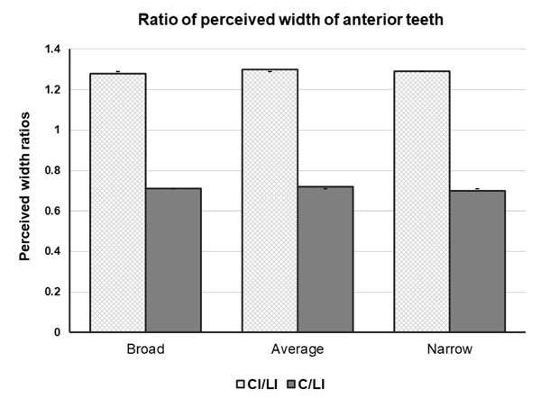
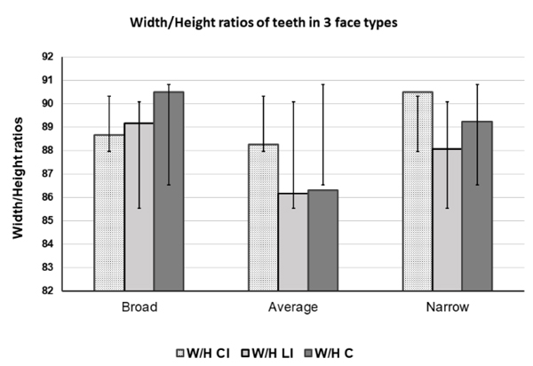
The studied teeth proportion showed significant differences with GP and GS (p<0.05). The mean teeth proportion in the total population included CI/LI 1.29 (0.13), C/LI 0.71 (0.08), W2CI/HCI 1.79 (0.11), CI W/H ratio 90.10 (5.39), LI W/H ratio 87.86 (6.71) and C W/H ratio 88.89 (5.74). All the data showed significant differences with GP and GS. No GP and GS were found in the studied population, as shown in Table 3 and Fig. (1). W/H ratio of the central incisor, lateral incisor, and canine showed no significant difference among 3 face types except W/H ratio between average and narrow face type (p<0.05) using One way ANOVA post-hoc: Gabriel Test as shown in Fig. (2).
| Facial Type | Gender | ||
| Male | Female | Total | |
| Broad | 6 (3%) | 2 (1%) | 8 (4%) |
| Average | 12 (6%) | 15 (7.5%) | 27 (13.5%) |
| Narrow | 49 (24.5%) | 116 (58%) | 165 (82.5%) |
| Total | 67 (34%) | 133 (66%) | 200 (100%) |
| Facial Measurement | Broad (n=8) |
Average (n=27) |
Narrow (n=165) |
p value (One-way ANOVA; post-hoc: Gabriel) |
||
| Mean (SD) |
Mean (SD) |
Mean (SD) |
Broad vs Average | Average vs Narrow | Broad vs Narrow | |
| Bizygomatic width (BZW) | 139.50 (2.95) |
130.40 (5.79) |
124.25 (6.56) |
.001* | <.001* | <.001* |
| Nasion-menton (NMH) | 114.28 (3.86) |
110.12 (5.65) |
113.10 (5.89) |
.182 | <.05* | .878 |
| Facial Index (FI): (NMH: BZW) |
81.90 (1.72) |
84.52 (1.71) |
90.92 (2.83) |
.035* | <.001* | <.001* |
| Teeth Proportion | Total studied Thai population (N=200) | ||
| Mean (SD) | p value (One-sample t-test with GPSL & GS) |
||
| CI/LI | 1.29 (0.13) | <0.001* | - |
| C/LI | 0.71 (0.08) | <0.001* | - |
| W2CI/HCI | 1.79 (0.11) | <0.001* | - |
| CI W/H (%) | 90.10 (5.39) | - | <0.001* |
| LI W/H (%) | 87.86 (6.71) | - | <0.001* |
| C W/H (%) | 88.89 (5.74) | - | <0.001* |
4. DISCUSSION
The Thai population is genetically diverse due to its different ethnicities and historical background. All the participating dental students of this present study came from different provinces of the country, representing the diverse Thai population. In this study, direct facial measurements were made, and FI was used to classify the facial types in other studies [29, 31, 32]. The predominant facial type in this study was a narrow face followed by an average face and broad face. This finding is similar to a study conducted in a Turkish population (173 subjects) where they found that 37.58% had a narrow face, 34.10% had a broad face and 28.33% had an average face [31]. Another similar study was conducted in a Nepali population, which showed 11 (7.33%) broad faces, 35 (23.33%), average faces, and 104 (69.33%) narrow faces [29]. A study conducted in a Bangladeshi population showed a narrow face was the most common facial type (56%), followed by an average face (44%) [33]. However, in their study, the broad face was not found. This all may indicate that a narrow face might be the most common facial type in Asian populations.
In this study, there was a significant difference in the BZW: IPD between broad and narrow, average, and narrow groups, because the BZW significantly differed between broad and average, average, and narrow and broad and narrow. A 3D electromagnetic study was conducted by Sforza et al. [34] to observe the size and form of faces of Italian adolescent boys and girls. Among all groups, a wider upper face than reference adolescents of the same age and sex was found; in both female groups and among young adolescent males significant differences were found. The maxilla was smaller among attractive girls than among their reference adolescents but larger among attractive boys relative to the mandible. Attractive adolescents had a smaller nose than reference subjects of the same age and sex. Ferrario et al. [35] found significant differences between television actresses and normal women while investigating the soft tissue facial landmarks. Larger foreheads, the larger middle third of the face, wider dimensions and deeper faces, smaller noses, and less facial convexity were observed in the television actress group. Longer lower one third and shorter upper two-thirds of the face were examined in the sample Thai population. The GP of the face was not found among normal people but was found in a special group of people who might have some history of surgical facial correction. According to other similar studies, no GP of the face was found in this study as we had excluded all kinds of esthetic facial correction in this research sample.
The neoclassical canon divides the face vertically into 5 equal spaces; all the studies conducted among white and Asian subjects used anthropometry and photogrammetric analyses, which manifested variations in these proportions, with the width of the eyes and nasal widths often being either less than or greater than the inner-canthi distance [36]. In this study, the vertical fifth of the face is equal in different types of faces except for the width of the eye on both sides showed significant differences in the average and narrow group and broad and narrow group. In addition, using the one-sample t-test, significant differences were found between the width of the nose and inner canthi distance, and between the width of the mouth and interpupillary distance (p<0.05).
In this present study, neither GP nor GS was found in teeth proportions similar to related studies [11, 18, 29, 30, 33, 37-39]. Beautiful smiles do not have these dental proportions, indicating some conflict with reality and the GP [38]. According to Mashid et al. [40] if we consider the occurrence of the GP in a broad range of 0.55 to 0.64, we would find that the GP in the average group was 2.5% and in the narrow group was 4.5% for CI/LI and, 0.5% in the broad group, 2.5% in the average group and 11% in the narrow group for C/LI. In the total studied population, GP was seen in 7% for CI/LI and 14% for C/LI. Regarding W2CI/HCI ratios, no GP was found in all three groups suggesting that Thai populations have wider central incisors than in the WCI using the GP. Similarly, for Nepalese, the GP and GS percentage were not found, and the Nepalese Esthetic Dental (NED) proportion results in an esthetically pleasing smile [29, 41]. The W/L ratio for CI, LI, and CN was found to be 90, 86, 86%, respectively, and a proportion of 66% for LI/CI and 70% for CN/LI were found. These can be used in the esthetic rehabilitation of the maxillary anterior teeth among Nepalese patients. In the present study, the obtained W/H ratios (90% for central incisor, 87% for the lateral incisor, and 88% for canine) could be helpful in clinical practice and in manufacturing artificial teeth.
Furthermore, the present study established a positive correlation between intercanine distance and intercanine tip distance with the BZW, interpupillary distance, nasal width, and mouth width, which was similar to the study conducted by Hasanreisoglu [32], where he found a positive correlation between the intercanine tip width and nasal width. Ellakwa et al. [42] found a weak positive correlation existed between intra-oral (WCI, canine to canine width) and extra-oral (intercanthal distance, interpupillary distance, interalar width, commissure of lip width) measurements that remained consistent when examined by gender. Similarly, al-El-Sheikh and al-Athel [43] in a Saudi population found a significant correlation between interalar, interpupillary and maxillary anterior teeth width for the entire sample and when the sample was divided by gender, correlation was found only among females. In our study, intercanine distance was 1/3.29 of the BZW, whereas intercanine tip distance was 1/3.48 of the BZW. These results were similar to the values given by Sears [44], reporting that the width of the total anterior teeth was 1/3.3 of the BZW. The results of our study also showed a linear positive correlation between WCI with the BZW revealing that the WCI was 1/14.02 of the BZW. This result was similar to the result obtained by Berry's biometric ratio method [45] and Pound's concept [46], where they proposed that the maxillary central incisor width was 1/16 of the width of the face or BZW. In addition, the WCI was 1/4.02 of the intercanine tip distance, 1/4.25 of the intercanine distance, 1/6.29 of the innerpupillary distance, 1/4.04 of the nose width and 1/5.11 of the mouth width. Thus, this could be useful in calculating the WCI.
Finally, in this study, some negligible amount of inaccuracy could have occurred during all kinds of measurements and cast fabrications.
CONCLUSION
Among the studied Thai subjects, 82.50% represented a narrow face, 13.50% represented average and 4% represented a broad face. No GP or GS was found compared with the 6 maxillary anterior teeth in the studied population. Anterior teeth measurements with different facial landmarks showed a significant correlation. However, no significant correlation was found in the WCI with interpupillary distance and alar width (p<0.05). The W/H ratio of central incisor, lateral incisor, and canine showed no significant difference with 3 face types except W/H ratios between average and narrow face types (p<.05). The size and shape of the maxillary anterior teeth were the most important factors to achieve pleasing dental and facial esthetics. The data obtained from this study may help to provide guidelines for prosthetic management with proper esthetic outcomes in Thai populations according to their facial proportions.
AUTHOR’S CONTRIBUTIONS
Dr. Asikul Wadud, Dr. M.L. Theerathavaj Srithavaj, and Dr. Amornrat Wonglamsam designed the research. Dr. Asikul Wadud performed the research, analyzed the data, and prepared the manuscript. Dr. M.L. Theerathavaj Srithavaj and Dr. Amornrat Wonglamsam supervised the research. Dr. Asikul Wadud analyzed the data. Dr. Jira Kitisubkanchana, Dr. Peerapong Santiwong, Dr. M.L. Theerathavaj Srithavaj, and Dr. Amornrat Wonglamsam reviewed the manuscript and gave an opinion.
ETHICS APPROVAL AND CONSENT TO PARTICIPATE
This study received approval from the Institutional Review Board of the Faculty of Dentistry, Mahidol University, Bangkok, Thailand.
HUMAN AND ANIMAL RIGHTS
No animals were used in this research. All human research procedures followed were in accordance with the ethical standards of the committee responsible for human experimentation (institutional and national), and with the Helsinki Declaration of 1975, as revised in 2013.
CONSENT FOR PUBLICATION
All participants signed the consent before participating in the study.
AVAILABILITY OF DATA AND MATERIALS
The authors confirm that the data supporting the findings of this research are available within the article.
FUNDING
None.
CONFLICT OF INTEREST
The authors declare they have no conflict of interest, financial or otherwise.
ACKNOWLEDGEMENTS
The authors would like to thank Associate Professor Nita Viwattanatipa, Associate Professor Amornrat Wonglamsam, Assistant Professor Sita Thaworanunta, Dr. Arunee Tirasriwat, Dr. Pokpong Amorvit, Dr. Dinesh Rokaya, Dr. Natdanai Chotprasert, Dr. Binit Shrestha, Dr. Ongart Puttipisitchet, Dr. Ornnicha Pooktuantong, and Phuangpaka Somtan, students and staff of Faculty of Dentistry, Mahidol University for their guidance and support in the conduct of this research. This research was supported by the Graduate Studies and Department of Prosthodontics, Faculty of Dentistry, Mahidol University, Thailand 10400, Thailand.

