All published articles of this journal are available on ScienceDirect.

Sodium Thiosulfate as a Neutralizing Agent: Implications for Dental Pulp Stem Cell Survival after Sodium Hypochlorite Irrigation
 iD
Arianti Amalia1
Dewa Ayu Nyoman Putri Artiningsih1
iD
Ike Dwi Maharti1
Romilda Rosseti1
iD
Valeria Widita Wairooy1
iD
Authors Info & Affiliations
Abstract
Introduction/Background
High concentrations of 5% sodium hypochlorite (NaOCl) reduce Human Dental Pulp Stem Cell (hDPSC) viability, despite its widespread use in endodontics. Sodium Thiosulfate (STS), an antioxidant with antimicrobial properties, may help reduce NaOCl-associated cytotoxicity. This study evaluated the effect of 5% STS following exposure to 1.5% and 5% NaOCl for different durations on hDPSC viability.
Materials and Methods
This in-vitro study was conducted at ProSTEM Laboratory (Prodia Stem Cell Indonesia), Jakarta, using primary hDPSCs from extracted human teeth. Groups were tested in triplicate and repeated twice by two operators. Cells (P3-P4,80% confluence) were starved for 24 hours and exposed to 1.5% and 5% NaOCl, followed by 5% STS for 30- and 60-second exposures. DMEM served as the control group, while the NaOCl and NaOCl-STS groups served as experimental groups. Cell viability was assessed using the MTT assay. Data were analyzed using one-way ANOVA (p < 0.05) with post hoc LSD test.
Results
The comparison between the 5% NaOCl group and the 5% NaOCl + 5% STS (60-second) group was not significantly different (p > 0.5). Descriptively, mean cell viability remained above 70% in all experimental groups.
Discussion
Within the limitations of this in vitro study, STS showed a possible trend toward higher hDPSC viability following 5% NaOCl exposure. All STS-treated groups maintained viability above the cytotoxicity threshold.
Conclusion
Although 5% STS demonstrated a slight numerical change in hDPSC viability, the difference was not statistically significant. These findings are a preliminary observation and require further study using optimized STS-to-NaOCl ratios.
1. INTRODUCTION
Modern inflamed pulp therapy prioritizes the regeneration of the pulp–dentin complex, aiming to restore pulp function, rather than merely replacing lost tooth structure [1, 2]. Effective disinfection of the root canal is essential, as pro-inflammatory cytokines such as IL-1 and TNF-α can inhibit the differentiation of stem cells. Human Dental Pulp Stem Cells (hDPSCs) play a crucial role in the regeneration process due to their capacity for proliferation and differentiation. In Regenerative Endodontic Treatment (RET), chemical irrigation is preferred over mechanical methods to minimize potential damage. Therefore, it is important that irrigants not only exhibit antimicrobial properties but also promote the viability and differentiation of stem cells [2-4].
Sodium hypochlorite (NaOCl) is commonly used as an endodontic irrigant, typically at concentrations ranging from 0.5% to 6%. It is a potent antimicrobial agent and is highly effective in dissolving organic tissue. However, its significant cytotoxicity to periapical tissues makes it important to use the lowest effective and biocompatible concentration [5, 6]. The American Association of Endodontists (AAE) recommends using a low concentration of 1.5% NaOCl for 5 minutes, as higher concentrations (5%–6%) have been shown to adversely affect the viability, proliferation, and differentiation of mesenchymal stem cells [1, 3, 4, 7]. However, the efficacy of low-concentration NaOCl in microbial elimination remains controversial. Interestingly, a study by Conde et al. [8] demonstrated successful outcomes in regenerative endodontic procedures using higher NaOCl concentrations of 2.5%, 5.25%, and 6% [3, 8, 9].
The success of regenerative endodontic treatment using high concentrations of NaOCl is likely attributed to its combination with adjunctive irrigation agents [10]. High-concentration NaOCl is known to provide superior antimicrobial effects compared to lower concentrations, such as 1.25%; however, its cytotoxic effects may compromise cell viability. In the study by Fouad et al. [11], 5% Sodium Thiosulfate (STS) was used for 5 minutes as part of the protocol to inactivate residual hypochlorite following irrigation with 5.25% NaOCl. Although its biological effects were not specifically evaluated, this approach suggests that STS may have a potential role in reducing the cytotoxic impact of NaOCl in regenerative endodontic procedures [12].
Sodium Thiosulfate (STS) is a water-soluble inorganic salt composed of two sodium ions and one thiosulfate ion. It possesses antioxidant, anti-inflammatory, and antihypertensive properties [13]. It has been increasingly investigated as an effective neutralizing agent for sodium hypochlorite (NaOCl) in microbiological and dental research [14, 15]. Nagata et al. [16] reported successful revascularization of immature teeth using 5% STS for 60 seconds following 6% NaOCl irrigation. As a potent antioxidant, STS neutralizes NaOCl by stabilizing reactive oxidants, with its effectiveness dependent on concentration and exposure time. Consistent with this, Yildiz et al. [17] demonstrated that 10-minute application of 5% STS effectively restored the bond strength of the sealer to NaOCl- treated dentin. Similarly, a laboratory investigation found that 5% STS reversed the adverse effects of 2.5% NaOCl on bonding and increased bond strength to pulp chamber dentin [18]. Collectively, these findings highlight the potential of STS to mitigate adverse effects of NaOCl in endodontic procedures. However, its effect on the viability of hDPSCs following NaOCl exposure remains unclear.
Chemical disinfection in regenerative endodontic treatment presents challenges due to the variability in irrigation protocols, which can impede tissue regeneration [1, 19, 20]. Persistent periapical pathosis, often associated with residual bacteria and biofilms, has been identified as a major cause of treatment failure. While strong antimicrobial action is crucial, preserving the viability of Dental Pulp Stem Cells (hDPSCs) is equally important. Due to the limitations of individual irrigants, combination strategies are often explored. However, the effect of Sodium Thiosulfate (STS) application following NaOCl irrigation on hDPSC viability remains unclear. This study tests the null hypothesis that STS exposure following NaOCl irrigation does not affect hDPSC viability.
2. SUBJECTS AND METHODS
This study was approved by the Dental Research Ethics Committee, Faculty of Dentistry, Universitas Indonesia, Indonesia, on February 13, 2023 (Approval No. 03/Ethical Approval/FKG UI/II/2023; Protocol No. 050050123). This research was carried out at the ProSTEM (Prodia Stem Cell Indonesia) laboratory in Jakarta, Indonesia. Two operators performed the experimental procedures.
2.1. Dental Pulp Stem Cell Culture
This study used primary hDPSCs obtained from extracted human third molars following ethical approval. The teeth were extracted due to impaction and were free of caries. The hDPSCs were isolated from nine healthy donors (male and female) aged 18-25 years. The donors met the inclusion criteria, which included good general health, the absence of systemic disease, and no history of smoking or alcohol consumption. All experimental groups were prepared in biological triplicate, and the experiments were conducted in three independent experiments and repeated twice, in accordance with ISO 10993-5.
The hDPSCs at passages 3-4 were incubated in a humidified 5% CO2 atmosphere at 37°C until reaching 80% confluence. Subsequently, the cells were subjected to starvation by replacing the cell culture supplement with DMEM (Thermo Fisher Scientific Inc., Waltham, Massachusetts, United States) supplemented with 5% fetal bovine serum (Thermo Fisher Scientific Inc.) for 24 hours. After serum starvation, the hDPSCs were divided into the following groups: (1) control group: hDPSCs + DMEM; (2) experimental groups: hDPSCs + 1.5% NaOCl, hDPSCs + 5% NaOCl, hDPSCs +1.5% NaOCl + 5% STS (30-second exposure), hDPSCs + 1.5% NaOCl + 5% STS(60-second exposure), hDPSCs + 5% NaOCl + 5% STS (30-second exposure), and hDPSCs + 5% NaOCl + 5% STS (60-second exposure).
The irrigation protocol was performed as follows: 0.1 mL of sodium hypochlorite (NaOCl; One Med; lot number: 3101238) at either 1.5% or 5% was applied to the hDPSCs for 30 seconds. Following NaOCl exposure, the cells were rinsed with PBS, after which 0.1 mL of 5% Sodium Thiosulfate (STS; Merck; catalog number 1.06516.1000; lot number: HC27403547) was applied to each well for either 30 or 60 seconds, according to the experimental group. The 5% STS solution was prepared by dissolving 5 grams of sodium thiosulfate pentahydrate in distilled water to a final volume of 100 mL. After the respective irrigation treatment, PBS was used as the final rinse. The hDPSCs were placed in a96-well tissue culture plate at 5 × 10^3 cells/well and incubated at 37°C in 5% CO2 for 24 hours. All treatments were carried out in a biosafety cabinet.
2.2. MTT Assay
After incubation, cell viability was assessed using the MTT assay, with absorbance measured at 450 nm using an ELISA plate reader. The resulting Optical Density (OD) values were recorded and used for quantitative analysis. In addition, a qualitative assessment was performed through microscopic observation of the cells at 100x magnification.
2.3. Statistical Analysis
The data were analyzed using one-way Analysis of Variance (ANOVA), at a significance level of α < 0.05, followed by the post hoc LSD test. In cases of non-normal data distribution, the non-parametric Kruskal-Wallis test was used, followed by the Mann-Whitney U post hoc test. All data were analyzed using IBM SPSS version 25.0 (IBM Corp., Armonk, NY, United States).
3. RESULTS
In this study, laboratory experimental tests were conducted to compare hDPSCs viability following the addition of STS to different concentrations of NaOCl used as endodontic irrigants. The study consisted of seven groups: Group I was the control group (DMEM); Groups II and III were treatment groups without STS (1.5% and 5% NaOCl solution); and Groups IV-VII were experimental groups exposed to 1.5% and 5% NaOCl followed by 5% STS for 30 or 60 seconds.
In this study, the OD value (Optical Density) was measured with a microplate reader at a wavelength of 450nm. After that, the cell viability value is calculated according to the following formula:

The results of the mean viability value are shown in Table 1, which shows that the highest mean viability was in the 1.5% NaOCl group and the lowest in the 5% NaOCl group. The mean viability in the 1.5% NaOCl treatment group appeared to decrease, while in the 5% NaOCl treatment group, it appeared to increase.
| Group | N | Mean Viability±SD (%) | P-value |
|---|---|---|---|
| DMEM NaOCl 1.5% NaOCl 1.5%+STS 5% 30s NaOCl 1.5%+STS 5% 60s NaOCl 5% NaOCl 5% + STS 5% 30s NaOCl 5% + STS 5% 60s |
6 6 6 6 6 6 6 |
100 ± 0 95.55 ± 11,41 92.82 ± 12,38 86.00 ± 6,77 76.50 ± 8,00 80.82 ± 7,84 84.02 ± 8,84 |
0.000* |
Test results of One-way ANOVA in Table 1 show that there are significant differences in the viability values of hDPSCs between groups. In Fig. (1E-H), the viability values are significantly different between the DMEM groups with NaOCl 5% (p < 0.01), NaOCl 1.5%-STS 5% 60-second exposure (p < 0.05), and NaOCl 5%-STS 5% 30-second exposure (p < 0.05). No statistically significant differences were observed between the 1.5% and 5% NaOCl groups and the corresponding groups treated with 5% STS. In this study, a qualitative assessment of the microscopic image was also carried out at a magnification of 100 µm. The results of the microscopic image are shown in Fig. (1).
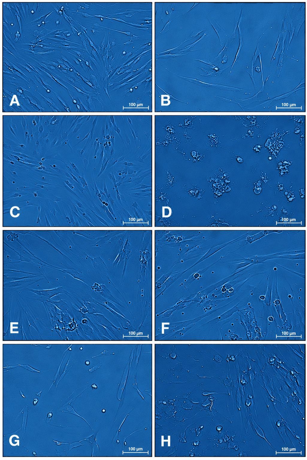
Microscopic image at 100 µm magnification: (A) DMEM, (B) DMEM after starvation, (C) NaOCl 1,5%, (D) NaOCl 5%, (E) NaOCl 1,5% + STS 5% 30 s, (F) NaOCl 1,5% + STS 5% 60 s, (G) NaOCl 5% + STS 5% 30s, (H) NaOCl 5% + STS 5% 60 s.
4. DISCUSSION
Root canal disinfectants are intended to eliminate microorganisms and neutralize their by-products while minimizing damage to host tissues. Although the combination of sodium hypochlorite (NaOCl) and Sodium Thiosulfate (STS) is recognized for its antimicrobial efficacy, its effects on host cell viability remain unclear. Given the known cytotoxicity of sodium hypochlorite (NaOCl), 5% STS has been used in previous protocols to inactivate residual hypochlorite following irrigation [11]. However, its effect on cell viability has not been specifically evaluated. Therefore, this study aimed to evaluate and compare the effect of 5% STS following irrigation with either 1.5% or 5% NaOCl on the viability of Human Dental Pulp Stem Cells (hDPSCs). The American Association of Endodontists (AAE) currently recommends using 1.5% NaOCl for 5 minutes, as higher concentrations (5–6%) have been shown to impair the viability, proliferation, and differentiation of mesenchymal stem cells [1, 21].
The results of this study demonstrated a numerical reduction in hDPSC viability following exposure to both NaOCl concentrations, with mean viability values of 95.6% and 76.5% for the 1.5% and 5% groups, respectively. Although the difference in viability between these two groups was not statistically significant compared to the control, the 5% NaOCl group exhibited a descriptive trend toward reduced cell viability. These findings are consistent with those of Ayoub et al. [3] and Li et al. [7], who also reported that higher concentrations of NaOCl negatively affect hDPSC viability [1, 3, 7, 22].
The results of this study showed that the mean cell viability in all groups remained above 70%, indicating that the tested materials were not cytotoxic. The 5% NaOCl group showed the lowest viability (76.5%), suggesting a greater reduction in cell viability than the other groups, although it remained above the threshold. The highest viability in the groups without STS was observed in the 1.5% NaOCl group (95.55%), while in the STS-treated groups, the highest viability was found in the 1.5% NaOCl + 5% STS 30-second exposure group (92.82%). According to ISO 10993-5, a viability value above 70% indicates that cells remain viable, confirming that the irrigation solutions used did not exert toxic effects [23, 24]. However, microscopic observations revealed morphological changes and a reduction in cell number in all test groups compared to the DMEM control group (Fig. 1). These changes may be attributed to NaOCl-induced cellular stress and metabolic disruption, potentially resulting in mitochondrial damage, cell shrinkage, and apoptosis in dental pulp stem cells [20]. Furthermore, NaOCl has been shown to decrease cellular metabolic activity, disrupt lysosomal integrity, disrupt cytoskeletal structures, and cause mitochondrial ultrastructural alterations, ultimately leading to apoptosis and necrosis in fibroblasts [25]. Although all test groups had hDPSC viability values lower than those of the DMEM control group, they remained above the 70% viability threshold. This indicates that while all tested irrigation materials had some negative impact on cell viability, they were not cytotoxic.
This study found no statistically significant difference in hDPSC viability between the NaOCl groups with and without STS. A 5% STS concentration was used based on previous studies- Yildiz et al. [17] demonstrated its ability to restore dentin bond strength after NaOCl exposure, and other studies reported no cytotoxic effects on fibroblasts [16]. An exposure time of 60 seconds was adopted from the study by Nagata et al. [16], which showed favourable outcomes in revascularization procedures. However, in the present study, using 5% STS for 30 or 60 seconds did not significantly neutralize the effects of 1.5% or 5% NaOCl. This outcome may be explained by volume limitations; according to Shelke et al. [26], a minimum of 3.5 mL of STS is required to effectively neutralize 5% NaOCl. In the present study, only 0.1 mL could be applied due to constraints imposed by the 96-well plate capacity. Nevertheless, evaluating this preliminary micro-scale setup provides initial insight into the baseline response of hDPSCs, offering a practical reference for future dosage optimization.
In this study, although overall changes were not statistically significant, distinct numerical trends were observed across the tested NaOCl concentrations. In the 5% NaOCl groups, mean viability increased numerically following the addition of STS, from 76.50% without STS to 80.82% and 84.02% following 30- and 60-second exposure to STS, respectively. In contrast, in the 1.5% NaOCl groups, mean viability decreased numerically from 95.55% without STS to 92.82% and 86.00% following 30- and 60-second STS exposure, respectively. This discrepancy can be explained by the stoichiometric interplay between NaOCl and STS. While 1.5% NaOCl yielded high baseline cell viability, the addition of 5% STS resulted in unreacted sodium thiosulfate. Exposure to this high concentration of antioxidant likely subjected the remaining cells to osmotic stress, leading to dehydration, shrinkage, and a consequent decline in viability [17]. These diverging responses demonstrate that the specific concentration ratio of STS to NaOCl is critical to maximizing the neutralizing efficiency of STS while avoiding unintended cytotoxic effects.
Based on the findings of this study, the null hypothesis stating that the addition of 5% Sodium Thiosulfate (STS) following NaOCl irrigation does not significantly affect hDPSC viability cannot be rejected. Although changes in mean cell viability were observed following STS treatment, these differences were not statistically significant. Notably, STS was associated with a slight numerical increase in viability in the 5% NaOCl group, whereas a decrease was observed in the 1.5% NaOCl group. The observed decrease in viability in the 1.5% NaOCl + STS group may be attributed to a relatively high STS concentration relative to the lower oxidative strength of 1.5% NaOCl, or possibly to prolonged exposure to STS, which may have resulted in unintended effects on cell viability. However, these possibilities require further investigation.
While STS is known to act as an antioxidant and NaOCl neutralizer, its impact in this study appeared limited, which may be attributed to methodological constraints. These include the fixed STS concentration and volume (restricted to 0.1 mL per well), limited variation in exposure times (30 and 60 seconds), and the absence of a standalone 5% STS control group. Furthermore, the absence of viability staining might have reduced the accuracy of qualitative assessments. Nevertheless, all test groups maintained cell viability above the cytotoxic threshold of 70% as defined by ISO 10993-5. Although the current STS protocol did not demonstrate a statistically significant enhancement in cell viability, numerical changes in viability were observed following 5% STS treatment after 1.5 and 5% NaOCl exposure for 30 and 60 seconds. These findings provide preliminary evidence supporting further investigation into the potential role of STS as a NaOCl-neutralizing agent in regenerative endodontic irrigation under optimized experimental conditions.
CONCLUSION
A numerical increase in hDPSC viability was observed in the 5% NaOCl groups after treatment with 5% sodium thiosulfate, suggesting a possible trend toward a beneficial effect of STS as a NaOCl-neutralizing agent in regenerative endodontic irrigation. Cell viability remained above the 70% threshold defined by ISO 10993-5 in all experimental groups. Future studies should explore optimized STS concentrations, volumes, and exposure times, alongside more detailed viability assessments to better support its use in regenerative endodontic protocols.
AUTHORS’ CONTRIBUTIONS
The authors confirm their contributions to the paper as follows: B.D.A., A. D.A.: Study concept or design; A.A.: Data collection; A.A.: Writing the paper; M.I.: Writing–original draft preparation; R.R., W.V.: Writing –reviewing and editing. All authors reviewed the results and approved the final version of the manuscript.
LIST OF ABBREVIATIONS
| hDPSCs | = Human Dental Pulp Stem Cells |
| NaOCl | = Sodium Hypochlorite |
| STS | = Sodium Thiosulfate |
| PBS | = Phosphate-Buffered Saline |
| DMEM | = Dulbecco's Modified Eagle Medium |
| MTT | = 3-(4,5-Dimethylthiazol-2-yl)-2,5-diphenyltetrazolium Bromide |
| ISO | = International Organization for Standardization |
ETHICS APPROVAL AND CONSENT TO PARTICIPATE
This research was approved by the Dental Research Ethics Committee, Faculty of Dentistry, Universitas Indonesia, Indonesia, on February 13, 2023 (Approval No. 03/Ethical Approval/FKG UI/II/2023; Protocol No. 050050123)
HUMAN AND ANIMAL RIGHTS
All procedures involving human participants were conducted in accordance with the ethical standards of the committee responsible for human experimentation (institutional and national), and with the Helsinki Declaration of 1975, as revised in 2013.
CONSENT FOR PUBLICATION
Informed consent was obtained from all participating adult subjects prior to the study.
AVAILABILITY OF DATA AND MATERIALS
The data sets used during the current study can be provided from the corresponding author [B.D.], upon reasonable request.
ACKNOWLEDGEMENTS
The authors would like to thank Dr. AC, S.Si, M.Farm., ANA, S.Si, and RA, S.Si from the Prodia Stem Cell Laboratory in Indonesia for their support.