All published articles of this journal are available on ScienceDirect.

Nanoparticle-modified Acrylic Resin Denture Bases for Controlling Candida Species Growth, Adhesion, and Biofilm Formation: A Systematic Review
 iD
Shahad Mohammed Shakir2
Mohammed Ali Faris Ahmed3
Fatanah Mohamad Suhaimi4
iD
Shahad Mohammed Shakir2
Mohammed Ali Faris Ahmed3
Fatanah Mohamad Suhaimi4
Authors Info & Affiliations
Abstract
Introduction/Objective
The colonization of Candida species on polymethyl methacrylate (PMMA) denture bases is a major contributor to denture stomatitis and continues to be a prevalent clinical problem. The purpose of this systematic review was to assess the efficacy of using organic and inorganic nanoparticles in acrylic resin denture bases to control the growth of Candida, its adhesion, and biofilm formation. This study aimed to determine whether adding nanoparticles would alter the material's mechanical and aesthetic properties.
Methods
In line with PRISMA, a systematic search was conducted across Web of Science (WOS), PubMed/MEDLINE, Scopus, Google Scholar, the Cochrane Library, and Embase for peer-reviewed in vitro studies published from January 2014 to February 2025. Combinations of words used in the search included “acrylic resin”, “denture base”, “nanoparticles”, “nanomaterials”, “Candida”, “antifungal,” and “biofilm”. The eligibility and selection of studies were then done tightly based on the PICO criteria.
Results
28 studies were identified after screening 1,430 records and met the strict inclusion criteria. Metallic particles (Ag and Cu), metal oxides TiO2, ZnO, ZrO2, SiO2, and CuO, and even bimetallic (Ag-ZnO) and carbon-based (nanodiamonds) were found to be antifungal and antibiofilm with a dose-dependent effect. The ability of silver nanoparticles (AgNPs) in general, and TiO2 in particular, to suppress the growth of Candida biofilm was fairly consistent at concentrations in the range of 0.5 to 1.0 wt%. With higher concentrations, metal nanoparticles sometimes caused decreased flexural strength, reduced surface hardness, and esthetic discoloration. Nevertheless, the use of low concentrations (0.5-1.0 wt%) of nanodiamonds and silanized ZrO2 was found to effectively inhibit fungal growth while not affecting the denture base structure.
Discussion
The main mechanisms of action appear to be the continuous release of ions, the generation of reactive oxygen species (ROS), and the disruption of the fungal cell membrane, which, in practice, physically limits biofilm adhesion. However, biocompatibility can be an issue; nanoparticles can aggregate, and there is an increased risk of cytotoxicity at high concentrations.
Conclusion
Adding a low concentration of silanized or hybrid nanoparticles (1 wt% AgNPs, TiO2, or ZrO2) would be a promising method to prevent denture stomatitis by limiting Candida growth on acrylic resin denture bases. For these nanocomposites to be routinely applied in dental care, standardized clinical trials are necessary, and long-term biocompatibility testing should be conducted.
1. INTRODUCTION
The oral cavity is a complex environment hosting hundreds of distinct microbial species essential for maintaining local physiological balance; disruptions, such as systemic immunodeficiencies, nutritional problems, or poor oral hygiene, can disturb this equilibrium, increasing the host's susceptibility to opportunistic infections [1]. Candida albicans is a commensal microfungus that is part of the normal human oral microflora and naturally resides on mucosal surfaces [2]. This opportunistic yeast is capable of assuming a pathogenic hyphal form and causing oral candidiasis in conditions where the local or systemic immune system is impaired [3].
Denture stomatitis is a localized, chronic inflammatory condition associated with oral candidiasis and is a common clinical manifestation [4]. The proportion of denture wearers is high, with epidemiological data showing that about 17% to 75% of wearers develop denture stomatitis, with a slight tendency for older females [5]. In Iraq, it is reported that 30% to 55% of complete denture users are affected [6]. The relatively high prevalence, both worldwide and in Iraq, is mainly attributable to inadequate denture cleaning, overnight wear without removal, and the formation of fungal biofilms on the porous, somewhat hydrophobic surfaces of polymethyl methacrylate denture bases [7, 5].
The physical surface features of the denture base, such as the average surface roughness and surface wettability, directly influence early microbial plaque formation and subsequent biofilm development. A few small bumps and grooves may be retained on rough PMMA surfaces, providing small hiding places where microorganisms are harder to remove with regular brushing or other mechanical hygiene, making biofilm removal much more challenging. Therefore, the clinicians typically seek a denture base roughness average of 0.2 μm or less to minimize microbial adhesion. Furthermore, if the surface is strongly hydrophobic, that is, low wettability, it helps the C. albicans cell wall to quickly attach to the acrylic surface and to begin to colonize the surface by fixing a first hydrophobic link between the two [8].
Topical or systemic antifungal medicines are often used to treat denture stomatitis. In practice, however, their efficacy is frequently poor due to rapid dilution in the saliva, noncompliance with drugs, and drug resistance by Candida strains [5, 9]. Therefore, recent developments in materials science have focused on incorporating organic and inorganic nanomaterials into PMMA to develop self-disinfecting denture bases [10].
Metallic nanoparticles such as silver (AgNPs) and copper (CuO) have been reported to exhibit broad-spectrum antimicrobial activity. The suggested mechanisms are the disruption of microbial cell membranes and the generation of oxidative stress within cells [9, 11, 12]. What's more, the use of metal oxide nanoparticles such as TiO2, ZnO, and ZrO2 has been widely studied. They have been recognized as beneficial for reducing biofilm formation and enhancing the mechanical properties of PMMA [13, 14].
There are also carbon-derived nanostructures, such as detonation nanodiamonds (NDs). These have received considerable attention for their stability and lack of cytotoxicity, and in many reports they do not adversely affect the integrity of the polymer matrix [15].
Although the use of these nanoparticles appears to be a promising way to control fungal biofilms, a balance needs to be struck between optimal antifungal activity and the mechanical, esthetic, and biocompatibility properties of the denture base. Therefore, this systematic review will synthesize recent literature on the in vitro efficacy of nanoparticle-reinforced acrylic denture bases against Candida species. Particular emphasis is placed on structural trade-offs, clinical translatability, and mechanisms of action.
2. MATERIALS AND METHODS
2.1. Main Question
The main question was whether adding nanoparticles (Intervention), compared to plain, unmodified acrylic denture bases (Population), reduces the growth and biofilm accumulation of Candida species (Outcome).
2.2. Outcome Measures
The effect of combining nanoparticles into acrylic resin denture bases on the antifungal property.
2.3. Searching Strategy
Relevant studies in English published between January 2014 and February 2025 were identified through searches of WOS, PubMed/MEDLINE, Scopus, Google Scholar, the Cochrane Library, and Embase. For the literature search, the following search phrases and Boolean operators were used: (“acrylic denture base” OR “denture base” OR “PMMA”) AND (“nanoparticles” OR “nanomaterials” OR “nano oxides”) AND (“Candida albicans” OR “antifungal effect” OR “biofilm inhibition”); the reference lists of eligible studies were manually screened to identify potentially relevant studies that may not have been retrieved through the electronic search.
2.4. Selection Standards
In vitro studies that added nanoparticles, nanofillers, or nano oxides to the acrylic denture base and compared their antifungal effect to plain acrylic denture base were included in this review.
2.5. Criteria for Inclusion
Studies involving the incorporation of nanoparticles into acrylic denture base in vitro, studies contrasting with plain acrylic denture base, and studies examining the impact of incorporating nanoparticles on the antifungal properties of acrylic denture base were all considered when choosing which studies to include. Studies that qualified had to be peer-reviewed and show academic indices. They also needed to publish quantitative findings on fungal colony-forming units (CFU/mL) or clearly report metabolic activity. On top of that, there had to be an explicit report of the nanoparticle concentration, given as wt%, and not just something vague.
2.6. Criteria for Exclusion
The exclusion criteria include articles comparing different brands of acrylic denture base material that are sold on the market, as well as articles examining the color stability and other biological as well as physical characteristics of acrylic denture base mixed with other fillers, coloring agents, and pigments. When reviewing the PRISMA screening process, about 1,200 articles were excluded because they did not focus on nanoparticle modifications, were duplicates, or focused on non-acrylic materials. In addition, 20 reports could not be retrieved because they were behind restricted-access abstracts or because the full text was not available online, even after direct outreach to the corresponding authors. Figure 1 shows the PRISMA diagram.
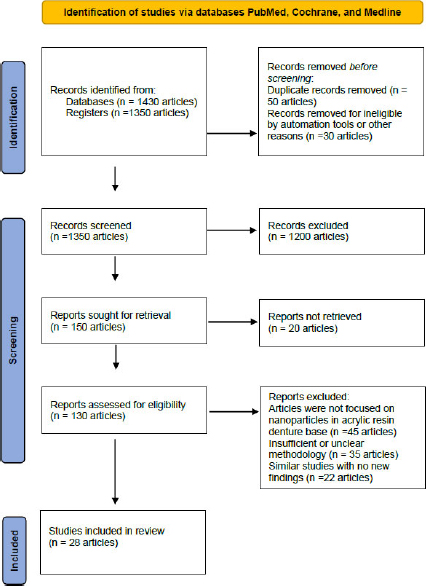
PRISMA diagram illustrating the screening, selection process, and workflow of the systematic review on nanoparticle-modified acrylic resin denture bases for controlling Candida species growth.
2.6.1. Risk of Bias Assessment
The methodological quality and risk of bias of the included in vitro studies were evaluated using a modified version of an established assessment tool for dental materials, adapted from earlier systematic reviews. The criteria examined a number of important parameters, including clarity of reporting sample size, standardization of specimen preparation, presence of a control group, standardized inoculation of Candida species, and blinding of the outcome assessors. For each parameter, the risk of bias was then categorized as low, high, or unclear.
2.7. Data Extraction
The first author carried out the data extraction, while the other authors reviewed it. Each full-text article that met the requirements for inclusion underwent independent data extraction, which was carried out in an electronic format following a predetermined process (Office Excel 2024 software, Microsoft Corporation).
3. RESULT
28 studies met the strict inclusion criteria, primarily focusing for how nanoparticle-based modifications could improve resistance to microfungal growth. Table 1 lists the key parameters, including nanomaterial type and concentration, denture base types, the specific target microorganisms, and the main findings from the included studies examining antifungal effects when nanoparticles are incorporated into acrylic denture bases.
| Ref. | Year | Nanomaterial Type and Concentration | Denture Base/microorganism | Findings and Mechanisms of Action |
|---|---|---|---|---|
| [16] | 2014 | Ag NPs | Heat-cured PMMA / C. albicans | For immunocompromised and elderly individuals, the antibacterial activity of silver might be employed in the denture base. |
| [17] | 2016 | Ag NPs AgVO3 | Acrylic resin / S. mutans, C. albicans | The suppression of the principal bacteria linked to dental prosthesis’ biofilm may be impacted by the addition of AgVO3 to dental acrylic resin. |
| [18] | 2017 | AgBr/cationic polymer nanocomposites | Heat-cured PMMA / C. albicans | Incorporating silver bromide/cationic polymer nanocomposites into heat-cured PMMA showed high contact-active antifungal properties against C. albicans without compromising essential mechanical characteristics. |
| [19] | 2017 | Silanized Zinc Oxide (ZnO) NPs (1.0-2.5 wt%) | Heat-cured PMMA / C. albicans | PMMA modified with 2.5 wt% silanized ZnO NPs exhibited stronger antifungal efficacy and less color change compared to non-silanized ZnO, while maintaining mechanical properties. |
| [20] | 2019 | Nanodiamonds (NDs) | PMMA resin / C. albicans | PMMA-ND composites may help prevent denture stomatitis, which is considered one of the most prevalent clinical issues among people who wear removable dentures. |
| [21] | 2025 | Fungal-mediated Ag NPs | Acrylic resin / Candida species | Silver nanoparticles biosynthesized via fungal pathways demonstrated potential broad-spectrum antifungal properties when integrated into denture bases, effectively inhibiting fungal biofilm growth. |
| [22] | 2021 | Ag NPs (0.05% and 0.5%) | Acrylic resin / C. albicans | It was determined that adding SNP to acrylic resin at concentrations of 0.05 and 0.5% had antibacterial effects against the C. albicans biofilm, did not affect the material's flexural strength, and could be regarded as biocompatible. |
| [23] | 2021 | Ag NPs | Denture base resin (DBRs) / Oral bacteria | DBRs include nanoparticles that inhibit bacterial growth. |
| [24] | 2021 | ZrO2 (1 wt%) | PMMA denture base / C. albicans | ZrO2 NPs were added to the polymethylmethacrylate denture base material to inhibit Candida albicans adherence and provide a long-lasting antifungal action. Surface roughness and contact angles reduced with the addition of ZrO2 NPs; 1% was the ideal concentration. |
| [25] | 2021 | Nano-ZrO2 | PMMA / C. albicans biofilm | When varying quantities of nano-ZrO2 particles were added to PMMA, surface roughness increased relative to the control; however, a negligible decrease in the C. albicans biofilm was observed. |
| [26] | 2021 | Nano-SiO2 | PMMA resin / C. albicans | C. albicans adherence to PMMA denture base resin was reduced with the addition of nano-SiO2. |
| [27] | 2021 | CuO and TiO2 (7.5 wt%) | PMMA / S. salivarius, S. sanguis, C. dubliniensis | Against S. salivarius, S. sanguis, and C. dubliniensis, both concentrations of CuO and TiO2 were efficient antibacterial agents, whereas the concentration of CuO was effective against S. mutans. Just 7.5% TiO2 demonstrated effective antibacterial action against C. albicans. |
| [28] | 2024 | Nystatin-coated CuO particles (1, 2, 4%) | PMMA-based denture material / C. albicans | Incorporation of nystatin-coated CuO particles into PMMA demonstrated antifungal activity against C. albicans, while also influencing the mechanical and thermal properties of the resulting denture-base composites. |
| [29] | 2022 | Ag NPs | Modified PMMA / C. albicans | The presence of infection at the site of contact between the denture and the denture-support mucosa may be greatly reduced with modified PMMA materials. |
| [30] | 2023 | Ag NPs and TiO2 (0.5% - 1%) | Acrylic resin discs / C. albicans clinical isolates | Both 1 wt% AgNPs and 1 wt% TiO2 NPs exhibited higher antimicrobial and antibiofilm effects, resulting in a notable reduction of the colony counts and biofilm biomass. |
| [13] | 2023 | AgVO3 (2.5%, 5%, 10%) | Heat-cured resin / S. mutans, C. albicans, C. glabrata | Incorporating 10% AgVO3 provided antimicrobial activity against S. mutans in a multispecies biofilm, but reduced the flexural strength of the heat-cured PMMA. |
| [11] | 2023 | CuO NPs (0.5 to 500 μg/mL) | Heat-polymerized PMMA / Gingival Fibroblasts | CuO NPs at lower concentrations (up to 50 μg/mL) were biocompatible, substantially increased the flexural strength of PMMA, and had no adverse effect on surface roughness. |
| [14] | 2025 | TiO2 NPs (0.10, 0.25, 0.50, 0.75 wt%) | 3D-printed resin / C. albicans | TiO2 NPs at 0.10-0.50 wt% significantly reduced C. albicans adhesion. Lower concentrations showed more homogeneous nanoparticle dispersion, whereas aggregation occurred at higher concentrations. |
4. DISCUSSION
Fungal infections have frequently increased among the elderly and immunocompromised populations. Two reasons include antimicrobial drug resistance and restrictions on their use due to adverse effects [31]. One example of an oral fungal infection brought on by Candida albicans is denture stomatitis. Many antifungal compounds and products have been developed in response to the growing demand for innovative antifungal options due to the increasing frequency of denture-induced stomatitis [4].
Denture adhesives, denture liners, and heat-cured and cold-cured acrylic resin have all been enhanced with a number of antifungal nanostructured polymers, according to many articles and studies in the literature. The inclusion of nanoparticles and their antibacterial qualities into acrylic base materials for removable dentures was identified in this study. Among the added nanoparticles are silver, zinc oxide, titanium dioxide, zirconium dioxide, silicon dioxide, copper oxide, and nanodiamonds. This made it possible to create a modified denture foundation with antifungal properties that would slow or prevent Candida albicans biofilm from forming on denture surfaces. Nanomaterials were shown to have a substantial inhibitory and reducing effect on Candida albicans accumulation and growth on denture acrylic resin. Because of their low cost and attractive appearance, heat-cure acrylic resins are often used to fabricate complete dentures and removable partial dentures [32].
The antibacterial performance of nanoparticles incorporated into acrylic denture base materials is strongly influenced by their composition and concentration. Usually used at 0.05-1 weight%, silver nanoparticles are metallic silver that releases Ag+ ions, thereby disrupting bacterial membranes, interfering with cellular respiration, and causing oxidative stress. Ag NPs penetrate bacterial biofilms and stop microbial attachment to denture surfaces, therefore displaying great antibacterial action [22]
When incorporated at 2-5 weight percent, zinc oxide nanoparticles are semiconductor metal oxides that generate reactive oxygen species (ROS), including hydrogen peroxide and superoxide anions, thereby compromising bacterial membrane integrity. Effective denture materials, ZnO NPs are well-known for their antibacterial, antifungal, and UV-shielding qualities [19]
Commonly utilized at 0.5-3 weight percent, titanium oxide nanoparticles (TiO2 NPs) are made of TiO2, a transition metal oxide with excellent photocatalytic performance. TiO2 generates hydroxyl radicals (OH2−) and superoxide anions (O2−) that destroy bacterial cell structures during UV exposure, therefore halting microbial colonization [27].
When added at 1-3 weight percent, zirconium oxide nanoparticles (ZrO2 NPs) consist of ZrO2, a ceramic oxide that reduces surface roughness and bacterial adhesion, rather than relying on ion release, thereby limiting biofilm development. Research shows that, despite preserving mechanical strength, ZrO2 NPs incorporated into denture bases exhibit long-term antifungal activity [24].
Used at 1-5 weight percent, copper oxide nanoparticles (CuO NPs) contain Cu2+ ions that damage bacterial proteins and enzymatic pathways, thereby generating oxidative stress and bacterial cell death. The strong bactericidal action of CuO NPs makes them a potential antibacterial ingredient in dental materials [27].
When included at 0.5-2 weight percent, nanodiamonds (NDs) are carbon-based nanoparticles with sp3-hybridized carbon structures and functional groups such as hydroxyl (-OH) and carboxyl (-COOH), which physically and electrostatically interact with bacterial cell walls, thereby reducing microbial adherence. Furthermore, improving the mechanical properties of denture bases is are ND, which provides better resistance to microbial colonization and durability [20].
These nanoparticles are carefully chosen and concentrated to provide antibacterial effects while preserving the mechanical and cosmetic integrity of the acrylic denture base material. These nanoparticles greatly enhance the antifungal and antibacterial activity of denture bases, thus helping to avoid diseases such as denture stomatitis [23].
Antimicrobial substances should not take away from the prosthesis's appealing appearance when they are covered. Due to scratches from mechanical cleaning, the denture surface may gradually lose its optical properties. These dents will serve as a breeding ground for germs, which will discolor the area. The ophthalmic properties of the acrylic resin denture material are affected differently by the introduction of antifungal drugs. Glass fillers and monomers change the color of the denture, while Ag NPs and natural extracts make it gray, and TiO2 nanofillers make it white [28]. These materials may be used to conceal the less-visible lower lingual and posterior denture base sections. The addition of nanomaterials to denture base materials substantially improved the development of denture base material, but the amount of the mentioned material has to be looked at to avoid endangering the optical, physical, and biocompatibility properties of the base denture material [33]. To determine the effects of adding more nanoparticles to various denture base materials, adhesives, and reline materials, more in-vivo and experimental research that mimics oral circumstances is required.
4.1. Pathogenesis of Candida albicans Adhesion to Denture Bases
The pathogenesis of denture stomatitis begins with the early adhesion of yeast cells to the PMMA denture base. This initial physical bond appears to be made by nonspecific interactions such as electrostatic attraction and hydrophobic forces, and by specific fungal adhesins, such as the Als and Hwp1 protein families, which bind to the salivary glycoprotein pellicle that forms over the acrylic resin, and is then followed by a change to a more aggressive hyphal phase. This switch facilitates mechanical penetration of tissues; it also promotes the secretion of extracellular polymeric substances, thereby allowing the biofilm to mature. The mature biofilm then acts as a shielding wall, a protective barrier that protects the fungal community from normal oral hygiene measures [5].
4.2. Detailed Antifungal Mechanisms and Subsections per Nanoparticle type
4.2.1. Silver Nanoparticles and Silver Zeolites
The antifungal activity of silver nanoparticles is broad-spectrum, primarily due to the release of silver ions [9]. These ions disrupt fungal cell membrane function, inhibit various intracellular enzyme activities, and inhibit DNA replication step by step [12]. A few studies [23] and [30] suggested that a biofilm control of approximately 0.5-1.0 wt % is optimal. Silver ions tend to aggregate, which diminishes the antifungal effectiveness; the flexural strength of PMMA declines, and its aesthetic qualities may suffer, leading to a gray or dull discoloration of the denture base. These issues pose a considerable challenge to its clinical use [34]. Similarly, silver-containing zeolites (SZ) rely on ion exchange to inactivate fungal enzymes, but at high concentrations, they can compromise the material's structural stability [35, 36].
4.2.2. Zinc Oxide and Titanium Dioxide Nanoparticles
When light is present, they form Reactive oxygen species (ROS), such as hydroxyl radicals and Superoxide anions, which is the photocatalytic effect of zinc oxide and Titanium dioxide nanoparticles. These ROS cause elevated oxidative stress and lipid peroxidation, ultimately leading to cell death in Candida biofilms [37]. Meanwhile, as for silanized ZnO, it appears to retain flexural strength at c. 2.5 wt% and also curtails this problematic color shift [19]. However, when using unmodified ZnO or TiO2 at levels above 3-5 wt%, a significant increase in surface roughness and opacity occurs, compromising both appearance and hygiene [19, 27].
4.2.3. Zirconium Dioxide and Silicon Dioxide Nanoparticles
Zirconium dioxide (ZrO2) plus Silicon dioxide nanoparticles is mostly achieved via physical and mechanical modification rather than strong photocatalytic action. Unlike numerous metallic nanoparticles, ZrO2 can reduce the mean surface roughness of PMMA; it also alters surface contact angles, making the acrylic base less attractive for the initial fungal cell attachment [24]. An auto-polymerized acrylic soft liner's C. albicans adhesion, surface roughness, and contact angle were reduced by the addition of 0.25% and 0.5% nano- SiO2 [28].
4.2.4. Copper Oxide Nanoparticles
Copper oxide nanoparticles (CuO) are relatively strong inorganic catalysts that act primarily by releasing copper ions, thereby inducing oxidative stress and disrupting fungal metabolic pathways [38]. More recently, Ayub et al. (2024) investigated the incorporation of nystatin-coated copper oxide particles into polymethyl methacrylate denture base materials to improve their functional properties. The modified PMMA demonstrated enhanced antifungal activity, indicating the potential of CuO-based particles to reduce fungal colonization associated with denture use [28].
4.2.5. Nanodiamonds
Nanodiamonds (NDs) are a nanocomponent of hybridized carbon frameworks, which are functionalized with hydroxyl and carboxyl groups on the surfaces. These moieties tend to repel microbes' cell walls electrostatically; thus, biofilm formation is physically blocked. Naturally, NDs are chemically stable and have no cytotoxic effects on human cells, making them biocompatible [15]. However, it may be challenging to maintain a uniform dispersion within the polymer matrix, and poor dispersion can lead to localized stress concentrations, which is undesirable [38].
4.3. Clinical Translation Challenges and Structural Trade-offs
While these nanocomposites show strong antifungal activity in laboratory tests, their use in clinical settings is limited. The current research reveals common obstacles to clinical adoption, as a key issue is balancing antifungal effectiveness with the material's flexibility and mechanical stability. Metallic additives such as Ag or Cu that release ions improve biofilm control but can also create microporosity and surface defects, weaken flexural strength, and discolor the denture base [36]. In vivo long-term release kinetics of these particles in the dynamic oral environment, involving salivation, thermal cycling, and chewing, are still poorly understood, and the potential for systemic cytotoxicity is challenging to address as hydrophobic nanoparticles uniformly dispersed inside a hydrophilic polymer matrix [39].
4.4. Proposed Mechanism of Action
The multi-directional mechanisms of action by which nanoparticle-modified PMMA controls Candida colonization and replication are schematically shown in Fig. (2). Depending on whether the integrated additives are active metallic nanoparticles or more passive ceramic- or carbon-based ones, they follow distinct routes to reduce fungal adhesion and slow biofilm development.
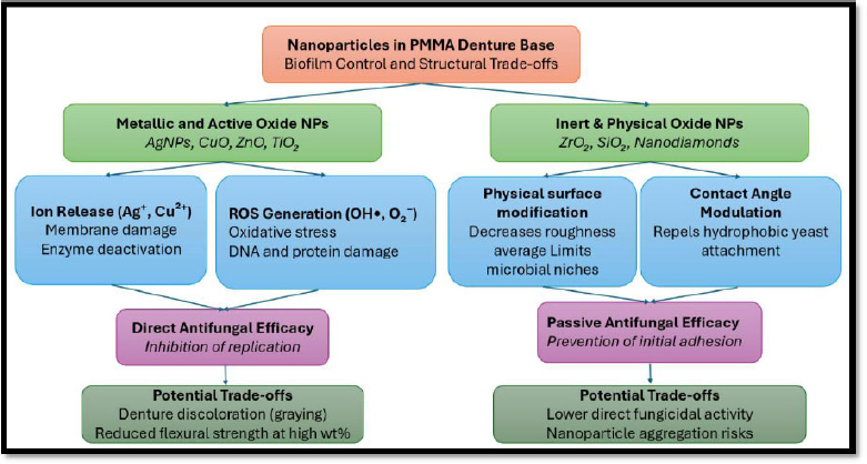
Schematic flowchart of the multidirectional pathways of nanoparticles in PMMA denture bases and their corresponding mechanical trade-offs.
As shown in Fig. (2), metallic nanoparticles Ag and CuO mostly follow active biological pathways. They continuously release active ions, such as Ag+ or Cu+, and generate reactive oxygen species, which in turn induce strong oxidative stress. This can cause cell wall degradation, lipid peroxidation, and inhibition of DNA synthesis. On the other hand, inert ceramic or oxide nanoparticles, such as ZrO2 and SiO2, rely on passive physical pathways. These materials primarily enhance the substrate surface by altering the average surface roughness and contact-angle wettability, rather than through chemical interactions. This reduction in adhesion lessens Candida cell attachment and decreases nonspecific hydrophobic interactions over time. Understanding these mechanisms aids in designing hybrid or bimetallic systems that effectively prevent biofilm formation while maintaining structural stability.
4.5. Limitations of the Study
There are limitations to this systematic review: the majority of included studies were in vitro and only partially recapitulated the dynamic conditions present in the oral cavity (salivation, chewing forces, thermal oscillations, and interactions among different biofilm species). There was also substantial variation between the studies in nanoparticle size, applied concentrations, and denture base preparation methods, making direct, clear quantitative comparisons difficult.
CONCLUSION
Adding a low concentration of silanized or hybrid nanoparticles (1 wt% AgNPs, TiO2, or ZrO2) would be a promising method to prevent denture stomatitis by controlling Candida growth on acrylic resin denture bases. For these nanocomposites to be routinely applied in dental care, standardized clinical trials are necessary, and long-term biocompatibility testing should be conducted. There is a mechanical and aesthetic balance problem: low levels may effectively slow fungal replication, while high levels often result in decreased flexural strength and discoloration. Standardization of nanoparticle surface treatment, such as salinization, is needed, and long-term dynamic clinical trials should be conducted before these nanoparticle-modified resins can be routinely employed in clinical practice.
AUTHORS’ CONTRIBUTIONS
The authors confirm contribution to the paper as follows: S.F.B., S.M.S.: Study conception and design; S.M.S.: Data collection; S.F.B., S.M.S., M.A.F.A.: Analysis and interpretation of results; S.M.S., F.M.S.: Draft manuscript preparation. All authors reviewed the results and approved the final version of the manuscript.
LIST OF ABBREVIATIONS
| AgNPs | = Silver nanoparticles |
| AgVO3 | = Silver vanadate |
| CFU | = Colony-forming units |
| CuO | = Copper oxide |
| DBR | = Denture base resin |
| NDs | = Nanodiamonds |
| NPs | = Nanoparticles |
| PICO | = Population, Intervention, Comparison, and Outcome |
| PMMA | = Polymethyl methacrylate |
| PRISMA | = Preferred Reporting Items for Systematic Reviews and Meta-Analyses |
| Ra | = Surface roughness average |
| ROS | = Reactive oxygen species |
| SiO2 | = Silicon dioxide |
| TiO2 | = Titanium dioxide |
| WOS | = Web of Science |
| ZnO | = Zinc oxide |
| ZrO2 | = Zirconium dioxide |
AVAILABILITY OF DATA AND MATERIALS
All the data and supporting information is provided within the article.
ACKNOWLEDGEMENTS
Declared none.