All published articles of this journal are available on ScienceDirect.

4D Digital Kinematic Workflow for the Full-mouth Rehabilitation of Severe Tooth Wear: A 5-Year Case Report
 iD
iD
Authors Info & Affiliations
Abstract
Background
Turner and Missirlian category 1 severe tooth wear involves loss of Occlusal Vertical Dimension (OVD) and may be accompanied by altered occlusal and neuromuscular function. Parafunctional habits may further increase biomechanical demands on restorations. This case report describes a Four-Dimensional (4D) digital kinematic workflow. The workflow integrates a Jaw Motion Analyzer (JMA), Virtual Articulator (VA), Electromyography (EMG) monitoring, and conventional analog verification.
Case Report
A 61-year-old male presented with multiple missing teeth and generalized severe tooth wear. He also had reduced clinical crown height and loss of OVD. The patient reported long-term Selective Serotonin Reuptake Inhibitor (SSRI) therapy, xerostomia, severe nocturnal bruxism, and predominantly left-sided chewing. Treatment comprised periodontal and endodontic therapy, an occlusal stabilization splint, implant placement, staged CAD/CAM provisional restorations, and definitive full-contour zirconia restorations. At the 5-year follow-up, bilateral centric contacts and prosthesis integrity were clinically maintained. Three minor occlusal adjustments had been performed, and no major biological or mechanical complications were observed. A study-specific, self-administered 10-point visual analog scale indicated high satisfaction with comfort, function, and esthetics.
Conclusion
In this patient, combining a 4D digital kinematic workflow with conventional analog verification was feasible, and the clinical findings remained stable over 5 years. These observations do not establish superiority over conventional methods, a causal effect, or broad clinical applicability. Larger prospective studies using standardized technical parameters and validated patient-reported outcome measures are needed.
1. INTRODUCTION
Tooth wear refers to loss of dental hard tissue that is not caused by dental caries, trauma, or developmental disorders [1, 2]. Its etiology is multifactorial and includes attrition, abrasion, erosion, and abfraction [3]. Turner and Missirlian classified severely worn dentitions according to OVD and available restorative space [4]. Category 1 involves excessive wear with loss of OVD. Category 2 involves excessive wear without loss of OVD but with adequate restorative space, whereas category 3 has limited restorative space. This classification concerns the worn dentition rather than dental caries.
Turner and Missirlian category 1 (also referred to as type I) is characterized by excessive generalized wear accompanied by loss of OVD [4]. Patients may also have missing posterior support and reduced clinical crown height. Unstable occlusal relationships can further complicate diagnosis and treatment planning [5-8].
Rehabilitation generally proceeds through disease control, reversible assessment of the proposed OVD, provisional treatment, and definitive restoration. The duration of each phase should be individualized according to symptoms, functional adaptation, restorative space, and treatment complexity [2, 5].
Conventional workflows can introduce errors during impression making, cast fabrication, jaw-relation transfer, and laboratory processing. Dimensional changes in impression, cast, and provisional materials may increase the need for chairside correction [9]. Long-span scans, tilted abutments, and reflective surfaces may also affect the accuracy of digital datasets [10]. Physical articulators remain clinically useful for verification but reproduce mandibular motion via programmed settings rather than directly recording the patient’s complete functional envelope.
Digital prosthodontic workflows can integrate an Intraoral Scanner (IOS), Jaw Motion Analyzer (JMA), and Computer-Aided Design and Manufacturing (CAD/CAM) [11-16]. Digital facebow and jaw-tracking systems can transfer maxillary orientation and patient-specific mandibular trajectories to a Virtual Articulator (VA). Digital occlusal analysis can also provide contact timing and relative force distribution information. Clinical performance, however, depends on acquisition quality, registration accuracy, operator experience, and appropriate cross-verification.
The originality of this case lies in the integration of several components within one full-mouth rehabilitation. These included patient-specific jaw-motion recording, virtual articulation, analog cross-verification, adjunctive Electromyography (EMG) monitoring, staged provisional therapy, and a 5-year follow-up. Selected related studies are compared in Table 2. The report is intended to illustrate feasibility and longitudinal clinical observations.
| Phase and Corresponding Section | Conventional Technique Pathway (Analog Validation) | Digital Technique Pathway (Data and Software) |
|---|---|---|
| Diagnostic Data and Virtual Patient Synthesis | Alginate/silicone impressions, dental stone casts, manual diagnostic wax-up. | Intraoral/extraoral scanning (STL), CBCT (DICOM), CAD-based multi-source data fusion. |
| Maxillomandibular Relationship and mandibular motion recording | GATD, mechanical facebow transfer, physical articulator mounting. | JMA 4D kinematics tracking, virtual patient, VA synchronization, EMG monitoring. |
| Transitional Phase and Implant Surgery | Chairside relining, GBR, manual occlusal splint adjustment. | CAD/CAM milled provisional splints, adjunctive EMG trend monitoring. |
| Provisional and Definitive Restorations | Physical articulator verification, articulating paper (tactile feedback), manual finishing. | DSD-guided lip-tooth analysis, CAD/CAM zirconia milling, digital occlusal analysis systems (T-Scan/Tee Tester). |
| Longitudinal Follow-up and Maintenance | Visual clinical examination, subjective feedback, articulating paper, nocturnal occlusal guard. | IOS-based wear tracking, digital occlusal stability assessment, longitudinal kinematic monitoring. |
| Study (Author, Year) |
IOS | JMA | EMG | VA | Analog Verification | Long-term Follow-up (≥5 Years) |
|---|---|---|---|---|---|---|
| Lepidi et al. 2022 [19] | Yes | NR | NR | Yes | No | NR |
| Feng et al. 2023 [20] | Yes | Yes | NR | Yes | No | NR |
| Park et al. 2023 [21] | Yes | Yes | NR | Yes | Yes | NR |
| Liu et al. 2024 [22] | Yes | NR | NR | Yes | NR | NR |
| Zhang et al. 2025 [13] | NR | Yes | Yes | NR | NR | NR |
| Vailati et al. 2013 [6] | NR | NR | NR | NR | NR | Yes (up to 6 years) |
| Ferrando Cascales et al. 2023 [25] | NR | NR | NR | NR | NR | Yes (5 years) |
| Present case | Yes | Yes | Yes | Yes | Yes | Yes (5 years) |
This case report describes an integrated 4D digital kinematic workflow combining a JMA, VA, and EMG with conventional facebow, articulator, and occlusal verification. The workflow included multimodal data acquisition and construction of static and dynamic virtual patient models. It then progressed through reversible splint therapy, staged CAD/CAM provisional restorations, definitive prosthetic rehabilitation, and long-term maintenance. Treatment phases and assessment points are summarized in Table 1 and Fig. 8. The report followed the CARE guidelines [17].
2. CASE PRESENTATION
2.1. Examination and Diagnosis
A 61-year-old male presented with multiple missing teeth and impaired mastication. He reported predominantly left-sided chewing, xerostomia, severe nocturnal bruxism, and long-term SSRI use. The temporal relationship between SSRI therapy and bruxism could not be established; therefore, no causal inference was made. Clinical examination showed eight missing teeth (#13, #23, #31, #34, #35, #36, #45, and #47). Generalized advanced tooth wear, reduced clinical crown height, and loss of OVD were also noted (Fig. 1). These findings were consistent with Turner and Missirlian category 1 severe tooth wear [4]. Periodontal examination showed deep pockets, particularly in posterior sites, generalized alveolar bone loss, and localized angular defects. Temporomandibular Joint (TMJ) imaging showed mild structural changes, although the patient was asymptomatic.
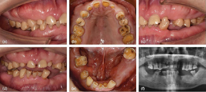
Pretreatment clinical and radiographic findings. (a) Right lateral intraoral view. (b) Maxillary occlusal view. (c) Left lateral intraoral view. (d) Frontal intraoral view. (e) Mandibular occlusal view. (f) Panoramic radiograph.
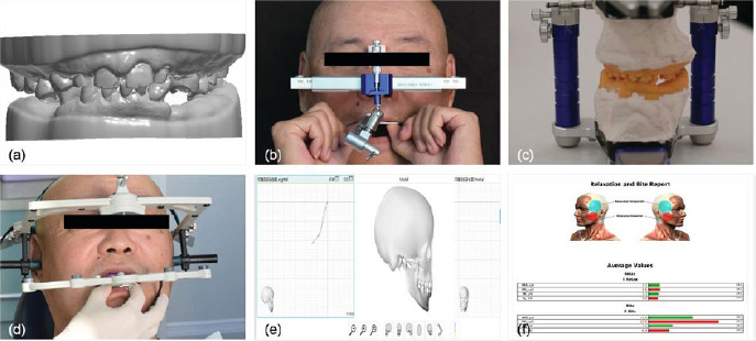
Recording and transfer of the maxillomandibular relationship. (a) Initial digital models in STL format. (b) Maxillary position transfer with a mechanical facebow. (c) Mounting on a semi-adjustable physical articulator. (d) Four-dimensional mandibular kinematic recording with a JMA. (e) Construction of the interactive virtual patient model. (f) Adjunctive neuromuscular assessment with an EMG.
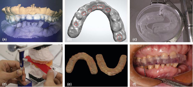
Design, fabrication, and delivery of the occlusal stabilization splint. (a) CAD design of the therapeutic splint. (b) Virtual occlusal adjustment. (c) Fabrication by CNC milling. (d) Extraoral verification on the physical articulator. (e) Finished splint. (f) Intraoral delivery and functional adaptation.
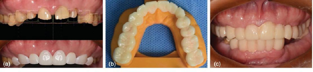
Esthetic planning and first provisional restorations. (a) Digital smile design based on facial reference lines and the lip-tooth relationship. (b) CAD/CAM design and milling of the first provisional restorations. (c) Intraoral placement for evaluation of esthetics, phonetics, and the planned incisal-edge position.
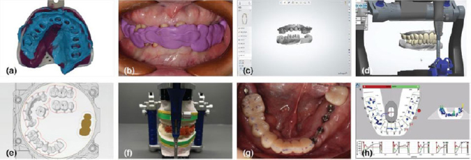
Fabrication and functional evaluation of the second provisional restorations. (a) Conventional impression after definitive tooth preparation. (b) Centric-relation record. (c) Digitization of the stone casts. (d) Virtual mounting in the virtual articulator. (e) CAD design of the second provisional restorations. (f) Verification on the physical articulator. (g) Intraoral try-in and clinical evaluation. (h) Dynamic occlusal analysis and adjustment.
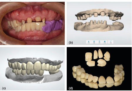
Fabrication of the definitive zirconia restorations. (a) Sectional removal of the second provisional restorations to preserve the verified jaw relationship. (b) Final digital master models. (c) Full-contour CAD design of the definitive restorations. (d) Milled monolithic zirconia restorations.
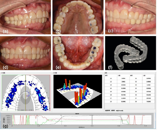
Delivery and verification of the definitive restorations. (a) Right lateral intraoral view. (b) Maxillary occlusal view. (c) Left lateral intraoral view. (d) Frontal intraoral view. (e) Mandibular occlusal view. (f) Custom nocturnal occlusal splint. (g) Digital occlusal analysis after adjustment, showing bilateral centric contacts.
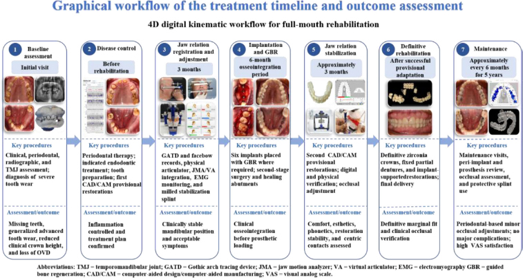
Graphical workflow of the treatment timeline and outcome assessment.
2.2. Therapeutic Strategy
A multidisciplinary plan included periodontal, endodontic, implant, and restorative treatment. Periodontal inflammation was controlled first, and endodontic treatment was completed where indicated to improve the prognosis of potential abutment teeth. Full-mouth rehabilitation then proceeded using a hybrid protocol that combined analog verification with patient-specific digital kinematic records (Table 1).
The therapeutic jaw position was established using Centric Relation (CR) records and a Gothic Arch Tracing Device (GATD). A mechanical facebow and semi-adjustable articulator were then used for cross-verification. JMA records were then imported into the VA to create a dynamic representation of mandibular movement. Digital and physical setups were compared for consistency of occlusal contacts and movement paths. EMG served as an auxiliary functional assessment for initial jaw position and splint evaluation. Serial EMG tests were not conducted systematically in subsequent treatment stages. All EMG data were analyzed descriptively and could not alone decide treatment progress. Treatment advancement depended on comprehensive evaluation of symptoms, comfort, mandibular stability, and occlusal conditions.
2.3. Treatment Process
2.3.1. Data Collection and Virtual Patient Construction
Alginate impressions were made to fabricate stone casts, which served as an analog reference. The casts and dental arches were digitized with an extraoral scanner (3Shape A/S, Copenhagen, Denmark) to generate STL datasets. CBCT imaging provided DICOM data for hard-tissue assessment. The datasets were imported into exocad DentalCAD (exocad GmbH, Darmstadt, Germany) and registered to create a static virtual patient (Fig. 2a).
2.3.2. Establishment of OVD and CR
The initial therapeutic OVD was established using facial reference lines. The spatial orientation of the maxilla was transferred to a semi-adjustable articulator with a conventional mechanical facebow for calibration of condylar and incisal guidance (Fig. 2b-c). CR was recorded with a GATD. A JMA (zebris Medical GmbH, Isny im Allgäu, Germany) recorded asymmetric functional trajectories. The archived zebris report used an “articulator & movement export” protocol. It included opening, protrusive, and right and left-laterotrusive movements, together with jaw-relation registrations at condylar and incisal reference points. Habitual, retral, mean-target, and selected-target positions were documented. Patient-specific Amann Girrbach Artex parameters were then derived. Sagittal condylar inclination was 27.4° on the left and 30.3° on the right. The Bennett angle was 13.0° left and 6.3° right. Immediate side shift was 0.0 mm bilaterally, and retrusion was 0.8 mm left and 1.7 mm right. Front-table inclination was 50.0° for both lateral paths and 80.0° for the sagittal path. The right shift angle was -20.0°. The movement data were exported in XML format. They were then imported into the exocad virtual articulator (exocad GmbH, Darmstadt, Germany) to generate an interactive virtual patient model (Fig. 2d-e). Surface EMG recorded masseter and temporalis activity bilaterally during rest, biting, protrusion, and lateral excursions (Fig. 2f). EMG was used to monitor adaptation to the target OVD, and no prespecified numerical threshold was defined as “neuromuscular tolerance.”
2.3.3. Transitional Therapy and Implant Surgery
The occlusal stabilization splint was designed in CAD software and milled with an X-MILL 500 unit (XTCERA, Shenzhen, China) (Fig. 3a-c). Intraoral adjustment was guided by occlusal contact assessment, patient symptoms, and qualitative EMG trends (Fig. 3d-f). After 3 months, the mandibular position remained clinically stable. The patient reported acceptable functional comfort without any elicited temporomandibular or masticatory muscle symptoms. Serial EMG assessment was not systematically repeated at this stage. Therefore, treatment progression was determined clinically rather than on the basis of EMG changes. After 6 months of osseointegration, second-stage surgery was completed, and healing abutments were placed.
2.3.4. Provisional Rehabilitation and Dynamic Occlusal Verification
The therapeutic maxillomandibular relation was re-evaluated with the JMA. Digital Smile Design (DSD; 3Shape A/S, Copenhagen, Denmark) was used to plan the incisal-edge position. Facial reference lines and the lip-tooth relationship were incorporated into this assessment (Fig. 4a). Endodontic treatment and post-and-core build-ups were completed on teeth #12, #22, and #46. The teeth were then prepared, and the first CAD/CAM provisional restorations were delivered (Fig. 4b-c).
After final tooth preparation, definitive impressions and CR records were obtained (Fig. 5a-b). The stone casts were digitized, virtually mounted, and used to design second-generation provisional restorations (Fig. 5c-e). Before intraoral placement, the restorations were verified on the physical articulator (Fig. 5f). Intraoral evaluation included fit, esthetics, phonetics, patient comfort, temporomandibular and muscle symptoms, and restoration stability (Fig. 5g). Occlusal contacts and timing were assessed with a digital occlusal analysis system (Tekscan, Inc., South Boston, MA, USA) and articulating paper. Posterior contacts and anterior guidance were adjusted as required (Fig. 5h). The provisional adaptation period lasted approximately 3 months.
2.3.5. Definitive Rehabilitation and Long-term Follow-up
After approximately 3 months of provisional adaptation, the patient reported acceptable comfort and function. Clinical and instrument-assisted assessment showed a reproducible mandibular position, bilateral centric contacts, and no elicited temporomandibular or masticatory muscle symptoms. The JMA recordings were obtained using predefined acquisition settings and yielded patient-specific kinematic parameters. However, no prospectively defined numerical decision thresholds were used to determine clinical acceptability based on JMA, EMG, or digital occlusal analysis findings. Therefore, these measurements were interpreted descriptively together with the clinical examination and the patient’s functional adaptation.
For the definitive phase, IOS data were registered to the verified interarch relationship. Implant abutment margins, gingival contours, and prosthetic design parameters were integrated in CAD software (Fig. 6a-c). The definitive rehabilitation comprised single crowns, fixed partial dentures, and screw-retained implant-supported bridges fabricated from full-contour zirconia (Upcera Co., Ltd., Shenzhen, China) (Fig. 6d). At try-in, occlusion was evaluated with the digital occlusal analysis system (Tee Tester, 3D-Digital, Beijing, China) and articulating paper. Contacts were adjusted to obtain bilateral centric contacts without clinically detectable eccentric interference. After cementation and screw retention as indicated (Fig. 7a-e, g), a protective nocturnal occlusal splint was delivered (Fig. 7f).
During maintenance, follow-up visits were scheduled approximately every 6 months. Each visit included oral-hygiene reinforcement, peri-implant examination, prosthesis inspection, and occlusal assessment. Over 5 years, three minor chairside occlusal adjustments were recorded. No implant loss, clinically diagnosed peri-implant mucositis, prosthetic screw loosening, ceramic chipping, restorative fracture, or visible mechanical fatigue was observed. These are reported as individual event observations rather than as a percentage-based complication rate. At the 5-year visit, the patient completed a study-specific, self-administered three-item 10-point VAS. The items assessed comfort, function, and esthetics. The scale ranged from 0 (completely dissatisfied) to 10 (completely satisfied). Scores were 9.5 for comfort, 9.7 for function, and 9.3 for esthetics. Because this study-specific instrument had not been formally validated, the results were interpreted descriptively.
3. DISCUSSION
For patients with significant tooth wear and complex occlusal instability, restorative rehabilitation aims to establish a functional relationship that is clinically tolerable. In this case, the planned jaw relationship was transferred sequentially from splint therapy to provisional restorations and then to definitive restorations. The favorable 5-year course remains an observation from one patient and does not demonstrate superiority over conventional treatment.
Patient-specific risk factors also need to be considered in complex rehabilitation. Long-term antidepressant use may be associated with oral adverse effects such as xerostomia and bruxism [18]. In the present patient, these factors, together with severe tooth wear and an unstable occlusal condition, supported the use of staged assessment and repeated functional verification rather than immediate definitive rehabilitation.
Previous studies have described individual components of similar digital workflows. Lepidi et al. used virtual articulator mounting in a fully digital complete-arch rehabilitation [19]. Feng et al. constructed a 4D virtual patient by integrating digital scans with jaw-motion tracing for maxillomandibular registration [20]. Park et al. combined intraoral scanning, JMA, VA, digital occlusal analysis, and semi-adjustable articulator verification in complete-mouth rehabilitation [21]. Liu et al. used a digital articulator to transfer a planned jaw relationship from a stabilization splint through provisional and definitive restorations in a patient with severe tooth wear [22]. These studies support the technical feasibility of digital functional workflows, but they differ in data acquisition, verification strategies, and follow-up duration. The distinguishing feature of the present case is the combined use of patient-specific jaw-motion recording, VA, adjunctive EMG monitoring, analog cross-verification, staged rehabilitation, and 5-year follow-up. A non-exhaustive comparison of selected studies is provided in Table 2.
The evidence supporting individual components of this workflow remains heterogeneous. A recent systematic review found that virtual articulators showed accuracy broadly comparable to that of conventional articulators, although substantial variation existed among software platforms, registration methods, and assessment protocols [23]. JMA and surface EMG have also been used together to characterize mandibular movement and masticatory muscle activity in patients with temporomandibular disorders [13]. These findings support their use as sources of physiologic information, but they do not establish numerical thresholds for restorative decision-making.
EMG only provided limited auxiliary data in this case. It analyzed masticatory muscle activity during the initial assessment, without regular, repeated tests during subsequent treatment. Thus, EMG results cannot prove long-term muscle relaxation or neuromuscular adaptation. No fixed EMG value was set to judge whether to advance treatment. Clinical symptoms, comfort, stable mandibular position, occlusal contacts, and restoration stability jointly determined treatment progress.
Digital occlusal analysis was used in a similar complementary manner. It provided information on contact timing and relative force distribution, while articulating paper remained part of the clinical verification process. Reich et al. reported moderate inter-observer reliability for digital occlusal analysis and demonstrated differences among occlusal indicators [24]. In the present patient, bilateral centric contacts were clinically maintained, three minor occlusal adjustments were recorded over 5 years, and no major biological or mechanical events were observed. The study-specific VAS also showed high patient satisfaction at the 5-year visit. These findings describe the clinical course of this patient rather than the efficacy of the workflow.
Long-term evidence in worn-dentition rehabilitation is better established for restorative outcomes than for digital kinematic workflows. Vailati et al. reported follow-up of up to 6 years after minimally invasive rehabilitation of severe erosive tooth wear [6]. Ferrando Cascales et al. also reported 5-year outcomes in a retrospective case series of patients with severe tooth wear treated with adhesive restorations [25]. These studies support the potential for durable restorative outcomes, but neither evaluated an integrated JMA-EMG-4D virtual-patient workflow. The present case therefore contributes longitudinal clinical observation of an integrated digital-analog workflow, while comparative benefit remains to be established.
The workflow also introduced practical demands. Successful implementation required coordination among the clinician, technician, and digital systems. Data acquisition, registration, software interoperability, and calibration created potential sources of error and required operator experience. Equipment cost and training requirements may further limit accessibility. To reproduce the core protocol in routine practice, several basic tools are required. These include digital scanning, jaw-motion recording, CAD software with virtual-articulator functions, and conventional articulator verification. Surface EMG and digital occlusal analysis can be added as adjunctive assessment tools. The clinical team also requires experience in jaw-relation recording, digital data registration, virtual articulation, and cross-verification between digital and physical records. These considerations should be balanced against the potential value of the workflow for visualization, interdisciplinary communication, and cross-verification.
3.1. Future Research
Future studies should evaluate this workflow prospectively in multicenter cohorts and controlled or randomized comparisons. JMA acquisition and EMG protocols should be standardized, with predefined clinical endpoints and validated patient-reported outcome measures. Future studies should use validated oral-health-related quality-of-life instruments, such as the 14-item Oral Health Impact Profile (OHIP-14). These measures can complement study-specific satisfaction scales and improve comparability across cohorts. Chairside time, laboratory time, and cost-effectiveness should be recorded prospectively. Automated or AI-assisted analysis of jaw-motion trajectories may also be explored, but such methods require external validation before they can inform treatment decisions.
3.2. Limitations
This report has several limitations. It describes one patient and has no comparator or control group, so causality and comparative effectiveness cannot be assessed. The workflow depended on the operator, laboratory, software, and hardware, which limits generalizability. In addition, the scanner, JMA, CAD/VA platform, and digital occlusal-analysis systems were supplied by different manufacturers. Their integration required compatible data formats, registration procedures, and software interoperability. This configuration may be difficult to reproduce in clinics using different hardware or software systems. The archived JMA reports did not display raw sensor sampling frequency or signal-filtering settings. No prospectively defined numerical acceptance threshold was used for JMA, EMG, or digital occlusal analysis. The three-item satisfaction VAS was study-specific and unvalidated. Follow-up observations were obtained during routine clinical care, and inferential statistical analysis was not appropriate.
CONCLUSION
This case shows that a 4D digital kinematic workflow can be combined with conventional analog techniques in complex full-mouth rehabilitation. Clinical outcomes remained stable during the 5-year follow-up, and the patient reported favorable functional and esthetic outcomes. The combined approach supported individualized planning and stepwise verification across treatment phases. Findings from one case cannot establish superiority, causality, or broader generalizability. Larger prospective comparative studies are needed.
CLINICAL SIGNIFICANCE
Patient-specific digital jaw-motion recording may support individualized treatment planning when used alongside conventional verification in complex full-mouth rehabilitation. In this case, its practical value was the transfer and cross-checking of the planned jaw relationship across treatment phases. No single digital metric was used as the sole basis for a clinical decision.
AUTHORS’ CONTRIBUTIONS
The authors confirm contribution to the paper as follows. Q.L.: Performed the clinical procedures; C.L., C.X., and Q.L.: Collected the case data; C.L., M.A., C.X., and Q.L.: Analyzed the case and interpreted the results; C.L.: Drafted the manuscript. All authors reviewed the results and approved the final version of the manuscript.
LIST OF ABBREVIATIONS
| IOS | = Intraoral Scanner |
| CBCT | = Cone-Beam Computed Tomography |
| STL | = Standard Tessellation Language |
| CAD | = Computer-Aided Design |
| CAM | = Computer-Aided Manufacturing |
| GATD | = Gothic Arch Tracing Device |
| 4D | = Four-Dimensional |
| JMA | = Jaw Motion Analyzer |
| VA | = Virtual Articulator |
| EMG | = Electromyography |
| GBR | = Guided Bone Regeneration |
| DSD | = Digital Smile Design |
CONSENT FOR PUBLICATION
Written informed consent was obtained from the patient for publication of the clinical details and all clinical and radiographic images.
FUNDING
This work was supported by the Research Fund of Nanfang Hospital, Southern Medical University (Grant No. 2023A023). It was also supported by the Guangdong Provincial Graduate Education Innovation Program (Grant Nos. 2024ANLK_016, 2026ANLK_041). Additional support was provided by the International Education Teaching Reform Research and Practice Special Project of Southern Medical University (Grant No. 2024GJG006). The study was also supported by the Higher Medical Education Research Project of Guangdong Pharmaceutical University (Grant No. GKP202503). The study received support from the 2026 Higher Education Teaching Reform Research Project under the 15th Five-Year Plan of the Guangdong Higher Education Teaching Management Association (Grant No. ZD200601008).
ACKNOWLEDGEMENTS
The authors would like to express their sincere gratitude to the dental laboratory technician for their outstanding skill and collaboration in fabricating the provisional and definitive restorations. The authors also extend their appreciation to the patient for his trust and assistance throughout the lengthy and complex treatment process.