All published articles of this journal are available on ScienceDirect.

Uncommon Restoration on Nonvital Premolar with Porcelain Inlay: A Case Report with Five-year Follow-up
Abstract
Introduction:
In fixed prosthodontics, the old paradigm on endodontically treated teeth leads to more fragilization due to the architectural preparation needed for a full coverage crown. Endodontically treated premolars with class II defects are common in daily practice. When only one proximal surface is affected, direct adhesive restorations are the most used. The use of porcelain inlay is quite uncommon in this situation despite its advantages.
Case Presentation:
This article illustrates a clinical case of a 31-year-old female with healthy teeth except for the non-vital first maxillary premolar, presenting a class II mesio-occlusal cavity. The occlusion exam revealed functional anterior guidance and canine guidance on both sides during lateral mandibular translation. Based on mini-invasive dentistry principles, the decision was to restore the nonvital bicuspid with a porcelain inlay. Although scientific literature did not report restoration of the premolars with a ceramic inlay in clinical practice, this option was retained. The realization of a porcelain inlay was described step by step. A follow-up up to 5 years was done.
Results:
The iatrogenic tooth fragilization was avoided by a mini-invasive approach.
At the end of this follow-up period, there was no complication or complaint. The patient was satisfied with the final result.
Conclusion:
Porcelain inlay seems to be a good option to restore the occluso-proximal cavity on nonvital premolars in a context of a favourable occlusion. Porcelain inlay is respectful of the principles of minimally invasive dentistry and allows to achieve the objectives of function, aesthetics and biocompatibility. However, clinical studies should be conducted to investigate the longevity of porcelain inlay on ETPs.
1. INTRODUCTION
According to several epidemiological studies, the majority of restorations on endodontically treated teeth (ETT) are performed at the posterior level [1]. It has been reported that Class-II cavity was the most common [2]. However, different approaches are available for their restoration. The old paradigms on the ETT claim an intrinsic fragility linked to endodontic treatment. The root post and the full coverage crown were considered as a reinforcement of the ETT [3]. This paradigm has been adopted systematically for a long period. Currently, it has been proven that the brittleness of ETT is essentially caused by loss of substance [4, 5]. It has been concluded that tooth strength is reduced in proportion to coronal tissue loss due to either caries lesions or restorative procedures [4, 5]. Furthermore, the authors claimed that the best current approach for restoring ETT seems to minimize tissue sacrifice, especially in the cervical area, so that a ferrule effect can be maintained [4]. Thus, peripheral preparation on a non-weakened ETT is considered as an unjustified mutilation. The use of partial restoration requires only a minimum of tooth preparation. It has been proved that partial coverage restoration preserved a significantly greater amount of tooth structure compared with all-ceramic crowns [6]. Therefore, partial restorations, which are minimally invasive, offer an alternative to the crown. However, the technical procedure to perform this partial restoration can be direct or indirect. In regard to the endodontically treated maxillary premolars (ETMPs), restoration using an inlay is not quite common compared to molars, which are rather quadrangular, while premolars are anatomically less resistant.
Moreover, it has been reported that direct restoration of ETMPs or indirect restoration using an inlay increased the fracture strength of teeth [7, 8]. Other authors concluded that indirect lithium disilicate inlay used to restore class II mesio-occlusal-distal cavity in posterior teeth generated lower stress levels than direct resin-based composite materials applied in multilayer techniques [9]. Thus, faced with a nonvital tooth that is not weakened by coronal destruction, dentists are asked to offer their patients therapeutic methods based on a mini-invasive approach. The aim of this work was to present, through a clinical case, a new approach to restore a nonvital premolar based on the new scientific data.
2. CASE REPORT
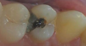
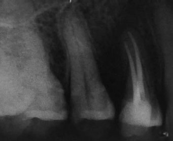
A 31-year-old female in good health consulted the Department of Fixed Prosthodontics at the Academic Clinic of Dental Medicine of Monastir for a food retention problem in the maxillary right first premolar (#14). The clinical exam revealed a fractured amalgam on tooth #14, as depicted in Fig. (1), while the interproximal papilla was inflamed by food impaction. The periapical radiograph showed recurrent caries, unfilled root canals and a widened periodontal ligament space. The level of coronal destruction was supra-osseous, as shown in Fig. (2). Furthermore, all the teeth were found to be of little height. The occlusion exam revealed functional anterior guidance and canine guidance on both sides during lateral mandibular translation. The patient was esthetically demanding, desiring natural tooth mimicry, and was reluctant to “reduce the entire tooth” for placement of a full-coverage crown. The choice of the type of restoration was established after initial treatments to properly assess residual tooth structures.

2.1. Initial Treatments
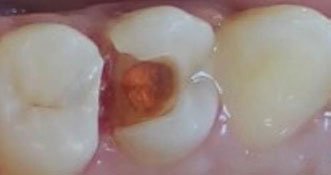
At this step, endodontic treatment and interproximal area conditioning are focused. After the removal of the defective amalgam, a caries curettage was achieved using an excavator in order to guarantee an ultraconservative approach. The tissue destruction level was subgingival, as depicted in Fig. (3).
The endodontic access cavity was prepared with a concern for dental tissue saving. A cold lateral condensation technique was performed using standard 6% gutta-percha cones as well as accessory cones. At this stage, a preliminary decision to perform an indirect restoration was taken. Therefore, the contact area was restored with a temporary inlay fabricated using a silicone matrix and methyl methacrylate resin. The sealing was carried out using a minimum quantity of non-eugenol provisional luting agent (Temp-Bond™ NE from Kerr) (Fig. 4). Due care was given to clean the interproximal area from any excess material using an interdental floss. After fourteen days of temporization, interdental soft tissue response was favorable without gingival bleeding or recession. A percussion test was performed by gently tapping the occlusal surfaces of teeth #14 and #15 with the end of the handle of a mouth mirror. It confirmed an asymptomatic treated tooth #14.
Based on a healthy tooth-periodontal environment, the indication of a resin-bonded restoration was confirmed. This treatment option of an occlusal distal porcelain inlay makes it possible to achieve the following treatment objectives:
• Function: porcelain is a mechanically resistant material.
• Esthetics: use of a material with excellent optical properties preserving natural dental color.
• Biocompatibility: use of a chemically, thermally and electrically inert material.
• Maximum preservation of sound tooth tissues.
2.2. Cavity Preparation
2.2.1. Deep Margin Elevation
A Deep margin elevation (DME) was performed under rubber dam isolation following the placement of a matrix. A resin-modified glass ionomer (Riva® from SDI) was applied, then light-activated. At the same time, the intracoronal undercuts were filled with the same material. The preparation consisted of shaping of occlusal cavity and proximal box according to well-defined criteria in the literature.
2.2.2. Occlusal Cavity Preparation
First, the antagonist teeth contacts were located to avoid placing the limits of the occlusal cavity at their level. A divergence angle of 6 degrees was sought for each cavity wall. Then, the preparation of the cavity floor led to its flattening with a depth of 2.5 mm. This provides correct seating and a homogeneous thickness for the ceramic inlay. In addition, all internal angles were rounded. The preparation margin was finished at an angle close to 90 degrees. Enamel prisms were broken without any beveling of the cavity margin. Subsequently, the preparation of the occlusal isthmus was carried out, resulting in a width of about 2.5mm.
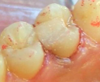
2.2.3. Proximal Box Preparation
The box cavity had a width of 2mm with a gingival floor of 1.5mm supragingival.
A rounded transition was achieved at the level of the gingival floor and the cavity walls. In order to preserve maximum sound tissues, the preparation of approximal walls of the cavity has not been extended (Fig. 5).
2.3. Impression and Laboratory Procedures
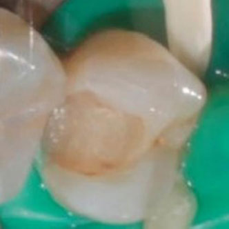
Putty and light body polyvinylsiloxane were used for impression. It was performed by single-step/double mix technique. The restoration was fabricated using computer-aided design/computer-aided manufacturing (CAD/CAM) technolo-gy. Firstly, the impression was poured in plaster type 4. Secondly, the master cast was scanned, and then the restoration was designed in the dental laboratory (InLab® 3D software from SIRONA). Then, a lithium disilicate glass-ceramic block was chosen corresponding to the patient’s tooth-color. The block was positioned in the milling machine along with the necessary cutters. The machining procedure was performed under continuous irrigation (InLab® MC XL from Dentsply Sirona). Finally, a crystallization heat treatment in a porcelain furnace (Multimat Cube Porcelain Furnace from Dentsply Sirona) provided the final mechanical characteristics of the inlay (Fig. 6).
2.4. Try-in and Bonding of the Inlay
During the try-in, the adaptation of the tooth-restoration interface was particularly checked and was proven to be satisfactory. This was done before and after isolation with a rubber dam.
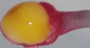
The procedure consisted of bonding the ceramic inlay to the cavity with an acid-etch technique. First, the inlay external surface was waxed and supplied by applicator handle that was used for prehension. Secondly, the internal surface was etched with 5% hydrofluoric acid for 20 seconds (Fig. 7). It was thoroughly rinsed and then dried with compressed air, which should be free from lubricants, followed by the application of a thin layer of adhesive (3M™ Scotchbond™ Universal Plus Adhesive from 3M).
It was well applied to the internal surface using a micro-brush, spread by an air spray and stored away from light. This precaution was taken to avoid excessive thickness, which could compromise the complete insertion of the restoration. The cavity preparation was etched with 37% phosphoric acid, always starting with the enamel. Application time was 30 seconds on enamel and 15 seconds on dentin. After rinsing and drying, the etched enamel should display a lightly frosted appearance.
Afterwards, a rigorous rinse with water was carried out. Then, the preparation was gently dried with compressed air. Using a microbrush, a thin amount of adhesive was applied to the prepared surface, followed by an immediate spreading with compressed air. Then, a second application optimized the formation of an adequate hybrid layer.
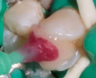
At this stage, the two substrates are ready for bonding. A dual-cure resin cement was used. It was applied to the intrados of the inlay and into the prepared cavity with a spatula. The previously designed pick-up stick facilitated the insertion of the small-size restoration, as shown in Fig. (8).
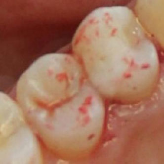
Next, the excess resin was removed with a disposable microbrush before exposing the inlay to the curing light. To ensure complete polymerization, the inlay should be cured for a minimum of 40 seconds each from occlusal and facial directions for a total exposure time of 80 seconds. After removing the rubber dam, a control of the occlusion with articulated paper was carried out (Fig. 9).
Occlusion was adjusted using fine-grained diamond points. At this step, canine guidance was preserved. Then, a porcelain polishing kit was used to restore a smooth ceramic surface according to a sequence of progressively finer abrasives.
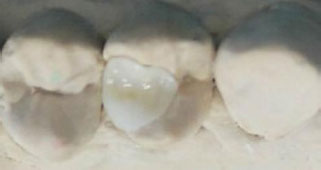
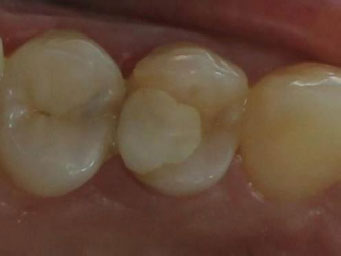
Control sessions were scheduled at 6 months, two years and five years after. At the end of a follow-up period of up to 5 years, there was no complication or complaint. The patient was satisfied with the final result (Figs. 10 and 11).
3. DISCUSSION
In the present case, a peripheral preparation for an all-ceramic crown would have resulted in an extensively damaged premolar because of the short clinical crown and the presence of a class II cavity. The most commonly used restoration for extensively damaged teeth with short clinical crowns involves a post-retained restoration. The use of post-retained restora-
tions has been questioned because of potential tooth weakening [10]. If a tooth is shortened excessively, a post and crown may cause excessive lateral forces on the root. This can result in a fracture of the root, bone loss, mobility, loss of the tooth, or any combination of these events [11]. In the present case, it was decided to avoid iatrogenic tooth fragilization due to peripheral preparation. Minimally invasive dentistry is the best approach to restore the affected nonvital premolar without any iatrogenic fragilization. The partial adhesive restorations (PAR) are currently the most respectful of the principles of minimally invasive dentistry. A direct partial adhesive restoration (DPAR) with composite resin could be the most convenient method for the present case. In general practice, the direct method is the most commonly used. Its main advantage is that restoration can be performed in one session. However, the DPAR does not perform well in terms of stability of colour, occlusion and proximal contacts. Moreover, a good quality of bonded joint is delicate to obtain due to polymerization shrinkage of the resin. Thus, direct restoration should be changed when detected a margin degradation, a lack of proximal contact, a colour change or unusual occlusal wear. The advantage of ceramic inlay is that it performs good stability in terms of colour, occlusion and proximal contacts. Inlay, as an indirect restoration, allows to avoid polymerization shrinkage of the resin. Therefore, better longevity is to be expected compared to direct composites. Moreover, it has been concluded in a systematic review and meta-analysis that ceramic partial coverage restorations (feldspathic porcelain and glass-ceramic) outperformed resin partial coverage restorations in terms of clinical performance both at 5-year and 10-year follow-ups [12]. Whereas the use of porcelain inlay is quite uncommon on ETPs. First, the old common practice was to perform a full-coverage crown. This was due to the old curricula, which had integrated the paradigm of the fragility of ETT for a long period. Dentists educated by these curricula and who are not up to date continue to indicate complete crowns on ETT. Second, in daily practice, class II cavities, either on premolars or on molars, are predominately restored with direct composite resin, considered easier and more convenient compared to Cl II ceramic inlay. Third, ceramic inlays are more indicated on molars than on premolars. This can be explained by the fact that premolars are smaller in volume and, thus, anatomically less resistant. As a result, cavity preparation requires more training. Any excess of the cavity preparation, even minimal, would lead to weakened residual walls. Besides that, the smaller dimensions of cl II porcelain inlays represent an additional difficulty during try-in and then bonding. Before dental bonding, a ceramic inlay is considered as brittle and should be carefully manipulated to avoid cracks due to unforeseen shocks. In fact, handling a very small ceramic restoration is not common in daily practice, and it requires special equipment and some changes in the organization and in dental practice. Finally, compared to direct composite resin, porcelain inlay has a higher cost. A more expensive restoration can only be preferred if it is clear to both the dentist and the patient that the cost-effectiveness ratio is better than the other alternatives. We believe that, in the current state, this condition is not met. In the future, ceramic adhesive restorations could be an interesting alternative to direct adhesive restorations. Through literature research, it has been noted that the majority of the studies investigating the longevity of ceramic inlays on endodontically treated maxillary premolars were of in vitro studies on class II MOD cavities. Very few studies have been conducted for class II cavities affecting one proximal surface cavities. In terms of fracture resistance of direct restoration versus porcelain inlay on ETMPs, it has been reported in an in vitro study that direct composite restorations of the occluso-mesial cavity on ETMPs showed the highest fracture resistance compared to CAD/CAM ceramic restorations. But there was no difference in the fracture mode [13]. In another in vitro study, it was concluded that ETPs with mesial-occlusal cavity preparation restored with Biodentine (Septodont) were more resistant to fracture than those restored with either Smart Dentin Replacement (SDR; Dentsply Sirona) material or ceramic inlays. There were no statistically significant differences between the groups in terms of fracture mode [14]. Despite these two previous studies, it remains unclear whether porcelain inlays on ETPs would or would not constitute an alternative to direct restoration methods in the future. In our opinion, porcelain inlays could be part of the restorative modalities for ETPs because of the better properties of dental ceramics compared to direct restorative materials, progress in dental bonding and the spreading of CAD/CAM in dentistry. The disadvantages of ceramic inlays are a higher cost for the patient and a more demanding implementation for the dentist. However, randomized controlled trials are needed to investigate the longevity of porcelain inlays on ETPs. It would also be necessary to determine the clinical conditions required to obtain the best results in terms of clinical performance. Dental parameters influencing the longevity of adhesive restorations on ETT, in terms of fracture resistance, are residual tooth walls, residual marginal ridges, wall thickness and occlusion stresses. In the present clinical case, the nonvital premolar presented a class II mesio-occlusal cavity. The defect was assessed as moderate since there were three residual walls (Buccal, distal and palatal) and an intact marginal ridge. In the following, wall thickness and occlusion stresses will be discussed. Wall thickness is a crucial parameter that influences the fracture resistance of ETT. It was found in an in vitro study that wall support of 2 mm was adequate, and support of 1 mm was completely insufficient. When the wall thickness was 1.5 mm, direct restoration with fiber at the occlusal level significantly improved resistance [15]. Recommendations have been developed stating that when the margins of inlay cavity preparations are closer than 1.5 mm to the functional cusp or when less than 2 mm of thickness remains, preparation for an onlay should be performed, with a 2 mm axial reduction [16]. Occlusion should be carefully checked during lateral translation. In the present clinical situation, the absence of parafunction and the presence of canine guidance were positive indices. Maintaining canine guidance is important to protect premolars from oblique forces, which are more exposed than molars because of their anterior position. If canine guidance is absent, an overlay will allow a better stress distribution thanks to cusp coverage [17]. When the buccal surface is worn, restored or discolored, a veneerlay should be preferred. The latter allows at a time occlusal and buccal coverage. Whereas, for this patient, tooth #14 was of normal color, and the cusps were sufficiently thick and did not require covering. The choice of the type of ceramic is guided by the translucency or opacity of the dental tissues, the required mechanical resistance and the bonding ability. In this case, a very high mechanical resistance is not required, because the inlay will not be stressed by significant forces. The translucency of the patient's dental tissue was medium. A good bonding ability is essential. Glass-matrix ceramics, including leucite- and lithium disilicate-reinforced glass ceramics, are etchable with hydrofluoric acid, and bonding strength can be optimized by an adequate surface treatment [18]. Glass-ceramics benefit from good clinical experience and remain relevant. For the present case, the characteristics of lithium disilicate glass-ceramic were suitable for an inlay on the maxillary premolar. Recent polymer-based CAD-CAM materials have been commercialized for inlay restorations, such as composite resin nanoceramics and polymer-infiltrated ceramic-network [19]. Mastery of the bonding protocol is essential for the success of a partial adhesive restoration. In fact, efficient bonding seems to provide the necessary cohesion for the restored tooth. The presence of a peripheral enamel band and supragingival limits are crucial for a perfect bonded interface. Bonding under a rubber dam provides the best isolation from saliva and buccal fluids. However, it should be checked that cervical margins are not covered by the rubber dam. Regarding the clinical situation, peripheral enamel was present except at the distal level, where the decay was subgingival. This problem was solved by the deep margin elevation technique (DME), which is considered a valid treatment option for clinicians in the case of subgingival margins [20]. Indeed, it has been reported that DME resulted in decreased ceramic fracture [20]. Several precautions were taken to ensure the success of the treatment in this case. Before preparing the cavity, the occlusal contacts had been marked (highlighted with an articulating paper) to avoid placing the limits of the occlusal cavity at the same level. This was essential to guarantee a durable bonding interface. During the preparation of the occlusal isthmus, there is always a compromise to be sought: that of not weakening the tooth by an excessive width of the isthmus and simultaneously providing sufficient space for the ceramic. Indeed, this area of the tooth is particularly stressed by occlusal forces, and the ceramic is resistant to thicknesses between 2 and 3 mm. However, this compromise is particularly tricky for a premolar, which is small in volume compared to a molar. In this case, an occlusal isthmus width of approximately 2.5 mm was considered suitable. In addition, bonding an inlay of small dimensions needs meticulous precautions for the best handling. A designed pick-up stick fixed on the center of the occlusal surface of the inlay seems to be useful in similar situations. Despite the slight color difference between the ceramic and the tooth, the patient was satisfied with the final result. Indeed, the ceramic inlay restored the occlusal and distal surfaces, which were invisible when smiling. This partial restoration kept the vestibular surface intact, and thus, the natural aesthetics of the tooth was preserved. The maintenance phase is essential, focusing on the quality of the bonded joint. The follow-up consists of a clinical assessment with a probe. In case of doubt, a bitewing radiograph would allow the control of the quality of the bonded joint especially at the level of DME.
CONCLUSION
In the case of an endodontically treated premolar, the conventional approach has consisted of peripheral preparation and possibly an intra-radicular post space. Unlike this invasive approach, porcelain inlay seems to be a good option to restore the occluso-proximal cavity on nonvital premolars in a context of a favorable occlusion. Porcelain inlay is respectful of the principles of minimally invasive dentistry and allows to achieve the objectives of function, esthetics and biocompatibility. Nevertheless, premolar restoration with a ceramic inlay remains an uncommon practice. According to a follow-up for more than 5 years, good results have been obtained. However, clinical studies should be conducted to investigate the longevity of porcelain inlay on ETPs.
LIST OF ABBREVIATIONS
| ETT | = Endodontically Treated Teeth |
| ETMPs | = Endodontically Treated Maxillary Premolars |
| PAR | = Partial Adhesive Restorations |
| DPAR | = Direct Partial Adhesive Restoration |
CONSENT FOR PUBLICATION
Written informed consent was obtained from the patient for the publication of this case report.
AVAILABILITY OF DATA AND MATERIALS
The data and supportive information are available within the article.
ACKNOWLEDGEMENTS
Declared none.

