All published articles of this journal are available on ScienceDirect.

Visual and Instrumental Color Match Evaluation of Single Shade Composites before and after Bleaching Procedures: A Pilot Study
Abstract
Background:
One of the major challenges in restorative dentistry is to obtain an adequate color match between composite restorations and the surrounding tooth. This match depends on several chromatic variables related to the composite itself but also to the teeth. Bleaching procedures are not to be able to modify the shade of composite restorations to a lighter color as it does on natural teeth.
Objective:
The objective of this study is to evaluate the visual and instrumental color match of two single-shade resin-based composites in human-extracted teeth before and after bleaching treatment.
Methods:
Six extracted human posterior sound teeth were used. Round-shaped V-class cavities (2x2x4mm) were prepared buccally 2mm away from the CEJ. Two single-shade resin composites (OM-Omnichroma and VE-Venus Diamond One) were used for the restorations. Tooth color was measured using an intraoral spectrophotometer. Visual analysis was carried out by 16 calibrated observers, and the differences were graded from 0 (excellent match) to 4 (huge mismatch). Teeth were then bleached using 40% H 2O2 (Opalescence Boost PF, Ultradent), and visual and instrumental evaluations were performed after 24 hours.
Results:
Before bleaching, visual analysis showed a mean value of 0.16 for OM and 0.24 for VE. After bleaching, the color match showed a mean value of 0.14 for OM and 0.22 for VE. Regarding the instrumental analysis, each restoration matched the tooth VITA scale grade before bleaching and followed the natural tooth VITA scale grade after bleaching procedures.
Conclusion:
Within the limits of this pilot study, both composites seem to have excellent color-match properties with the surrounding tooth structure.
1. INTRODUCTION
Resin composites are the restorative materials of choice due to their aesthetic and handling properties, for being able to show good mechanical characteristics and for their good adhesion performance
One of the most complex challenges in restorative dentistry is achieving a correct color match between composite restorations and the surrounding teeth [ 2]. In fact, color matching depends on various chromatic variables related to both composites and teeth, which are responsible for the difficulty of obtaining this match [ 3].
The variability of the color of natural teeth leads to the development of different composite systems that include many different shades, following as a reference, in most cases, the VITA Classical shade guide [ 4]. Composites present several opacities (Enamel = translucent, dentin = opaque/body) that are useful to obtain similar optical properties to natural enamel and dentin [ 1]. These different composites of opacities and chromas are clinically employed in several layers through a layering technique in order to achieve a restoration with the same optical properties as the natural tooth [ 5]. This technique is able to obtain acceptable results in color matching but is complex; it requires increased operative timing and is much more skill–dependent than other one- or two-shade procedures [ 1].
The potential of a resin to take on the color of the surrounding tooth structure via reflection is called the blending effect (BE). This effect is the basis of the latest generation composites, designed to blend with the surrounding tooth, using the lowest possible number of shades [ 4, 6]. Furthermore, the composite blending effect contributes significantly to achieving a proper shade match and improving the aesthetics [ 4]. Recently, innovative single-shade resins have been introduced, supposedly able to match all VITA classical shades from A1 to D4 [ 1, 7].
The choice of using filler morphology for shade matching is a modern technological innovation named “structural color”, where shade matching is obtained by resin composite light absorption and emission [ 4].
The structural color phenomenon is based on the discrimination of different wavelengths by the interaction of incident light with nanostructures like diffraction gratings, photonic crystals, or thin films. As structural colors are the result of fundamental optical processes of interference, scattering, and diffraction, they do not fade compared to conventional colors, which originate from light absorption by pigments [ 8].
The match between teeth and composite is of fundamental relevance when performing the reconstruction of a tooth in the aesthetic area. In this clinical situation, it is not possible to endure aesthetic compromises, and the reconstruction must perfectly blend with the surrounding dental structures [ 2].
It has been shown that composite resins suffer from intrinsic or extrinsic staining over time, caused by pigments from drinks or food or smoking habits [ 9, 10]. Whitening procedures for bleaching teeth at home or in the office are often proposed to modify the color of natural teeth toward a desired aesthetic appearance [ 11, 12]. These products are sometimes able to remove stains from composites but seem not to be able to modify the shade of composite restorations to a lighter color as it does on natural teeth [ 11]. Since composites seem to be unable to change to a lighter shade, bleaching is usually recommended before a new composite filling in order to obtain a match between the restoration and the new lighter tooth shade [ 12].
The aim of this pilot study is to evaluate the instrumental and visual color match of two different single-shade resin-based composites with the surrounding tooth before and after professional bleaching procedures on human-extracted teeth. The null hypotheses are that (1) there is no color match between single-shade composites and the surrounding tooth and (2) that single-shade composites are not able to follow tooth color after bleaching.
2. MATERIALS AND METHODS
Six human extracted posterior sound teeth (molars) without caries, restorations, and endodontic treatment, stored for 24 hours in distilled water at 37°C in specific individually sealed compartments, were used in this pilot study [ 13, 14]. Extracted teeth were used according to the Ethical Committee of the University of Bologna approval (protocol No:71/2019/OSS/AUSLBO).
On the tooth buccal side, 2 mm above the cemento-enamel junction (CEJ), a standardized V° class cavity (2 mm depth, 2 mm high, and 4 mm width) was performed using a water-cooled dedicated round-shaped diamond burr (#6801314029, Komet Dental, Lemgo, Germany) [ 15].
Two different one-shade resin composites were selected, Omnichroma (OM) (Tokuyama Dental, Tokyo, Japan) and Venus Diamond One (VE) (Kulzer, Hanau, Germany), with their correspondent adhesive systems, Universal Bond (Tokuyama) and iBond Universal (Kulzer). Details about the tested resin composites are given in (Table 1).
| Material | Monomer | Filler Type |
|---|---|---|
| Omnichroma | UDMA, TEGDMA | SiO2, ZrO2 & CF (68 vol.-%; 79 wt%; 0.2 μm – 0.4 μm in ø) |
| Venus Diamond One | UDMA,TCD-DI-HEA, TEGDMA | BaAlF, SiO2 (64 vol.-%; 81 wt%; 5 nm – 20 μm in ø) |
The six extracted teeth were randomly divided into two groups, with three teeth in each group. Group OM included teeth restored with OM (teeth OM1, teeth OM2, and teeth OM3), while group VE included teeth treated with VE (teeth VE1, teeth VE2, and teeth VE3).
Teeth were restored through a single increment of the correspondent one-shade composite resin. Bonding procedures were performed according to the manufacturer. Composite restorations were then polymerized at 1400 mW/cm2 with a middle-intensity blue-led light-curing unit (Mectron Starlight Pro, Italy) for 40 seconds, as indicated by the manufacturer.
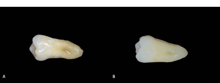
After curing procedures, the restorations were dry polished with a dedicated composite finishing and polishing system (Clearfil Twist DIA, Kuraray), using a slow-speed handpiece at 4000 rpm for 30 seconds per step and were stored in distilled water at 37°C for 24h [ 13] (Fig. 1). Instrumental and visual color measurements were performed after the restoration, as described further.
2.1. Bleaching Procedures
Teeth were bleached through a 40% hydrogen peroxide (Opalescence Boost PF 40%, Ultradent, South Jordan, USA). Teeth were fixed on a wax plate before the application of the bleaching gel [ 11]. This gel was applied to cover the whole tooth surface except the composite restoration because bleaching agents at high concentrations could promote a physical deterioration on the surface roughness of composite resins [ 16, 17]. Two consecutive simulated bleaching sessions, 20 minutes each, were performed for each specimen according to the manufacturer [ 18].
After both applications, the bleaching agent was gently removed using gauze soaked in distilled water, and then the specimen surfaces were washed out and dried with absorbent paper. Samples were not air-dried with any system that could dehydrate the samples [ 11].
Instrumental and visual color measurements were performed after bleaching, as described further.
The specimens were always stored separately in distilled water at 37°C [ 11].
2.2. Instrumental Color Measurements
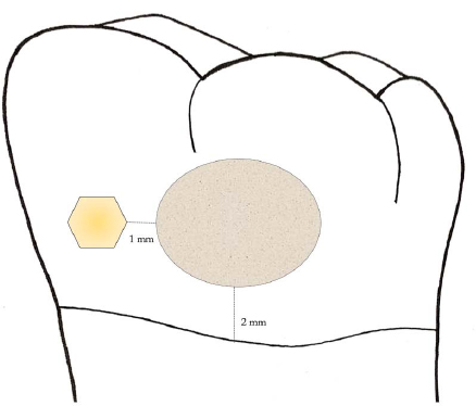
Using an intraoral spectrophotometer (VITA Easyshade V, VITA Zahnfabrik, Bad Sackingen, Germany), by placing its measuring tip perpendicular to the vestibular side, VITA color was evaluated. A neutral grey paper was used as a background during the measurements [ 6, 18]. Measurements were taken (24 hours after the restoration, T0, and 24 hours after professional bleaching, T1) in the center of the restoration and on the teeth, 1 mm away from the margin of the restoration [ 13] (Fig. 2).
2.3. Visual Color Measurements
After instrumental evaluation, visual color assessments were carried out by 16 dental professionals [ 6]. These observers were both male and female dentists with at least 5 years of experience in restorative dentistry, attending the Restorative Department of the Dental School of the University of Modena and Reggio Emilia between January and June, 2022. All evaluators passed the test for color-matching competence in dentistry according to ISO/ TR28642 [ 19].
Under the illumination of a D65 light source and using a 0°/45° viewing geometry, the observers performed blind visual evaluations of all specimens [ 1] before (T0) and after bleaching (T1). The color differences between teeth and restorations were graded from 0 to 4, using the scale based on a previous study where a level “0” means excellent match, 1: very good match, 2; not so good match (border zone mismatch), 3: obvious mismatch, and 4: huge (pronounced) mismatch [ 1]. The mean values of these observations were calculated.
3. RESULTS
At baseline (T0), OM1 and OM3 showed the same VITA color registration both on teeth and on the restorations (OM1: A3, OM3: A3.5). OM2 showed an A3 tooth VITA color grade and A3.5 on the restoration. At T1, after bleaching procedures, OM1 and OM3 showed the same VITA color registration both on tooth and restoration (OM1: A2, OM3: A2). OM2 showed a B2 VITA color grade on the tooth and an A2 VITA color grade on the restoration.
At T0, VE2 showed the same VITA color registration (A3) both on teeth and restorations, whereas VE1 and VE3 showed a tooth VITA color grade of A3.5 and a restoration VITA color grade of A3. At T1, VE3 showed the same VITA color registration (A2) both on teeth and restoration. VE1 and VE2 showed an A2 VITA color grade of the tooth and a B2 VITA color grade of the restoration (Table 2).
| Resin Composites | VITA Tooth(T0) | VITAComposite(T0) | VITA Tooth(T1) | VITAComposite(T1) | VisualAnalysis(T0) | Visual Analysis(T1) |
|---|---|---|---|---|---|---|
| OM1 | A3 | A3 | A2 | A2 | 0.18 | 0 |
| OM2 | A3 | A3.5 | B2 | A2 | 0.18 | 0.25 |
| OM3 | A3.5 | A3.5 | A2 | A2 | 0.12 | 0.18 |
| VE1 | A3.5 | A3 | A2 | B2 | 0.3 | 0.25 |
| VE2 | A3 | A3 | A2 | B2 | 0.18 | 0.25 |
| VE3 | A3.5 | A3 | A2 | A2 | 0.25 | 0.18 |
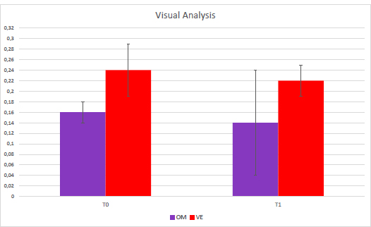
The visual analysis showed a mean value of 0.16 (± 0.02) for OM and 0.24 (± 0.05) for VE at T0. After bleaching (T1), the color match showed a mean value of 0.14 (± 0.1) for OM and 0.22 (± 0.03) for VE (Table 3 and Fig. 3).
| Resin Composites | VisualAnalysis(T0) | Visual Analysis(T1) |
|---|---|---|
| OM | 0.16 (± 0.02) | 0.14 (± 0.1) |
| VE | 0.24 (± 0.05) | 0.22 (± 0.03) |
4. DISCUSSION
The blending effect in dentistry is helpful since it neutralizes or minimizes color mismatches and/or the absence of adequate shade in the selected restorative material [ 20]. Recent research has focused on filler morphology in order to influence the color match between teeth and composites. This structural color technology is based on the interaction of reflection from supra-nano particles and light refraction. This phenomenon leads to single-shade universal composites, which intend to match all shades of the VITA color guide [ 7].
In 2021, Iyer et al. used three different composites (Tetric EvoCeram, TPH Spectra ST, and Omnichroma) to restore 15 occlusal cavities on resin teeth [ 6]. Color differences between composites and teeth (∆E00) were calculated for all reconstructions with the VITA Easyshade V spectrophotometer, and 30 evaluators gave their consideration regarding the color match using a visual scale from 1 (best match) to 3 (poor match). Tetric EvoCeram showed similar values in all cases, independently from the VITA color of the resin teeth. Omnichroma and TPH Spectra ST instead showed lower values on teeth with brighter VITA colors. The visual analysis showed the best color matches of TPH Spectra ST with brighter resin teeth and Tetric EvoCeram with darker resin teeth (VITA C2 and D3) [ 6]. These visual analysis results are difficult to compare with those of the present pilot study since, in the study by Iyer et al., the visual scale was divided into three points (Best, intermediate, and poorest match), while in this case, a more precise and detailed scale, made up of 5 evaluation degrees, was used [ 1].
Another recent in vitro study tested 120 resin teeth, with shades A1 to A4, filled with occlusal restorations performed with three different composites (Supra-nano-filled, Micro-hybrid filled and Clustered-nano-filled). Differences in color between teeth and resins appeared significantly lower when the supra-nano-filled composite was used to restore A2, A3, and A4 teeth shades [ 4].
In order to improve aesthetics, dental bleaching has become a common treatment [ 10]. Bleaching is not able to change the shade of composite restorations to a lighter color as it does on natural tooth structures; however, it can sometimes remove stains on composites and return them to their original shade [ 12, 21].
Pecho et al., in 2019, evaluated the influence of a professional bleaching gel on the color changes of resin composites. Ten discs were created from three different composite resins (Durafill, IPS Empress Direct, Amelogen Plus), and differences between tooth and resin color were obtained before bleaching and after the first and second bleaching application through a spectrophotometer (VITA Easyshade). Lightness values of all resins did not change after bleaching, and there were no statistically significant differences in color variations among all tested materials. This study reported that bleaching with 35% hydrogen peroxide is able to influence restoration color, but this influence is not perceived clinically [ 11].
Another study investigated color variations after bleaching using a universal one-shade composite (Omnichroma). Ten extracted sound teeth were prepared with a cervical cavity and then restored with Omnichroma. Instrumental analysis was carried out through a spectrophotometer on 5 points of the composite restoration, and visual analysis was performed by two examiners. After professional bleaching, lightness and color coordinate values were registered after 24 hours, 1, 2, and 4 weeks after bleaching procedures and then compared to the initial ones. There was no significant difference between these coordinates between restorations and teeth at each time interval, and the examiner's scores showed a perfect match between teeth and composites at all intervals [ 12]. This study presented interesting visual analysis results, but also, in this case, they were difficult to compare with those of the present pilot study. In a study by Mohamed et al., the visual scale was divided into three not-very-specific points (complete match, somewhat match, and non-match), while in this pilot study, a more impartial and detailed scale, made up of 5 evaluation degrees (Excellent, very good, not so good match, and obvious and huge mismatch), was used [ 1].
Recently, a study by Kobayashi et al. evaluated the effect of the structural color phenomenon on the color adjustment of restorations by investigating their color reproduction performance in cervical cavities of human incisors of various shades. Cavities were filled with Omnichroma, Estelite Σ Quick, Clearfil AP-X, and two experimental resin composites. The first (R1) was characterized by 5-50 nm fumed silica fillers, and the second (R2) with 100 nm spherical fillers. Color parameters (L, C, h) were measured along the centreline of the restorations and registered using a CIE camera. These data were used to calculate the color difference (∆E 00) between the restoration and the intact tooth. Omnichroma resulted in the material, which exhibited significantly lower ∆E 00 than other tested composite resins. Moreover, Omnichroma showed a higher color adjustment potential [ 8].
CONCLUSION
Within the limitations of this pilot study, it can be concluded that with both tested composites, an excellent color match between restoration and teeth can be obtained, both before and after tooth bleaching. The positive trend obtained in this pilot study should work as an incentive for a more well-structured study with a greater number of samples and tested materials.
If these results could be generalized and applied to all single-shade composites, an essential clinical breakthrough could be achieved. In fact, by using these types of composites, the operative timing could be shortened by avoiding the stratification of several composite masses, costs could be reduced, and the final result would be less skill-dependent than a layering technique. Furthermore, these composites seem to follow the physiological aging of natural teeth, and there could be a possibility of performing tooth bleaching without issues related to the aesthetic discomfort caused by the discoloration of previous restorations.
LIST OF ABBREVIATIONS
| BE | = Blending Effect |
| CEJ | = Cemento-enamel Junction |
| OM | = Omnichroma |
| VE | = Venus Diamond One |
ETHICS APPROVAL AND CONSENT TO PARTICIPATE
Extracted teeth were used according to the Ethical Committee of the University of Bologna approval (protocol No:71/2019/OSS/AUSLBO).
HUMAN AND ANIMAL RIGHTS
No animals/human were used in this research.
CONSENT FOR PUBLICATION
Informed consent was obtained from all participants.
AVAILABILITY OF DATA AND MATERIALS
The data supporting the findings of the article is available, upon request, directly from the corresponding author [V.C].
FUNDING
None.
CONFLICT OF INTEREST
The authors declare no conflict of interest, financial or otherwise.
ACKNOWLEDGEMENTS
The authors are extremely grateful to Dr. Shaniko Kaleci of the University of Modena & Reggio Emilia for data handling and processing.

