All published articles of this journal are available on ScienceDirect.

Tooth Color Differences between Digital Photography and Spectrophotometer in both Genders: An In vivo Study
Authors Info & Affiliations
Abstract
Background:
Tooth color is one of the most concerning issues in cosmetic dentistry. Different lighting, materials, and genders have influenced tooth color, hence, influencing the esthetic appearance. Knowing the differences between different materials to measure tooth color and differences in tooth color between genders could help in better understanding and choosing artificial tooth color.
Objective:
This study aims to compare two color-measuring methods, spectrophotometer and standardized digital photography, and gender differences in terms of tooth color.
Methods:
An evaluation of 300 vital maxillary central and lateral incisors was carried out on 150 adult participants. Tooth color was measured using the CIEL*a*b* color system on digital photography images and the Vita Easyshade spectrophotometer. The settings for digital photography were standardized, and the reliability of the method was tested. An independent sample t-test was used to compare the two color-measuring methods and gender differences in terms of tooth color, with a significant level set at p = 0.05.
Results:
There were statistically significant differences between Vita Easyshade Spectrophotometer and digital photography in all color spaces (L*, a*, and b*) for central and lateral incisors. There was a significant difference between males’ and females’ L* color space in digital photography for central (p = 0.04) and lateral (p = 0.05) incisors. The reliability of the digital photography method used in this study was high.
Conclusion:
Tooth colors were found to be lighter and more yellowish in VITA Easyshade compared to the digital camera. Females had lighter teeth compared to males in digital photography. The digital photography method showed excellent reliability.
1. INTRODUCTION
There are two methods to determine tooth color: conventional methods using the naked eye and shade guides [1] or digital methods, like spectrophotometer, colorimeter, and digital photography. Conventional methods using the naked eye are subjected to bias as many factors can affect the measurements [2-4]. However, for more reliable measurements, digital methods can be used as they are more reliable and standardized compared to conventional methods [5]. Several factors can affect color measurements, such as light sources, settings, and calibration processes [6, 7].
Several studies have compared spectrophotometer and digital photography [8-10]. These studies found a high proportion of agreement and a statically significant correlation between the two methods [8-10]. However, some of these studies used sunlight during photography [9, 10]. However, the direction of light, the size of the window [11], and the clarity of the sky can affect the amount of sunlight, hence, affect the glare. Very high sunlight (increased glare) or low sunlight (darkness) will affect the quality of photos and affect the color appearance. Another study [8] used standard and measurable light and photography settings and calibration, but the study was an in vitro study. Another study [12] used almost the same digital photography technique as used previously [8] for skin and tooth color measurement. However, this study did not compare spectrophotometer and digital photography.
The quality of light determines an object’s color during color measurement [13]. Therefore, color measurement should be done under daylight at a color temperature ranging between 5000K to 6500K [14]. The color’s perception would change due to different lighting conditions [15]. Therefore, in order to get more reliable results, light conditions during color measurement and photography should be controlled.
Moreover, some studies [9, 10] converted the color CIELab values of Adobe Photoshop in digital photography using a mathematical formula for the CIELab system. Adobe Photoshop website states that the Adobe Photoshop software uses the CIELab color system, and there is no need to convert it again to CIELab [16].
Color calibration is important to achieve accuracy in color measurement using digital photography [17]. The calibration decreases the color differences between the real tooth color and the photograph color to a small difference of ∆E = 2.1 [7]. Grey cards [18] and calibration boards with different color ranges [19] have been used for calibration in dentistry and medicine. This is in accordance with the belief that the human body, including the oral cavity and teeth, consists of a different range of colors [7, 19]. A study found that colors can be replicated with a color difference of ∆E = 2.1 between the original color and the photograph if an appropriate calibration protocol is used [7].
Moreover, previous research measured the color differences (ΔE) between a digital camera and a spectrophotometer but did not explain the color characters [8-10, 20, 21].
Battery-operated devices tend to have their battery efficiency decrease every time they are used. The strength of illumination (flash) that is powered by the battery is subjected to the charged level of the battery. Therefore, as digital photography color measurement accuracy depends on the light condition during photography, a stable illumination power that is functioned by electricity, such as strobe lights with soft boxes, could be used.
According to our limited knowledge, no studies have been conducted to compare tooth color between males and females using a digital camera. Most of the studies were carried out using subjective visual shade matching or spectrophotometer.
On that account, the objectives of this study are to evaluate anterior tooth color differences between the digital photography method used by [12] Moazam et al. in a study and spectrophotometer and between males and females. The null hypotheses of this study are that there is no statistically significant difference in tooth color between the Vita Easyshade spectrophotometer and digital photography, as well as no statistically significant gender differences.
2. MATERIALS AND METHODS
2.1. Sample Size Calculation and Recruitment
The minimum sample size for this in-vivo cross-sectional study was calculated using the single mean formula: n (minimum required sample size) = (1.96σ (Standard deviation from a previous study)/∆ (margin of error))2. By referring to a previous study [22], the standard deviation was set at 35%, and the margin of error was 6. Therefore, n= ((1.96×35)/6)2, n =131. By considering a 10% of non-response rate, the minimum sample size for this study was 144 respondents.
Convenience sampling was used to recruit participants. The sampling and data collection were done between April, 2021, to June, 2021, at the School of Dental Sciences, Health Campus, USM, Malaysia.
2.2. Eligibility Criteria
The inclusion criteria for the study were: systemically healthy Malaysians with sound anterior teeth. The exclusion criteria were participants wearing dental braces, overlapped, rotated, or tilted teeth, anterior fillings, caries, or prostheses, and participants with missing anterior teeth.
All participants were asked to brush their teeth for two minutes and remove any lipstick before the color-measuring process.
2.3. Ethics Approval and Consent to Participate
The ethical approval was obtained at the School of Dental Sciences at the Health Campus, Universiti Sains Malaysia. Ethical approval was obtained from the Human Research Ethics Committee No. USM/JEPeM/20110552 and in accordance with the Helsinki Declaration.
All participants signed a written consent form for their voluntary participation before the study, with the ability to withdraw without any consequences.
2.4. Digital Photography
The light position and photography settings were done according to a previous study [12]. Participants were seated in an upright position looking forward to the camera. The photography was done in a completely dark room to prevent the interference of outside light sources, such as sunlight. Two Lastolite lumen 8 F400 light strobes with soft boxes were used with color temperatures ranging between 5300 to 5600k during the photo-taking session. The camera’s flashlight was used for triggering the strobe lights, and the camera’s batteries were ensured to be fully charged to obtain the full intensity of the flashlight.
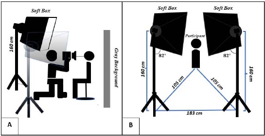
A digital single lens reflex (DSLR) camera (Nikon D90) (Malaysia) and a Nikon N AF-S MICRO NIKKOR 105 mm 1:2.8 G ED lens were used for tooth photography. The camera settings were set at RAW for photo format, 200 for ISO, 20 for aperture, and 1/125 for shutter speed, and the white/black balance was set to Auto. The photography settings were fixed at all times, as shown in Fig. (1).
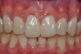
The distance between the participant and the camera was set according to the appearance of all anterior teeth and the mesial side of the first premolar only on the camera screen, and teeth photos were taken, as shown in Fig. (2).
2.5. Calibration and Color Measurement Process of the Digital Photos
A photo was also taken for the calibration board ColorChecker Digital SG(semi-gloss) (X-Rite PANTONE), which was placed in front of a patient’s face. The calibration board photo was used as the reference photo for creating the calibration profile on ColorChecker Camera Calibration Software V. 2.0. The calibration profile was uploaded to Adobe Lightroom 2020 V.5.RU-EN software and was applied to all photos. Then, Adobe Photoshop 2020 V.5.RU-EN was used for CIELab color measurement of the middle third of the right central and lateral incisors.
2.6. Spectrophotometer Color Measurement
Tooth CIELab color measurements were also done on the middle third of the right central and lateral incisors using VITA Easyshade ®Advance 4.0 (VITA Zahnfabrik, Bad Säckingen, Germany). The device was calibrated using the calibration block in the device before every color measurement. The manufacturing instructions were followed.
2.7. Statistical Analysis
An independent sample t-test was used to compare the CIELab color readings between the two color measurement methods and between genders for central and lateral incisors in IBM SPSS Statistics 26.0. The Intra Class Correlation ICC was used to test the reliability of the digital photography method. 30% of the participants' teeth were photographed twice and calibrated, and colors were measured. The means of the two readings (the first and second photos) were used for the reliability test using correlation in the reliability statistic test in IBM SPSS Statistics 26.0. A p-value of 0.05 was considered significant. The normality assumption was fulfilled, and CI was set at 95%. Missing data were excluded from the analysis.
3. RESULTS
A total of 150 Malaysian participants were included in this study (75 males and 75 females) and were evaluated for 150 right central incisors and 150 right lateral incisors. It was more than the minimum sample size of the study that was calculated earlier. No missing data was recorded. Participants aged between 20 to 55. Out of 150 participants, 139 participants (92.7%) were in their 20s, 7 (4.7%) in their 30s, 3 (2%) in their 40s, and 1 (0.7%) in their 50s.
3.1. Reliability of the Method
The reliability test showed a significant relationship between the two readings with p = 0.000, with an excellent intraclass correlation value [23]. For single means, it was 0.999 and for average measures, it was 1.000.
| Tooth | Colour Space | Gender | Vita | Camera | t | df | Mean difference | Mean difference(95% CI) | p-value | |||
| - | - | - | Mean | (SD) | Mean | (SD) | - | - | - | - | - | - |
| (Central) | L | Male | 84.74 | 4.33 | 68.80 | 3.98 | 23.48 | 146.96 | 15.94 | (14.60, 17.28) | 0.00* | |
| Female | 85.10 | 3.64 | 70 | 2.91 | 28.5 | 141.23 | 15.10 | (14.04, 16.17) | 0.00* | |||
| a | Male | -1.41 | 0.54 | 4.24 | 4.88 | -9.98 | 148 | -5.65 | (-6.77, -4.53) | 0.00* | ||
| Female | -1.53 | 0.41 | 3.41 | 1.46 | -28.20 | 148 | -4.95 | (-5.29, -4.60) | 0.00* | |||
| b | Male | 19.34 | 4.80 | 13.16 | 3.50 | 9.01 | 148 | 6.18 | (4.82, 7.54) | 0.00* | ||
| Female | 18.15 | 4.02 | 12.61 | 3.77 | 8.70 | 147.40 | 5.53 | (4.28, 6.79) | 0.00* | |||
| (Lateral) | L | Male | 82.60 | 4.89 | 63.44 | 4.52 | 24.93 | 147.08 | 19.16 | (17.64, 20.68) | 0.00* | |
| Female | 82.77 | 4.06 | 64.75 | 3.32 | 29.78 | 142.39 | 18.02 | (16.82, 19.21) | 0.00* | |||
| a | Male | -1.10 | 0.85 | 5.64 | 1.71 | -30.65 | 148 | -6.74 | (-7.18, -6.31) | 0.00* | ||
| Female | -1.30 | 0.51 | 5.15 | 1.84 | -29.20 | 148 | -6.45 | (-6.89, -6.01) | 0.00* | |||
| b | Male | 21.36 | 4.48 | 16.28 | 7.62 | 4.98 | 119.70 | 5.08 | (3.06, 7.10) | 0.00* | ||
| Female | 21.29 | 4.11 | 15.00 | 4.17 | 9.29 | 147.97 | 6.28 | (4.95, 7.62) | 0.00* | |||
3.2. Color Differences between Vita Easyshade Spectrophotometer and Digital Photography
3.2.1. Color Space L*
The results in Table 1 show significant differences in tooth color between the Vita Easyshade spectrophotometer and digital photography, as Vita Easyshade showed lighter teeth color than the digital camera in males and females for incisors.
The p-value was statistically significant between the two methods in color space L* for central incisors in males [ t (23.48) = 146.96, 95% CI (14.60, 17.28), p =0.000] and in females [t (28.5) = 141.23, 95% CI (14.04, 16.17), p = 0.000]. It was also statistically significant in color space L* for lateral incisors in males [t (24.93) = 147.08, p = 0.000] and females [t (29.78) = 142.39, p = 0.000] (Table 1).
3.2.2. Color Space a*
For color space a*, the Vita Easyshade spectrophotometer means were in a negative sign for males and females in the incisors. On the other hand, the means of digital camera were positive. This indicates that tooth color readings for the Vita Easyshade spectrophotometer were green, and they were red for digital photography.
The p-value was statistically significant between the two methods in color space a* for central incisors in males [ t (-9.98) = 148, p = 0.000] and females [t (-28.20) = 148, p = 0.000]. It was also significant for lateral incisors in males [ t (-30.65) = 148, p = 0.000] and females [ t (-29.20) = 148, p = 0.000], as mentioned in Table 1.
3.2.3. Color Space b*
Vita Easyshade spectrophotometer presented tooth color as more yellowish than digital photography.
The differences were significant in central incisors for males [mean(Vita) (SD): 19.34 (4.80)], [mean(Camera) (SD): 13.16 (3.50)] with [ t (9.01) = 148, p = 0.000] and females [mean(Vita) (SD): 18.15 (4.02)], [mean(Camera) (SD): 12.61(3.77)] with [t (8.70) = 147.40, p = 0.000]. The differences were also significant in lateral incisors for males [mean(Vita) (SD): 21.36 (4.48)], [mean(Camera) (SD): 16.28 (7.62)] with [t (4.98) = 119.70, p = 0.000] and females [mean(Vita) (SD): 21.29 (4.11)] [mean(Camera) (SD): 15 (4.17)] with [t (9.29) = 147.97, p = 0.000], as mentioned in Table 1.
3.3. Color Differences between males and Females’ Tooth Color in Vita Easyshade Spectrophotometer and Digital Photography
There was a significant difference in tooth color between males and females for the L* color spaces for central incisors [ t (-2.12) = 135.61, 95% CI (-2.33, -0.07), p = 0.04] and lateral incisors [ t (-2.02) = 135.82, 95% CI (-2.59, -0.03), p = 0.05] in digital photography (Table 2).
Table 2 shows no significant differences between the tooth color of males and females on the Vita Easyshade spectrophotometer in all color spaces (L*, a*, and b*) for central and lateral incisors. Moreover, Table 2 also shows no statistically significant difference between males' and females’ tooth color for a* and b* color spaces for central and lateral incisors.
4. DISCUSSION
In general, the first null hypothesis of the study was rejected, and the second null hypothesis was partially rejected. The differences between the Vita Easyshade spectrophotometer and digital photography are pronounced. Vita Easyshade spectrophotometer showed lighter and more yellowish tooth color than the camera readings. Vita Easyshade spectrophotometer also showed the teeth colors range in green-yellow dimension, while the camera presented tooth color in red-yellow dimension. The results also showed that males had darker teeth color while females had lighter teeth upon digital camera color measurement. Moreover, the results showed that the method used for tooth color measurement using digital photography is reliable.
| Device | Tooth | Colour space | Males | Females | t | df | Mean Difference | Mean difference (95% CI) | p-value |
|---|---|---|---|---|---|---|---|---|---|
| Mean (SD) | Mean (SD) | - | - | ||||||
| Vita Easyshade Spectrophotometer | Central | L | 84.74 (4.33) | 85.10 (3.64) | -0.55 | 143.75 | -0.36 | (-1.65, 0.93) | 0.58 |
| a | -1.41 (0.54) | -1.53 (0.41) | 1.54 | 138.17 | 0.12 | (-0.03, 0.28) | 0.13 | ||
| b | 19.34 (4.80) | 18.15 (4.02) | 1.65 | 143.58 | 0.10 | (-0.24, 2.62) | 0.17 | ||
| Lateral | L | 82.60 (4.89) | 82.76 (4.06) | -0.22 | 143.11 | -0.16 | (-1.61, 1.29) | 0.83 | |
| a | -1.10 (0.85) | -1.30 (0.51) | 1.75 | 121.65 | 0.20 | (-0.03, 0.43) | 0.08 | ||
| b | 21.36 (4.48) | 21.28 (4.11) | 0.11 | 146.93 | 0.08 | (-1.31, 1.46) | 0.91 | ||
| Digital Camera | Central | L | 68.80 (3.98) | 70.00 (2.91) | -2.11 | 135.61 | -1.20 | (-2.33, -0.07) | 0.04* |
| a | 4.24 (4.88) | 3.41 (1.46) | 1.41 | 87.198 | 0.83 | (-0.34, 2.00) | 0.16 | ||
| b | 13.16 (3.50) | 12.61 (3.77) | 0.92 | 147.21 | 0.55 | (-0.63, 1.72) | 0.36 | ||
| Lateral | L | 63.44 (4.52) | 64.75 (3.32) | -2.02 | 135.82 | -1.31 | (-2.59, 0.03) | 0.05* | |
| a | 5.64 (1.71) | 5.15 (1.84) | 1.70 | 147.12 | 0.49 | (-0.08, 1.07) | 0.09 | ||
| b | 16.28 (7.62) | 15.00 (4.17) | 1.28 | 114.68 | 1.28 | (-0.71, 3.27) | 0.20 |
The differences between the Vita Easyshade spectrophotometer and digital photography are usually because of the different lighting settings during measurements [13] and the calibration system [19]. Likewise, the calibration system can affect color [19].
Vita Easyshade spectrophotometer and digital camera showed lighter central incisors than lateral incisors in the present study. Lateral incisors were more yellowish than central incisors in both methods. This was supported by another study [4]. This could be due to the anatomy of central and lateral incisors since the central incisors have thicker enamel compared to lateral incisors [9].
Even though there was no significant difference in tooth color between males and females using digital photography for color spaces a* and b*, the means of tooth color readings for females were less reddish and less yellowish than males for central and lateral incisors. These differences in lightness, redness, and yellowness could be due to the difference in dentine thickness, as males have thicker dentine than females by 6.5% [24].
In contrast, another study [4] used a VITA Easyshade V spectrophotometer to measure tooth color. This study showed no significant differences between males' and females’ tooth color in lightness, while there was a significant difference in a* color space (redness). They also reported that the mean L* value of central incisors was higher than lateral incisors and canines. This means that the central incisor was lighter than the lateral incisor. It was also found that central and lateral incisors had high values of the color space b*. The differences between this present study and the previous study could be due to the different models of Vita Easyshade spectrophotometer used and different populations.
4.1. Limitation of the Study
The study was done only on Malaysian participants, mostly undergraduate students. The results may vary in different age groups and different races. Moreover, this study was done on the central and lateral incisors only. A wider range of examined teeth and a larger population would provide more information.
4.2. Scope of Further Studies
Digital photography has a lot of benefits. Additional to the common advantages of dental photography, it can help in reliable color measurement, and it saves clinical time. Further studies can be conducted on different races, teeth, and wider age groups. Moreover, further studies can be done to measure the accuracy of using this method to print colors and to measure other structures in the oral cavity, such as lips, gingival, and mucosa colors. Digital smile designers, dentists, researchers, and dental technicians could take advantage of this method to get better results.
CONCLUSION
Within the limitations of this current study, it was concluded that there were statistically significant differences in tooth color measurements between the Vita Easyshade spectrophotometer and digital photography for males and females in central and lateral incisors. Furthermore, there were no statistically significant differences in tooth color between males and females in the Vita Easyshade spectrophotometer, while there were partially statistically significant differences in tooth color between males and females in digital photography of central and lateral incisors. The standardized digital dental photography method used in this study for color measurement is highly reliable.
ETHICS APPROVAL AND CONSENT TO PARTICIPATE
The ethical approval was obtained at the School of Dental Sciences at the Health Campus, Universiti Sains Malaysia. Ethical approval was obtained from the Human Research Ethics Committee No. USM/JEPeM/20110552.
HUMAN AND ANIMAL RIGHTS
No animals were used in this research. All procedures performed in studies involving human participants were in accordance with the ethical standards of institutional and/or research committee and with the 1975 Declaration of Helsinki, as revised in 2013.
CONSENT FOR PUBLICATION
All participants signed a written consent form for their voluntary participation before the study, with the ability to withdraw without any consequences.
STANDARDS OF REPORTING
STROBE guidelines were followed.
AVAILABILITY OF DATA AND MATERIALS
The data supporting the findings of the article is available in the Zenodo Repository at https://zenodo.org/record/ 8250872, reference number DOI 10.5281/zenodo.8250872.
FUNDING
The study is funded by the Universiti Sains Malaysia, School of Dental Sciences (RUI Grant 1001/PPSG/8012371.
CONFLICT OF INTEREST STATEMENT
All authors disclose that there are no conflicts of interest in any financial and personal relationship with other people or organizations that could inappropriately influence or cause bias to their work or in the companies whose materials are included in this article.
ACKNOWLEDGEMENTS
The authors would like to thank the participants who participated in this study. They would also like to thank Universiti Sains Malaysia, School of Dental Sciences, for granting this study and providing the materials and place to conduct it.

