All published articles of this journal are available on ScienceDirect.

Assessing The Proximity of the Roots of Maxillary Molars and Premolars to the Maxillary Sinus in UAE Residents
Abstract
Background:
The proximity of the roots of the maxillary posterior teeth (MPT) to the maxillary sinus (MS) may result in an increased risk of the development of MS pathology. Moreover, surgical procedures for the MPT, which are close to the MS can result in several complications like oroantral communication and displacement of roots into the MS.
Objective:
This cross-sectional study evaluated the MPT's proximity to the MS using digital panoramic radiography in a sample of individuals living in the United Arab Emirates (UAE).
Methods:
A total of 141 panoramic radiographs showing the presence of all MPT were retrieved from the Oral Radiology Department of Ajman University, Fujairah Campus. Thus, the proximity of 1410 MPT to the MS was evaluated on a scale of zero to three.
Results:
The roots of the first and the second molars are the closest to the MS (χ2 = 374.612; p 0.000). The distance between the roots of MPT and MS did not differ statistically between males and females (χ2 = 2.44, p= .124), nor between the right and left sides (χ2 = 0.46, p= .872). However, there was a highly significant negative correlation between the root-sinus proximity and age. (rho= -.201, p < .001).
Conclusion:
In UAE residents, the roots of the first and second molars are closer to the MS than other MPT. Therefore, more precautions should be taken when doing a surgical procedure in this region.
1. INTRODUCTION
The proximity of the roots of maxillary posterior teeth (MPT) to the maxillary sinus (MS) may result in an increased risk of the development of MS pathology like sinusitis [1]. Moreover, surgical procedures for the MPT, which are close to the MS can result in several complications like oroantral communication and displacement of roots into the MS. Therefore, studying the relationship of MPT and the MS floor is important forendodontics, dental prosthetics, periodontal surgery, exodontia, implantology, and orthodontic treatment [2, 3].
Located in the posterior maxilla, the MS is the largest of the paranasal sinuses and is closely associated with the roots of the MPT. As a result of pneumatization, the floor of the MS extends into the posterior alveolar bone, causing root apices to protrude into the MS [4]. Furthermore, the topography of the MS floor differs greatly, which influences the development of different sinus-root relationships [3, 5].
Clinically, panoramic radiography and computed tomography, like cone-beam computed tomography (CBCT) are used to assess the relationship between MPT and MS. While CBCT is known to be more accurate, the most common radiographic technique in dentistry is still panoramic radiography. It has been shown that for the assessment of dental implants, panoramic radiography is the only imaging procedure used by 64% to 95% of dental practitioners [6]. The Council on Scientific Affairs of the American Dental Association recommends using CBCT only when it can benefit patient care, improve patient safety, or improve clinical outcomes significantly. Moreover, CBCT should not be performed for screening purposes [7]. The panoramic radiograph provides comprehensive information about dentition, multiple adjacent structures, and their relationships bilaterally. However, it shows only two dimensions and has certain disadvantages, such as magnification, superimposition, blurring, ghost images, and spreading of curved structures [3]. As differences in population characteristics may contribute to anatomical variations among individuals, we conducted this study to evaluate the proximity of the MPT to the MS, for the first time, among UAE residents.
2. MATERIALS AND METHODS
In this cross-sectional, retrospective study, 141 panoramic radiographs with their dental records were obtained from the Department of Oral Radiology at Ajman University, Fujairah Campus, between 2016 and 2018. The study was performed following the Declaration of Helsinki and approved by the Research Ethics Committee of Ajman University, College of Dentistry (Ref. SSF-2017/18-02-S). Panoramic radiographs were selected within the inclusion criteria. These radiographs showed the root apices of the MPT and the floor of the MS. These digital panoramic radiographs were taken by a Planmeca system (Planmeca Promax® 3D, Finland), operated at 8mA and 66kVp with an exposure time of 19 seconds and stored digitally.
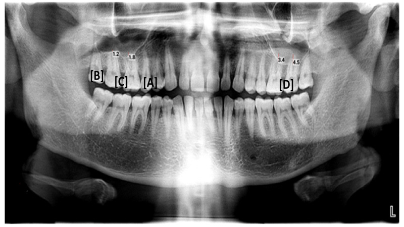
Included patients were of both genders over 18 years old, fully developed MS, and a UAE residents who attended Ajman University, Fujairah Campus Clinic. The images used in this study included only teeth with clearly visible root apices showing bilateral MPT without missing teeth. Records and images that show MS or nasal surgery, the extraction of the MPT, orthodontic treatment in the upper arch, features of infection or pathology in the examined area, artifacts in the MS, or unclear were excluded from the study. Two observers independently examined the images on the same monitor in a dim room. The images were adjusted for brightness, contrast, and magnification using the Planmeca software's tools (Planmeca Promax® 3D, Finland). If there were discrepancies in the individual assessments, the images were re-evaluated till reaching a consensus. On the images, measurements were taken from the related deepest point of the floor of the MS to the tips of the root of each MPT using built-in measurement tools. The criteria used before [8] have been modified to be as follows: 0: No roots are in contact with the cortical borders of the MS; 1: One or more roots are in contact with the MS; 2: One or more root apexes are projecting on the MS cavity for a distance less than 2 mm; 3: One or more root apexes are projecting on the MS cavity for a distance more than 2 mm (Fig. 1). The scores were further categorized as to whether the roots of the teeth project onto the MS (scores 2 and 3) or not (scores 0 and 1).
Statistical package for the social sciences (SPSS) 28 software was used for the statistical analyses. A descriptive statistics was used to describe the relationship between each posterior maxillary tooth and the MS. Variables, tables, figures, and numerical values were included. In addition, the Chi-square test was used to find the differences between different variables. Differences were deemed statistically significant if (p<0.05).
3. RESULTS
This cross-sectional retrospective study analyzed 141 panoramic radiographs of different individuals. Among them, 109 (77.3%) were males, and 32 (22.7%) were females, ranging in age from 18 to 60, with a mean age of 31.4 years. In total, the proximity of 1410 MPT to MS was assessed (705 teeth on each side).
Figs. (2 and 3) illustrate the relationship between the MPT and the floor of the MS according to the criteria used and whether roots protrude into the MS, respectively. The maxillary first and second molars are closest to the MS, while the maxillary first premolar is the furthest from the MS (Table 1 and Fig. 4). A statistically significant difference was seen in the relationship between the MPT and the MS (χ2 = 374.612; p 0.000).
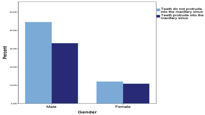
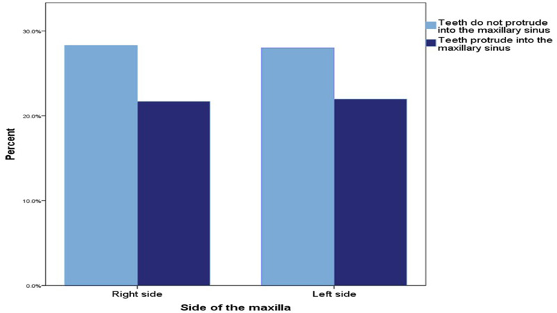
The roots of the MPT in this study were closer to the MS in females than in males, but this difference was not statistically significant (χ2 = 2.445, p= .124) (Fig. 5). Additionally, there was no difference between the two sides (χ2 = 0.46, p= .872) (Fig. 6). However, there was a highly significant negative correlation between the proximity of MPT to the MS and age (rho= -.201, p < .001).
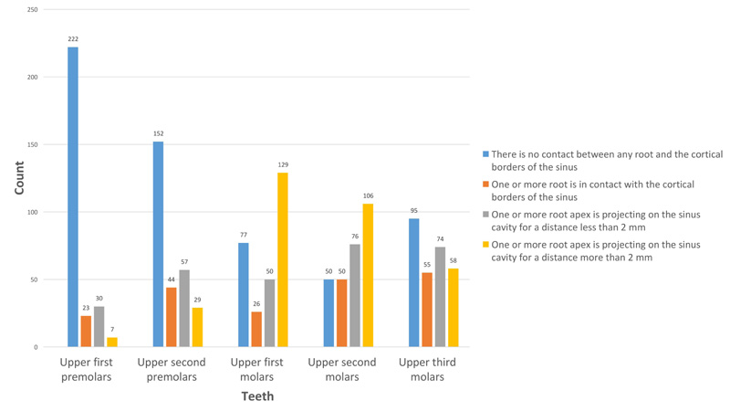
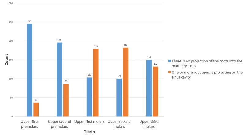
Table 1.
| - | There is no Contact between any Root and the Cortical Borders of the Sinus | One or More Root is in Contact with the Cortical Borders of the Sinus | One or More Root Apex is Projecting on the Sinus Cavity for a Distance Less than 2 mm | One or More Root Apex is Projecting on the Sinus Cavity for a Distance more than 2 mm | Total | |
|---|---|---|---|---|---|---|
| Upper Right Third Molar | 50 | 24 | 38 | 29 | 141 | |
| Upper Right Second Molar | 33 | 20 | 37 | 51 | 141 | |
| Upper Right First Molar | 45 | 9 | 22 | 65 | 141 | |
| Upper Right Second Premolar | 76 | 22 | 29 | 14 | 141 | |
| Upper Right First Premolar | 106 | 14 | 16 | 5 | 141 | |
| Upper Left First Premolar | 116 | 9 | 14 | 2 | 141 | |
| Upper Left Second Premolar | 76 | 22 | 28 | 15 | 141 | |
| Upper Left First Molar | 32 | 17 | 28 | 64 | 141 | |
| Upper Left Second Molar | 17 | 30 | 39 | 55 | 141 | |
| Upper Left Third Molar | 45 | 31 | 36 | 29 | 141 | |
| Total | 596 | 198 | 287 | 329 | 1410 | |
4. DISCUSSION
The fact that MPT is close to MS contributes to multiple complications during disease progression and dental treatment of these teeth. There have been several studies looking at the relationship between MPT and the MS floor, but none dealt with populations living in the UAE. Since differences in population characteristics may lead to variations in this relationship, assessing it in the UAE population was essential. So, we examined this proximity in UAE residents. Indeed, it is possible for the MS to expand into the alveolar process and to extend between roots, so that the root apex encompasses the MS floor, covered by only a thin bone plate or a layer of the MS membrane. Additionally, in many cases, the roots penetrate the MS [9]. Therefore, if the tooth has a pathological condition, the disease can easily spread to the MS, causing infection and inflammation. Additionally, inexperienced tooth extraction can result in an oro-antral communication and foreign material in the MS. A variety of radiographic techniques can be used to investigate the relationship between the roots of the MPT and the MS, including two-dimensional techniques, such as panoramic radiographs. A panoramic radiograph can provide a general idea of further precautions to be taken during the extraction of MPT [8].
The relationship between MPT and MS was analyzed using panoramic images, which fulfill this study's requirement for large samples. The panoramic radiograph can provide the surgeon with signs of possible root extension into the MS. Although panoramic radiography has some shortcomings, such as reduced sharpness and image distortion, it is still a viable option for routine clinical situations. It is also crucial to determine whether a CT scan is necessary before surgery [10]. Comparing panoramic radiography with CBCT, very high and high agreement was found when roots were distant from the MS and when roots protruded into the MS, respectively [5]. However, in borderline cases where the roots are very close or contacting the floor of the MS, the differences between the two imaging modes are evident. Accordingly, the panoramic radiograph may show a larger portion of the root protruding inside the MS than the CT image. It is also possible that the roots were lateral to the MS cavity, but they were mistakenly viewed as protruding into the sinus on the panoramic radiograph [11]. However, the roots extending into the MS cavity caused interruptions of the MS floor on the panoramic radiograph [3, 12]. Hence, a previous study concluded the decision to request CBCT should only be considered when the panoramic radiographs reveal that the root is protruding into the MS [10].
Inclusion criteria for the current experiment included samples containing all maxillary teeth. The extraction of maxillary molars may cause pneumatization of the MS, leading to root apices being projected onto the MS [12]. This may occur due to the absence of chewing stimulation for the alveolar process, resulting in higher osteoclastic activity and reconstruction MS and alveolar bone [13].
According to the current study, the upper first and second molars are the closest teeth to the MS, while the upper first premolar is the farthest one. The findings of this study are consistent with several previous studies that have shown that the roots of the maxillary first and second molars are the most proximate to the MS [8, 14, 15] and the second molar was the nearest to the MS [13, 16-18]. Meanwhile, the furthest root from the MS was that of the upper first premolar [14, 16, 18]. These results indicate that MS in this region might be at higher risk of developing dental infections. It also highlighted the need for exceptional care when treating or extracting upper first and second molars. A previous study suggested that the general dentist should avoid removing upper molars when these teeth are shown to be projected onto the MS on the panoramic radiograph, and to consult with an oral surgeon to avoid further complications from such extractions [8]. In contrast to our findings, a previous study found that the maxillary third molar was the second nearest maxillary posterior tooth to the MS after the maxillary second molar [17]. This controversy can in part be explained by the different types of populations studied in various reports and supports the clinical relevance of conducting our study among UAE residents.
Although the roots of MPT in females look closer to the MS than in males in the current study, this difference was not statistically significant. This is consistent with other studies which found that there were no differences in the proximity of MPT to MS between males and females [13, 15, 16, 18]. In contrast, different studies found that the protrusion of roots of the upper teeth into the MS is much more common in males than in females [1, 17, 19]. This may be attributed to differences in growth patterns between genders and longer roots found in males [19]. In agreement with other previous studies [13, 15-18], we do not find a significant difference in MPT proximity to MS between the left and right sides.
The study found that the distance between MPT roots and the MS floor increases with age, indicating that younger adults had a higher probability of developing odontogenic sinusitis due to iatrogenic factors and developing oroantral communication after MPT extraction than older age groups. The results are consistent with previous studies [1, 13, 15]. It has been suggested that the functional attrition of the teeth is increased with aging. As a result of the clinical crown reduction, secondary cementum formed at the root apices, increasing the distance between the floor of MS and apices of the roots of the MPT [13]. Additionally, another study concluded that the MS volume will decrease after the MS has been fully formed unless something interferes, such as a tooth extraction, which causes pneumatization of it [20]. The anatomical fact may also explain this negative relationship that the physiological volume increase of the MS by pneumatization typically stops with the upper third molar eruption by age ~21–30, after which the MS volume starts to decline [21]. Radiographs of individuals from a variety of countries who reside in the UAE are included in our database. A wide range of patients was included, ranging from 18 to 60 years of age, and 1410 MPT were well evaluated. Studying panoramic radiographs to evaluate the relationship between MPT and MS was a limitation of this study. A study using CBCT would be more accurate. However, a study such as this, with its exclusion of any extracted MPT, requires a larger sample size, which is difficult to achieve by CBCT. Knowing that it is not ethically acceptable to use CBCT with its radiation hazards unless it is indicated.
CONCLUSION
In UAE residents, the roots of the first and second molars are closer to the MS than other MPT. Therefore, more precautions should be taken when doing a surgical procedure in this region. There is no difference in this relationship based on gender or the side of the jaw. Meanwhile, the root proximity of the MPT to the MS is reduced with age.
LIST OF ABBREVIATIONS
| UAE | = United Arab Emirates |
| SPSS | = Statistical package for the social sciences |
| MPT | = Maxillary posterior teeth |
| MS | = Maxillary sinus |
AUTHORS’ CONTRIBUTIONS
MAA was responsible for the study design. MAA, RAA and RMA did the analyses. RAA and RMA helped in the study design, data collection, and interpretation. MAA, RAA and RMA draft and revise the manuscript. All authors have read and approved the final manuscript.
ETHICS APPROVAL AND CONSENT TO PARTICIPATE
The Research Ethics Committee approved the study of Ajman University, College of Dentistry (Ref. SSF-2017/18-02-S). Date: 27.1.2018
HUMAN AND ANIMAL RIGHTS
No animals were used in this research. All research procedures on humans were followed following the ethical standards of the committee responsible for human experimentation (institutional and national), and with the Helsinki Declaration of 1975, as revised in 2013 (http://ethics.iit.edu/ecodes/node/3931).
CONSENT FOR PUBLICATION
All participants’ parents signed an informed consent regarding their participation in this research and their agreement regarding the publication of the data gained in the current research.
STANDARDS OF REPORTING
STROBE guidelines were followed.
AVAILABILITY OF DATA AND MATERIALS
The data supporting the findings of the current research are available on Mendeley, V2, DOI: 10.17632/7wck3nczbp.2
FUNDING
None.
CONFLICT OF INTEREST
The authors declare no conflict of interest, financial or otherwise.
ACKNOWLEDGEMENTS
Declare none.

