All published articles of this journal are available on ScienceDirect.

Effect of Orthophosphoric Acid and Er:YAG Laser Etching on Micro-shear Bond Strength to Enamel: An In Vitro Pilot Study
Authors Info & Affiliations
Abstract
Background:
Dental conditioning is one of the most important phases during enamel bonding procedures to obtain clean surfaces, smear layer removal with collagen active sites and hydroxyapatite exposure.
Objective:
The aim of this study was to compare the micro-shear bond strength (µSBS) of different adhesive systems after two different etching techniques: 37% orthophosphoric acid (H3PO4) and Er:YAG laser.
Methods:
Ninety permanent extracted molars were embedded into epoxy resin blocks and sectioned longitudinally. Specimens were randomly assigned to one of the following groups (n=30), depending on the etching protocol: 37% H3PO4 for 30 s (Group 1), Er:YAG laser 100mJ-10Hz (Group 2), and Er:YAG laser 100mJ-10Hz followed by 37% H3PO4 for 30 s (Group 3). Each group was further divided into two subgroups depending on the bonding agent used on enamel (n=15): A) EE-Bond (Tokuyama) and B) Peak universal (Ultradent). A two-way analysis of variance (two-way ANOVA) was conducted and the level of significance was set to p=0.05.
Results:
The etching procedure was a significant factor influencing the results (p=0.006), while no differences were observed for the two adhesive systems tested (p>0.05). Group 3 recorded the highest bond strength values, according to the following sequel: Group 3 < Group 2 < Group 1 < 0.05.
Conclusion:
The combination of phosphoric acid etching with Er:YAG laser provided the most favourable bond strength to enamel. Further morphological studies are currently ongoing.
1. INTRODUCTION
Dental conditioning is one of the most important phases during enamel bonding procedures to obtain clean surfaces, smear layer removal with collagen active sites and hydroxyapatite exposure. Consequently, a direct interaction between the bonding agent and the dental hard tissue is enhanced by creating an increased contact surface [1]. The bonding mechanism is then completed with the polymerization of the resins into the enamel micro-porosities [2].
Multi-step bonding systems rely on a previous application of 35-37% orthophosphoric acid (H3PO4) gel for 30 s to condition the enamel surface. Acid etching removes up to 10 µm of enamel surface and creates a layer with 5-50 µm micro-porosities [3]. Water rinsing after etching is mandatory for completely cleaning the enamel from the etchant’s chemicals (mainly silica) as they can jeopardize the bond strength [1].
In modern dentistry, lasers are often used due to their favorable tissue interaction and their efficacy in reducing invasiveness and patient discomfort. Erbium-doped Yttrium Aluminium Garnet (Er:YAG) laser presents a 2.940 nm wave length that is adsorbed electively from the water present inside the tissues and this feature makes it ideal for hard tissues treatment [4]. This laser operates on water molecules, heating them and determining their transition from liquid to steam status; this causes a pressure increase with successive micro-explosions and organic matrix removal. Er:YAG laser was proposed as an enamel surface conditioner before bonding procedures as it can act selectively on enamel hydroxyapatite crystals, producing an irregular surface that permits correct micro-mechanical retention [4, 5]. The use of these lasers has several advantages, such as the low invasiveness and the surface’s decontamination. Moreover, no heat is expected to be produced, and this aspect reduces post-operative complications and relapses [6, 7]. in vitro studies showed that enamel surfaces exposed to laser radiations are more resistant to acids attack [8, 9].
Nowadays, several adhesive systems are available to the clinicians, of which universal adhesives represent the last introduced to the market. While the etching step with phosphoric acid is mandatory for the multi-step total-etch systems, universal adhesives have the versatility to be used with/out a previous etching step, according to the dental substrate to be bonded [10-12]. Despite the interest in the use of Er:YAG laser in adhesive dentistry, there are still not so many reports on its effects on bond strength of different adhesive systems to enamel [4, 6].
Therefore, the aim of this study was to compare the micro-shear bond strength (µSBS) of two adhesive systems, after two different etching techniques: 37% orthophosphoric acid and Er:YAG laser. The null hypotheses tested were that (1) the etching mode and (2) the type of adhesive system does not influence the µSBS to enamel.
2. MATERIALS AND METHODS
Ninety permanent sound human molars, extracted for periodontal or orthodontic reasons, were stored in 0.5% Chloramine T water solution at 4 °C for no longer than one month until used for the study. The teeth were donated after informed consent was obtained from the patients under a protocol reviewed and approved by the Ethical Committee of the University of Bologna.
The roots of the teeth were cut two mm below the cemento-enamel junction (CEJ) using a low-speed diamond saw under water-cooling (Microremet, Remet, Bologna, Italy). The teeth were embedded in a self-curing epoxy resin (Epofix, Struers, Westlake OH, USA) and the convex outermost buccal enamel surface was ground with a #180-grit silicon carbide (SiC) abrasive paper until a flat enamel surface was obtained. A standardized smear layer was created with #240-grit SiC paper.
Specimens were equally and randomly assigned to one of the following enamel surface conditionings (n=30): 1) Etching with 37% H3PO4 for thirty seconds (Scotchbond Etchant, 3M, Seefeld, Germany) followed by water rinsing for fifteen seconds and gently air-drying; 2) Etching with Er:YAG laser (Smart 2940D, DEKA/ElEn Group, Calenzano, Italy) with the following irradiation parameters: 2940mm, 10 Hz, 100 mJ pulse energy (equivalent to 19.89 J/cm2 laser energy density), ten seconds in a non-contact mode at a one-two mm focal distance, with a 2.5 mm spot size; 3) Er:YAG laser etching followed by 37% H3PO4 etching applied as in Group 1.
Two adhesive systems were applied on enamel after the experimental etching procedures (n=15 for each group):
(A) EE-Bond adhesive (Tokuyama Dental Corp., Tokyo, Japan): the adhesive was applied on the etched enamel and left undisturbed for ten seconds, slightly air-dried for five seconds, and light-cured for ten seconds (LED curing lamp, output>800 mW/cm2; Ultra Lume 5 LED-Ultradent Products Inc., South Jordan, UT, USA);
(B) Ultradent Peak universal adhesive (Ultradent Products) used in the selective etching mode.
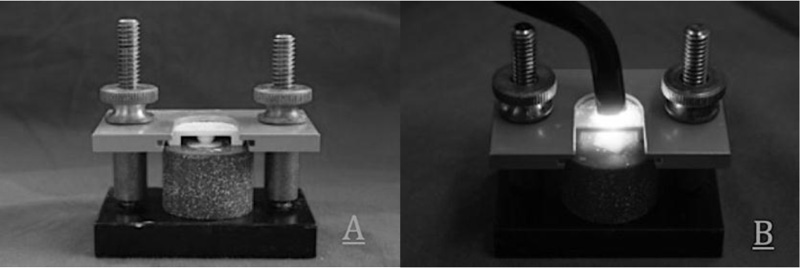
Following the adhesive application, a cylindrical-shaped plastic matrix (internal diameter 1.5 mm and height three mm; Ultradent Products) was placed over the bonded area. Two 1.5 mm layers of a light-curable nanohybrid resin composite (Filtek Z250, 3M, St. Paul, MN, USA) were packed into the hole, being careful not to incorporate air, and each layer was light-cured for forty seconds with an irradiance of 1.200 mW/cm2 maintaining the tip of the lamp in contact with the plastic mold (Fig. 1).
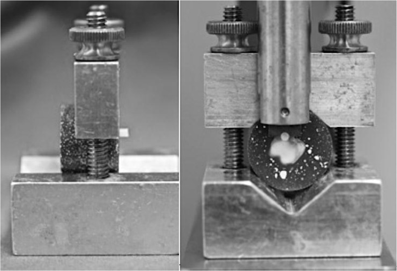
The bonded specimens were inserted in a mounting jig to provide the parallelism between the bonded enamel surface and the downward blade of the testing device. The blade had a hemispherical notch with a diameter close to that of the composite rod. Shear forces were performed until failure with a universal testing machine (Ultradent Testing Device, Ultradent Products; cross-head speed: 1 mm/min) (Fig. 2).
After checking the normal (Kolmogorov-Smirnov test) and equal (Levene’s test) distribution of the data, a two-way analysis of variance (two-way ANOVA) was conducted (SPSS, Version 19; IBM Corporation, New York City, NY, USA). Differences between groups have been evaluated by means of T- test. The level of significance (alfa) was set to p=0.05. IBM SPSS Statistics, version 19 (IBM Corporation, New York City, NY, USA) has been used. Based on the ANOVA results, the null hypotheses that (1) the etching mode and (2) the type of adhesive system do not influence the µSBS to enamel were refused.
3. RESULTS
Mean µshear bond strength ± standard deviations (MPa) are presented in Table 1.
| Groups | EE Bond | Ultradent Peak |
|---|---|---|
| H3PO4 | 18.8±0.7 C1 | 18.3±0.8 C1 |
| Er:YAG | 22±0.8 B1 | 21.4±0.9 B1 |
| Er:YAG + H3PO4 | 24.5±0.7 A1 | 25.7±0.9 A1 |
Regarding the etching procedures, the higher bond strength values were obtained in Group 3, followed by Group 2 and Group 1 and the differences were statistically different (p=0,006). No differences were observed between the two tested bonding agents (p>0.05).
4. DISCUSSION
In the present study, the combined use of laser etching plus 37% H3PO4 determined statistically higher bond strength than the two etching procedures alone. Accordingly, the first null hypothesis must be rejected. However, no differences were found in the shear bond strength of the two adhesive systems tested, independent of the etching protocol. The second null hypothesis has to be, therefore, accepted.
Phosphoric acid etching is commonly used for dental conditioning before multi-step adhesive application. Higher bond strength values have been related to the use of multi-step bonding systems when compared to simplified adhesives. However, increased bond strengths were found when simplified adhesives were used on acid etched enamel [1, 13].
The Er:YAG laser, with or without phosphoric acid, has been used to promote enamel surface changes to increase restorative material retention. It has been shown that both laser and acid conditioning are able to increase the etching depth, not significantly damaging the enamel subsurface. Analysis of the laser etched enamel subsurface revealed a small reduction in mineral concentration suggesting an increase in porosity and allowing greater acid penetration [14]. The fact that the combination of Er: YAG laser with orthophosphoric acid yielded highest bond strength values can be explained by the fact that acid could reach and etch parts of enamel inside the irradiated area [15, 16].
An in vitro study compared enamel resistance to demineralization after etching with phosphoric acid or Er:YAG laser for orthodontic brackets bonding. The mentioned study compared two groups: in the first group, enamel was etched with 37% phosphoric acid for fifteen seconds; in the second group, Er:YAG laser (wavelength, 2.940 nm; 300 mJ/pulse, 10 pulses per second, 10 s) was used for tooth conditioning. Teeth were subjected to four-days PH-cycling process in order to induce caries-like lesions. Teeth were then sectioned and the surface area of the lesion was calculated. There was no significant difference in lesion area between the two groups, showing that Er:YAG laser does not reduce enamel demineralization when exposed to acid challenge [17].
Nevertheless, this laser is able to rough enamel surface, increasing the amount of exposed dental surface, there are no unanimous opinions on the efficacy of Er:YAG laser in terms of adhesion. Few studies demonstrate that the micro-retentive surface produced by laser irradiation is favorable for adhesion procedures [7-9]; on the contrary, recent articles showed that laser application alone is not an effective etchant agent if compared with acid alone or in combination with laser [18, 19].
Martinez-Insua et al. cemented orthodontic brackets on extracted premolar teeth after enamel etching with orthophosphoric acid at 37% (for fifteen seconds) or with Er:YAG laser (4 impulses per second at 200 mJ). Statistical analysis showed that adhesion to enamel after laser etching was weaker than the one obtained through acid etching; moreover. Scanning Electron Microscope (SEM) analysis showed wide fissurations between resin-enamel interface after laser etching [13]. Another in vitro study evaluated the enamel tensile bond strength (TBS) of an adhesive system applied after different etching techniques: orthophosphoric acid etching at 37%, Er:YAG laser (80mJ-2Hz) and Er:YAG laser followed by orthophosphoric acid etching at 37%.
An in vitro study in 2005 tested the effectiveness of an Er:YAG laser in etching the enamel surface for orthodontic indications. Incisors were acid-etched (37% phosphoric acid) or laser-treated and an orthodontic bracket was attached to each treated surface using a one-step adhesive and self-curing resin. Tensile bond strength was evaluated, showing that specimens after Er:YAG laser ablation showed statistically similar tensile bond strength (9.9 +/- 1.3 MPa) to that of phosphoric acid-etched specimens (11.8 +/- 1.7 MPa) [20].
Lasmar et al. (2012) evaluated the effects of different combinations of laser treatments and bonding agents. The tensile bond strength of metallic and ceramic brackets using different bonding agents was tested using acid etching, laser treatment or a combination of both. The bond strength with laser was weaker than with acid, and stronger when combining both. Therefore, it was shown that the combination of laser and acid could produce the best retention results, again being in line with the results of our study [21].
Usually, adhesion strength on pre-treated enamel is evaluated through mechanical tests as micro-shear and micro-tensile tests [22-24]. Micro-shear is a very versatile test: it is useful for evaluation of bond strength between mineralized dental tissues and composites due to the small dimensions (0.4 mm2) of bonded surfaces and permits to test various samples on a single selected surface of enamel or dentin [23, 25].
Also Hosseini et al. sought to compare shear bond strength of orthodontic brackets bonded to enamel prepared by Er:YAG laser and conventional acid-etching. The authors investigated the following groups: conventional etching with 37% phosphoric acid; irradiation with Er:YAG laser at 1 W; irradiation with Er:YAG laser at 1.5 W. Metal brackets were bonded on prepared enamel using a light-cured composite. After thermocycling process, shear bond strength was measured using a universal testing machine. The mean shear bond strength obtained with an Er:YAG laser operated at 1W or 1.5W was approximately similar to that of conventional etching [25].
CONCLUSION
Further studies are currently ongoing to evaluate the morphological effects of etching procedures on enamel and to investigate the influence on the longevity of the bonds.
In conclusion, enamel etching with Er:YAG laser followed by 37% orthophosphoric acid can be recommended in clinical settings since it provides higher bond strengths than laser or acid etching alone.
LIST OF ABBREVIATIONS
| H3PO4 | = Orthophosphoric acid |
| Er:YAG | = Erbium-doped Yttrium Aluminium Garnet |
| µSBS | = Micro-shear bond strength |
| CEJ | = Cemento-enamel junction |
| SiC | = Silicon carbide |
ETHICAL STATEMENT
This study was approved by the ethics committe of the university of Bologna.
CONSENT FOR PUBLICATION
Informed consent was obtained from the patients.
AVAILABILITY OF DATA AND MATERIALS
The data supporting the finding of the study are available within the article.
FUNDING
None.
CONFLICT OF INTEREST
Vittorio Checchi is the Editorial Board Member of The Open Dentistry Journal.
ACKNOWLEDGEMENTS
Authors would like to thank the Medical Center of San Patrignano (Coriano, RN, Italy) for providing hospitality and facilities during the present research. Furthermore, the authors thank dr. Giacomo Bruzzesi and dr. Paolo Calvani for the logistic and didactic help in the use of the laser.

