All published articles of this journal are available on ScienceDirect.

Reconstruction of Pulpotomized Primary Molar and Retention of Stainless-steel Crowns: An In-vitro Study
Abstract
Background:
Stainless steel crowns are proposed to restore severely decayed teeth in pediatric dentistry. Yet, their retention is still debatable.
Objective:
This study aimed to investigate whether dental tissue reconstruction before placement of stainless steel crown on pulpotomized first primary molar affects the retention of stainless steel crowns.
Methods:
This experimental in-vitro study was performed on 30 extracted first primary molars with carious occlusal and distal surfaces. Dental caries and the pulp chamber roof were respectively removed. The stainless steel crowns were placed after tooth preparation. The samples were randomly divided into two groups (n=15) i.e, ZoE core buildup, and ZoE liner. Stainless steel crowns were cemented with self-curing glass ionomer cement. The crown retention was tested with the Instron testing machine. The two groups were compared by using SPSS software through the t-test (α=0.05).
Results:
The mean crown retention in the liner group (291.45±43.196 N) was significantly higher than that of the core buildup group (202.00±63.515 N) (P=0.001).
Conclusion:
Based on the results of this study for restoring the teeth with extensive tissue loss by SSC, ZoE should be used as lining without a core on teeth.
1. INTRODUCTION
Dental caries is the most common chronic disease of childhood [1, 2]. Extensive dental caries is among the main causes of premature loss of primary molars, especially the first primary molar [3, 4], which is associated with complications like the ectopic eruption of the teeth, the space loss, disrupting the inter jaw relationships, and aesthetic problems [5]. Thus, the primary molar teeth should be preserved as far as possible, even in the loss of extensive dental tissue [6, 7]. Primary molars can be repaired through various types of restorations, namely Stainless Steel Crowns (SSCs), which is one of the desired options for pulpotomized teeth with extensive caries [8-10]. Studies investigating the retention and longevity of stainless steel crowns reported this feature to be affected by several factors such as the cement type, preparation type, the remaining tooth tissue, crown influence on the gums, and choice of the proper size [4, 11-15]. The occluso-gingival length of the prepared tooth is said to determine the retention of the crown; however, some studies showed that the retention of primary crowns mostly relies on the cervical area, indicating that the rest of the tooth length could only slightly increase the retention [3, 16]. The question is whether to increase the longevity of the prepared teeth, they should be repaired in an access cavity or to increase the contact area with cement the walls must be left without repairing. Since this subject has not been investigated so far, the present study was designed to evaluate the effect of dental tissue reconstruction with cement in pulpotomized first primary molars on the retention and longevity of stainless steel crowns.
2. MATERIALS AND METHODS
In this experimental in-vitro study, 30 freshly extracted first primary molars with carious distal surface were selected through an in-access method. The teeth with caries in surfaces other than the distal surface, restoration, or developmental defects were excluded from the study. The sample size was determined based on a previous similar study [3].
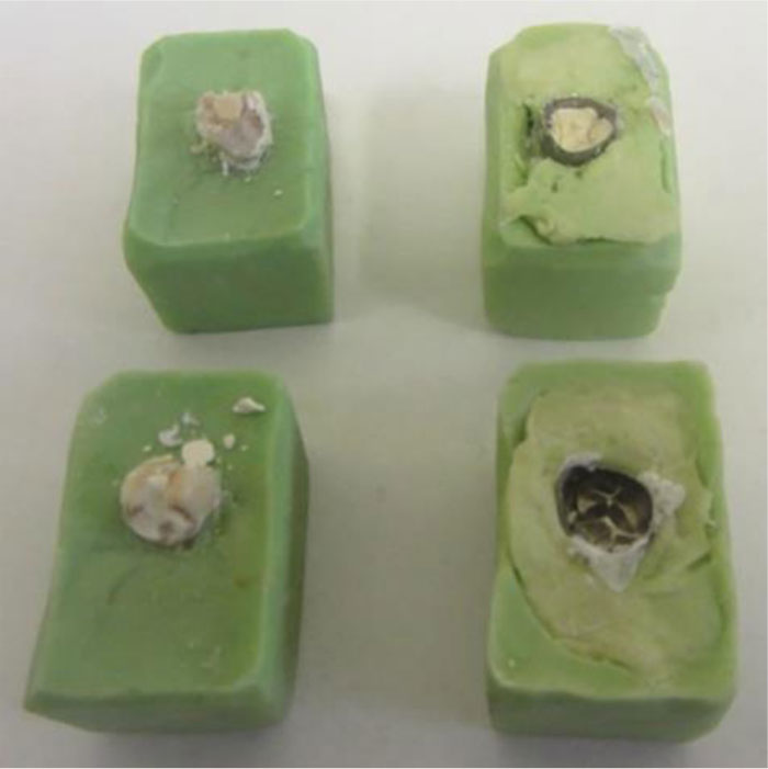
To reduce the effect of different surface areas on the resistance of the crowns under mechanical forces, 30 teeth were selected, fitting to the size 4 of SSC (3M, ESPE, St. Paul, MN, USA). The teeth were embedded in acrylic resin blocks to facilitate handling. Carious lesions were removed by using a high-speed handpiece, a standard pulpotomy procedure was performed [15], and the pulp chamber was filled with hard setting ZoE (Kemdent, UK). Half of the pulpotomized sample teeth (n=15) were randomly selected to receive ZoE core build-up.
To prepare the samples for SSC restoration, the occlusal third of the lingual and buccal surfaces were uniformly reduced to approximately 1 mm by using a high-speed handpiece; and the proximal surfaces were parallelly flattened with a 69L bur. Stainless steel crowns (size 4) were placed on the teeth with similar perimeters fitting properly for this size of the crown. Uniform contouring and crimping were performed by a single operator if necessary. The crowns were examined by using an explorer to ensure optimal marginal fit. Then, they were completely filled with self-curing glass ionomer cement (type III, GC America Inc, Alsi), and placed on the prepared teeth by using finger pressure. A mechanical load of 5 kg was applied for 10 minutes on each crown until the cement hardened. Excess cement was removed by using a dental explorer. Cemented SSCs were kept in distilled water at room temperature for 24 hours.
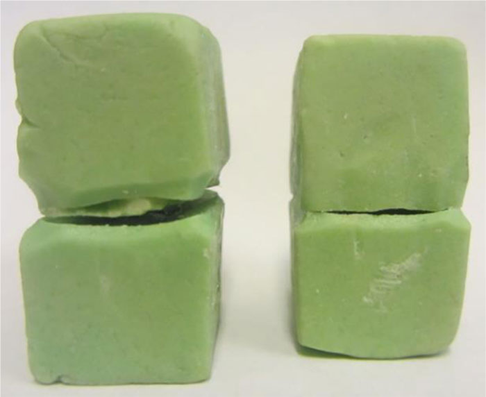
The upper surface of the acrylic block was covered with a layer of dental wax as a barrier. The crowns were embedded in an acrylic resin cap, which was removed upon setting and adhered to the occlusal surface of the crown by using epoxy glue (UHU, epoxy ultra-strong, Germany) (Fig. 1). The specimens were tested via a mechanical universal testing machine (8502, Instron, USA) at a crosshead speed of 0.05 inch/min Figs. (2 and 3). The force leading to the first dislodgement of the crown was recorded in Newton (N) as the retentive force. Data were statistically analyzed with SPSS software (version 16.0, SPSS for Windows, SPSS Inc., Chicago, USA). The groups were compared by using an independent sample t-test (α=0.05).

3. RESULTS
The mean ±standard deviation (SD) of retentive force was 202.00±63.515 N in ZoE core build-up group, and 291.45±43.196 N in the ZoE liner group (Table 1). The retentive strength of group A ranged from 123 N to 314 N, with a mean value of 200.18 N. The retentive force values for group B varied from 203 N to 333 N, with a mean value of 291.36 N. The results of the independent sample t-test revealed significant differences between the groups in terms of retentive force (P=0.001). Accordingly, the SSCs cemented onto the teeth with ZoE liner had significantly greater retention compared with the crowns administered on the teeth crowns completely reconstructed with ZoE (Table 2).
| Groups | Mean | Std. Deviation | Std. Error Mean |
|---|---|---|---|
| ZoE core build up | 200.18 | 63.515 | 19.151 |
| ZoE as a liner | 291.36 | 43.196 | 13.024 |
| t | df | Sig. (2-tailed) | Std. Error Difference | 95% confidence interval of the difference | ||
|---|---|---|---|---|---|---|
| Lower | Upper | |||||
| Equal variances assumed | -3.863 | 20 | .001 | 23.160 | -137.765 | -41.144 |
4. DISCUSSION
Dental caries is among the most common reasons for premature loss of primary teeth. The stainless steel crowns were introduced in 1950 as the most effective and most durable restoration in primary molar teeth [9]. Although they are highly successful, their retention is still debatable [16]. The present study investigated the effect of dental tissue reconstruction before repairing pulpotomized first primary molars crown on the retention of SSCs.
The results showed more retentive crowns when only the orifice was filled with ZoE, compared with those crowns which were completely filled with ZoE. It can be attributed to the anatomy of the primary molar in the cervical part that includes a prominent ridge helping the crown retention in the retention zone [17]. This finding indicates that the preparation of ZoE core before the placement of SSCs decreases the retention, rather than improve.
The occluso-gingival length is known to imperatively contribute to the retention of the crown through preventing the rotation and axial movement of the crown [18]. The cervical surface of primary teeth is the most determining factor of the crown retention, i.e., if the cervical surface of primary molar teeth is healthy and intact, other dental tissues do not interfere with achieving the maximum crown retention through optimal cutting and minimum tissue removal [16, 18].
The present study was the first to investigate the effect of reconstruction of dental tissues in pulpotomized primary molars crown on the retention of SSCs. No similar study was found in the literature, although different studies evaluated retention of SSC cemented with different cementing agents [13, 14]. Studies also evaluated micro-leakage and marginal adaptation of SSCs [3, 12].
Shillingburg et al. [18] reported the restoration of the crown before cover. It was different from the present study probably due to different dentition evaluated in these studies, which were permanent teeth in Shillingburg’s study and primary teeth in the present study. The current results were consistent with the findings of Seraj et al [3]; however, they evaluated the microleakage of SSCs placed on sound and extensively destroyed primary molars.
Among the limitations of the present study were the samples being the first primary molars that met the inclusion criteria. Moreover, this study only used first primary molar teeth with cover size 4 as one of the most common sizes used in restoring primary molars [15], which also precluded the potential bias of different crown sizes on retention forces [4]. This in-vitro study could not identically simulate oral conditions; further studies are recommended to investigate the reconstruction of oral conditions. Besides, the crowns in this study were cemented with glass ionomer; therefore, evaluation of other cement types is suggested.
CONCLUSION
Based on the results of this study, for restoring the teeth with extensive tissue loss by SSC, it is better to use ZoE as lining without core on the teeth.
ETHICAL APPROVAL AND CONSENT TO PARTICIPATE
This study was approved by the Ethics Committee of Shahed University, Iran (IR.Shahed.REC.1394.305).
HUMAN AND ANIMAL RIGHTS
Not applicable.
CONSENT FOR PUBLICATION
Not applicable.
AVAILABILITY OF DATA AND MATERIALS
The datasets analyzed during the current study are available from the corresponding author [R.H.] upon reasonable request.
FUNDING
None.
CONFLICT OF INTEREST
The authors declare no conflicts of interest, financial or otherwise.
ACKNOWLEDGEMENTS
Deep appreciations are expressed to Ms. Farzaneh Rasouli for reviewing, proofreading, editing, and improving the language of this manuscript.

