All published articles of this journal are available on ScienceDirect.

Associations of Sinus Width, Septa, and Schneiderian Membrane Thickness with Bone Formation After Lateral Window Sinus Augmentation
 iD
Nhan Van Vo4
iD
iD
Nhan Van Vo4
iD
Authors Info & Affiliations
Abstract
Introduction/Objective
Anatomical variations of the maxillary sinus may affect surgical access and graft distribution after lateral window sinus augmentation, but their association with histologic bone regeneration remains uncertain. This study aimed to evaluate associations between CBCT-derived sinus width, Schneiderian membrane thickness, and sinus septa and histomorphometric outcomes six months after lateral window sinus augmentation in severely atrophic posterior maxillae.
Methods
This prospective secondary analysis included 60 biopsy sites from 42 patients with residual bone height ≤4 mm. Sinus width was measured 10 mm above the sinus floor, and membrane thickness and sinus septa were assessed on clinically indicated CBCT examinations. Newly formed bone (NB) and residual graft material (R) were quantified in core biopsies obtained at six months. Site-level associations were explored using non-parametric methods, and patient-clustered linear mixed-effects models with a patient-specific random intercept were additionally fitted to account for multiple sites contributed by the same patient. Analyses were interpreted as exploratory because of the fixed cohort size and limited power for small associations.
Results
Sinus width was greater in molar than premolar sites (19.75 ± 3.44 mm vs 15.63 ± 4.40 mm; p = 0.005). Sinus width was not significantly associated with NB or R. Septa were not associated with NB (p = 0.897), R (p = 0.327), baseline membrane thickness (p = 0.715), or post-augmentation membrane thickness (p = 0.941). Three membrane perforations (5.0%) were managed intraoperatively; no postoperative infections were recorded.
Discussion
The data did not identify a moderate or larger association between the evaluated anatomical variables and six-month histomorphometric outcomes. The null findings should not be interpreted as proof of no effect because the study was a secondary analysis with clustered observations and limited power for small associations.
Conclusion
CBCT-defined sinus morphology was not significantly associated with NB or R in this cohort. CBCT remains important for surgical planning, while its value for predicting histologic bone formation requires confirmation in larger, cluster-aware studies.
1. INTRODUCTION
Loss of posterior maxillary teeth is commonly followed by alveolar ridge resorption and changes in maxillary sinus dimensions, which can reduce the bone available for implant placement [1]. Restoratively driven assessment of residual bone and local anatomy is therefore central to treatment planning in the posterior maxilla [2]. In cases of severe vertical deficiency, lateral window sinus floor elevation is a well-established and predictable technique for increasing bone volume and enabling implant-supported rehabilitation [3, 4].
Beyond vertical bone height deficiency, anatomical variations of the maxillary sinus may influence the mechanical environment and spatial distribution of graft materials during healing [5]. Cone-beam computed tomography (CBCT) allows detailed evaluation of sinus morphology, including sinus width, membrane thickness, and the presence of sinus septa, all of which may potentially affect surgical access, clot stabilization, and vascular supply within the grafted compartment [6, 7].
Bone regeneration after sinus augmentation is influenced by multiple clinical and biological factors, including defect morphology, vascular supply, graft stability, and the selected augmentation approach, which may collectively affect angiogenesis and osteogenesis within the augmented compartment [8-10]. Similarly, sinus septa represent a frequent anatomical variation that may complicate membrane elevation and influence graft distribution. Although septa have traditionally been associated with increased surgical difficulty and risk of membrane perforation, their influence on biological healing outcomes remains uncertain [10, 11].
Previous studies have extensively evaluated graft materials and biologic adjuncts in maxillary sinus augmentation, whereas sinus anatomy has received comparatively less attention in regenerative outcomes [11, 12]. Contemporary reviews emphasize that anatomical characteristics can influence surgical access, membrane management, and treatment planning [13]. Importantly, prospective histomorphometric evidence following lateral sinus augmentation has reported an association between bucco-palatal sinus width and the amount of newly formed mineralized tissue, supporting sinus width as a biologically plausible predictor warranting further study [14]. A Vietnamese interventional pre-post study has also evaluated maxillary sinus augmentation using PRF with immediate implant placement, providing relevant regional clinical evidence. However, its surgical protocol and baseline bone conditions differed from those of the present study [15]. Understanding whether anatomical parameters influence bone regeneration is clinically relevant because CBCT-derived measurements are routinely obtained during implant treatment planning [16]. Sinus anatomical characteristics have also been investigated as potential determinants of implant-related outcomes following sinus augmentation [17].
Therefore, the present study aimed to evaluate the association between CBCT-derived anatomical characteristics of the maxillary sinus and bone regeneration following lateral window sinus augmentation in patients with severely atrophic posterior maxilla. Specifically, this study investigated whether sinus width, Schneiderian membrane thickness, and the presence of sinus septa were associated with histomorphometric outcomes obtained six months after augmentation. In severely atrophic posterior maxillae, CBCT-based assessment is important not only for evaluating bone availability and sinus anatomy but also for guiding reconstructive strategies and surgical planning [18, 19].
2. MATERIALS AND METHODS
2.1. Study Participants
2.1.1. Inclusion Criteria
Patients presenting with posterior maxillary edentulism and a residual bone height (RBH) ≤ 4 mm who required lateral window sinus floor augmentation (LWSFA) before implant placement were considered eligible for participation. All patients provided written informed consent before enrollment. Participants were consecutively recruited from the Department of Oral and Maxillofacial Surgery, Can Tho University of Medicine and Pharmacy Hospital.
2.1.2. Exclusion Criteria
Patients were excluded if they presented with acute or chronic maxillary sinusitis, metabolic bone disorders, uncontrolled systemic diseases, malignancy, or conditions known to affect bone healing. Individuals receiving medications influencing bone metabolism or coagulation (e.g., corticosteroids, anticoagulants, or long-term NSAIDs) were also excluded. Smokers and pregnant women were not eligible for participation.
2.2. Study Design
This prospective secondary analysis included 60 sinus augmentation sites from 42 patients with severely atrophic posterior maxillae (RBH ≤4 mm) treated between January 2022 and February 2024. All procedures followed a standardized lateral window sinus augmentation protocol. Ethical approval was obtained from the Ethics Committee for Biomedical Research at Can Tho University of Medicine and Pharmacy (Decision No. 504/PCT-HĐĐĐ).
Sample-size sensitivity analysis: This study was a secondary analysis of a fixed prospective cohort. Because the primary outcomes (NB and R) were continuous percentages, a single-proportion formula was not appropriate. For a two-sided correlation test with α=0.05 and 80% power, 59 independent observations are required to detect an absolute correlation of 0.36 using Fisher’s z transformation. Accordingly, 60 sites provide approximately 81% power to detect |r|=0.36 before accounting for within-patient clustering. The analysis therefore had sensitivity for moderate associations but could not exclude smaller effects.
The biopsy site was the observational unit. Because some patients contributed more than one site, site-level observations may be correlated. Primary analyses were therefore interpreted as exploratory. This cohort partially overlaps with a previously published descriptive histological study; the present analysis addresses a distinct question concerning CBCT-derived anatomical predictors [12].
2.3. Radiographic Assessment of Maxillary Sinus Anatomy
Preoperative cone-beam computed tomography (CBCT) scans were used to evaluate anatomical characteristics of the maxillary sinus. All CBCT images were obtained using standardized imaging parameters and analyzed using digital imaging software.
Measurements were performed on cross-sectional images perpendicular to the alveolar ridge at the planned implant location. The following radiographic variables were assessed:
- Residual bone height (RBH): vertical distance from the alveolar crest to the sinus floor
- Sinus width (SW): horizontal distance between medial and lateral sinus walls measured 10 mm above the sinus floor
- Schneiderian membrane thickness at baseline (S0)
- Membrane thickness changes after sinus augmentation and during follow-up (S1–S5)
- Presence or absence of sinus septa
Two calibrated examiners independently performed the radiographic measurements following a calibration exercise using CBCT sections that were not included in the principal analysis. Disagreements were resolved by consensus. Inter-examiner reliability was assessed in a randomly selected subset of 20 sinus sites. Continuous measurements were evaluated using a two-way random-effects intraclass correlation coefficient for absolute agreement based on single measurements [ICC(2,1)], whereas agreement for sinus septa detection was evaluated using Cohen’s κ and percentage agreement.
2.4. CBCT Acquisition Protocol
CBCT examinations were acquired using an ORTHOPHOS XG 3D system (Sirona Dental Systems GmbH, Bensheim, Germany). Images were obtained using an 8 × 8 cm field of view, an isotropic voxel size of 0.20 mm, a tube voltage of 90 kVp, a tube current of 8 mA, and an acquisition time of 17 seconds. Images were reconstructed and evaluated using SIDEXIS software. Patients were positioned according to the manufacturer’s instructions, and all measurements were performed on cross-sectional reconstructions perpendicular to the alveolar ridge at the planned implant location. CBCT imaging was performed using the smallest diagnostically acceptable field of view in accordance with ALADA/ALADAIP principles.
Sinus width was measured on a cross-sectional image perpendicular to the alveolar ridge at the planned implant site, as the horizontal distance between the medial and lateral sinus walls at a line 10 mm superior to the sinus floor, following the approach used in previous sinus-dimension studies [20, 21]. Figure 1 illustrates the measurement landmarks.
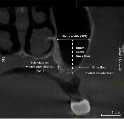
Representative CBCT cross-sectional image illustrating measurement of sinus width 10 mm superior to the sinus floor and Schneiderian membrane thickness at the planned implant site.
2.5. Surgical Procedure
All sinus augmentation procedures were performed using a standardized lateral window approach under local anesthesia and antibiotic prophylaxis. A lateral window was prepared using a piezoelectric device, followed by Schneiderian membrane elevation and graft placement according to the previously published protocol [12]. Three intraoperative membrane perforations occurred (5.0%); all were managed during surgery. No postoperative infections or graft-related complications were recorded during healing [13].
Autologous platelet-rich fibrin was prepared using a standardized centrifugation protocol. The use of a uniform surgical approach across all patients aimed to minimize procedural variability and allow evaluation of anatomical factors as primary predictors of healing outcomes.
After a 6-month healing period, bone core biopsies were obtained from the planned implant sites immediately before implant placement, which was performed in accordance with the manufacturer’s instructions.
2.6. Histomorphometric Outcome Assessment
Biopsy processing and histomorphometric procedures followed the previously published protocol [12]. Briefly, specimens were fixed in buffered formalin, decalcified in EDTA, embedded in paraffin, sectioned at approximately 5 μm, and stained with hematoxylin and eosin.
Histomorphometric analysis was conducted using ImageJ software (National Institutes of Health, USA) to quantify the proportion of newly formed bone (NB) and residual graft material (R). For each specimen, three non-consecutive sections corresponding to coronal, middle, and apical regions were analyzed to improve representativeness.
Areas not identified as mineralized tissue or residual graft particles were categorized as connective tissue or marrow-like structures. All measurements were performed independently by two blinded examiners to ensure reproducibility.
Histomorphometric variables were considered outcome parameters for evaluating the relationship between sinus anatomical characteristics and bone regeneration.
A representative hematoxylin and eosin–stained histological section obtained six months after lateral window sinus augmentation, showing newly formed bone, residual graft particles, and intervening connective tissue/marrow-like spaces. Scale bar = 100 μm (Fig. 2).
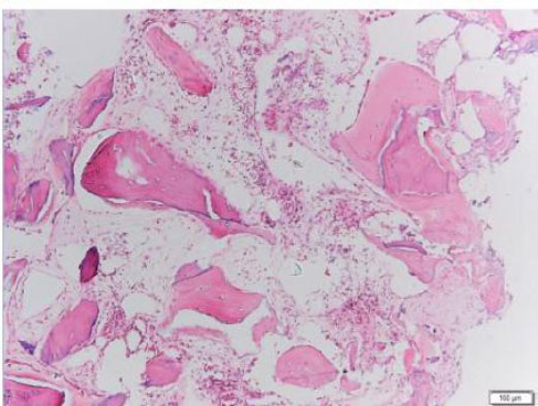
Representative histological appearance of regenerated tissue six months after lateral window sinus augmentation.
2.7. Statistical Analysis
Statistical analyses were performed using SPSS version 26.0 (IBM Corp., Armonk, NY, USA). Continuous variables were summarized as mean ± standard deviation and range, and categorical variables as counts and percentages. Normality was evaluated using the Shapiro-Wilk test.
Because several variables were not normally distributed, exploratory two-group comparisons were conducted using the Mann-Whitney U test. Site-level associations between continuous CBCT-derived predictors and the histomorphometric outcomes NB and R were explored using Spearman's rank correlation coefficient (ρ), with approximate 95% confidence intervals derived via Fisher's z-transformation.
Because some patients contributed more than one biopsy site, site-level observations were potentially correlated. Therefore, linear mixed-effects models with a patient-specific random intercept were additionally fitted for NB and R to account for within-patient clustering. Continuous anatomical predictors were entered per 1-mm increase, and sinus septum status was entered as present versus absent; adjusted β coefficients with 95% confidence intervals were reported. Intraclass correlation coefficients were calculated from the random-intercept variance components. These cluster-aware models were interpreted as exploratory because this was a secondary analysis of a fixed cohort with a limited number of patients. NB and R are compositional percentages and are mathematically dependent on the remaining tissue component, which further supports cautious interpretation of inferential results.
All statistical tests were two-tailed, and p < 0.05 was considered statistically significant.
Inter-examiner reliability was excellent for sinus width, baseline Schneiderian membrane thickness, and post-augmentation membrane thickness, with ICC (2,1) values of 0.94, 0.91, and 0.93, respectively. Agreement for sinus septa detection was almost perfect, with a Cohen’s κ of 0.89 and an overall percentage agreement of 96.7%. All reliability estimates were statistically significant, and the corresponding 95% confidence intervals indicated good to excellent reproducibility.
3. RESULTS
Data are presented as median and interquartile range (IQR). Comparisons between sites with and without sinus septa were performed using the two-sided Mann–Whitney U test-S0, baseline Schneiderian membrane thickness; S1, membrane thickness after sinus augmentation.
In this cohort, the evaluated CBCT variables did not show statistically significant site-level associations with histomorphometric outcomes. The findings do not demonstrate equivalence and should not be generalized to effects beyond moderate or larger magnitudes.
3.1. Study Population and Distribution of Implant Sites
A total of 42 patients (60 sinus augmentation sites) were included in the study. The mean patient age was 53.57 ± 12.90 years (range: 23–72 years). Sixteen patients were male (38.1%), and 26 were female (61.9%). The majority of implant sites were located in the molar region (78.3%), whereas 21.7% were positioned in the premolar region. All sites presented residual bone height ≤ 4 mm and were therefore considered appropriate for lateral window sinus augmentation. Baseline characteristics of implant location are summarized in Table 1.
| Characteristic | Value | |
|---|---|---|
|
Implant position n (%) |
Premolar group | 13 (21.7%) |
| Molar group | 47 (78.3%) | |
CBCT analysis revealed variability in the anatomical morphology of the maxillary sinus among implant sites. The molar region demonstrated a significantly greater sinus width measured 10 mm above the sinus floor than the premolar region (19.75 ± 3.44 mm vs 15.63 ± 4.40 mm, p = 0.005).
No statistically significant differences were observed between premolar and molar regions in baseline Schneiderian membrane thickness (S0) or post-augmentation membrane thickness (S1). Furthermore, changes in membrane thickness at follow-up time points (S2–S5) did not differ significantly between anatomical regions (all p > 0.05).
Although sinus width differed between premolar and molar sites, no statistically significant differences in membrane-thickness measures were detected (Table 2). These findings were interpreted as exploratory due to unequal group sizes and site clustering.
| Variable |
Premolar group, n = 13 Mean ± SD |
Molar group, n = 47 Mean ± SD |
Mann–Whitney U | Z | p |
|---|---|---|---|---|---|
| Sinus width at 10 mm (SW) | 15.63 ± 4.40 | 19.75 ± 3.44 | 149.50 | −2.801 | 0.005 |
| Baseline membrane thickness (S0) | 1.11 ± 1.81 | 1.90 ± 2.69 | 224.50 | −1.474 | 0.140 |
| Post-augmentation membrane thickness (S1) | 3.70 ± 2.93 | 3.81 ± 2.62 | 289.50 | −0.288 | 0.773 |
| S1 − S0 | 2.59 ± 2.34 | 1.91 ± 2.78 | 275.50 | −0.540 | 0.589 |
| S2 − S1 | −1.60 ± 1.44 | −1.84 ± 2.10 | 296.00 | −0.171 | 0.864 |
| S3 − S2 | −0.93 ± 1.38 | −0.37 ± 0.91 | 260.50 | −0.827 | 0.408 |
| S4 − S3 | −0.02 ± 0.21 | −0.14 ± 0.79 | 285.00 | −0.392 | 0.695 |
| S5 − S4 | 0.05 ± 0.17 | −0.07 ± 0.52 | 262.00 | −0.812 | 0.417 |
| S5 − S0 | 0.09 ± 0.60 | −0.52 ± 2.27 | 252.00 | −0.963 | 0.336 |
Abbreviations: SW, sinus width measured 10 mm above the sinus floor; S0, baseline membrane thickness; S1, after sinus augmentation; S2, after implant placement; S3, after prosthetic loading; S4, 6 months after loading; S5, 12 months after loading. Differences are expressed as the later time point minus the earlier time point.
Sinus septa were identified in a subset of implant sites. Comparison of histomorphometric outcomes between sites with and without septa showed no statistically significant differences in newly formed bone (NB) or residual graft material (R) (NB: p = 0.897; R: p = 0.327).
No statistically significant difference in NB or R was detected between sites with and without septa (Table 3). Because this was a null result in a modest, unbalanced sample, it should be interpreted as failure to detect an association rather than evidence that septa have no biological effect.
| Outcome |
Septum Absent n = 48 Mean Rank |
Septum Present n = 12 Mean Rank |
Mann–Whitney U | p |
|---|---|---|---|---|
| Newly formed bone (%) | 30.35 | 31.08 | 281.00 | 0.897 |
| Residual graft material (%) | 29.40 | 34.92 | 235.00 | 0.327 |
Abbreviations: NB, newly formed bone; R, residual graft material.
Baseline Schneiderian membrane thickness was comparable between sites without and with sinus septa [median: 1.00 mm (IQR, 0.40–1.53) versus 1.00 mm (IQR, 0.50–1.35); Mann–Whitney U = 268.50; p = 0.715]. Similarly, no significant difference was observed in post-augmentation membrane thickness [2.80 mm (IQR, 2.08–5.10) versus 3.20 mm (IQR, 2.33–4.13); U = 284.00; p = 0.941]. (Table 4).
| Variable |
Septum absent, n = 48, median (IQR), mm |
Septum present, n = 12, median (IQR), mm |
Mean rank: absent | Mean rank: present | Mann–Whitney U | p |
|---|---|---|---|---|---|---|
| Baseline membrane thickness (S0) | 1.00 (0.40–1.53) | 1.00 (0.50–1.35) | 30.09 | 32.13 | 268.50 | 0.715 |
| Post-augmentation membrane thickness (S1) | 2.80 (2.08–5.10) | 3.20 (2.33–4.13) | 30.42 | 30.83 | 284.00 | 0.941 |
3.2. Associations between CBCT-derived Variables and Histomorphometric Outcomes
At the biopsy-site level, sinus width was not significantly correlated with newly formed bone percentage (Spearman ρ = 0.064; 95% CI: −0.193 to 0.312; p = 0.629) or residual graft material percentage (ρ = 0.106; 95% CI: −0.152 to 0.351; p = 0.420) (Table 5).
| Predictor | Outcome | Spearman ρ | Approximate 95% CI | p |
|---|---|---|---|---|
| Sinus width | Newly formed bone | 0.064 | −0.193 to 0.312 | 0.629 |
| Residual graft material | 0.106 | −0.152 to 0.351 | 0.420 | |
| Baseline membrane thickness | Newly formed bone | 0.252 | −0.002 to 0.476 | 0.052 |
| Residual graft material | −0.062 | −0.311 to 0.195 | 0.637 | |
| Post-augmentation membrane thickness | Newly formed bone | 0.106 | −0.152 to 0.350 | 0.420 |
| Residual graft material | −0.056 | −0.305 to 0.201 | 0.672 |
Baseline Schneiderian membrane thickness demonstrated a weak positive association with newly formed bone that approached, but did not reach, statistical significance (ρ = 0.252; 95% CI: −0.002 to 0.476; p = 0.052). Baseline membrane thickness was not significantly associated with residual graft material (ρ = −0.062; 95% CI: −0.311 to 0.195; p = 0.637) (Table 5).
Post-augmentation membrane thickness was not significantly associated with newly formed bone (ρ = 0.106; 95% CI: −0.152 to 0.350; p = 0.420) or residual graft material (ρ = −0.056; 95% CI: −0.305 to 0.201; p = 0.672). These site-level findings were considered exploratory because multiple biopsy sites may have originated from the same patient (Table 5).
In patient-clustered mixed-effects models, sinus width was not significantly associated with newly formed bone percentage (β = 0.22 percentage points per 1-mm increase; 95% CI: −0.53 to 0.97; p = 0.560) or residual graft material percentage (β = 0.19; 95% CI: −0.30 to 0.68; p = 0.443) (Table 6).
| Predictor | Outcome | adjusted β | 95% CI | p-value |
|---|---|---|---|---|
| Sinus width, per 1-mm increase |
Newly formed bone (%) | 0.22 | −0.53 to 0.97 | 0.560 |
| Residual graft material (%) | 0.19 | −0.30 to 0.68 | 0.443 | |
| Baseline membrane thickness, per 1-mm increase | Newly formed bone (%) | 1.36 | −0.08 to 2.80 | 0.064 |
| Residual graft material (%) | −0.31 | −1.27 to 0.65 | 0.521 | |
| Post-augmentation membrane thickness, per 1-mm increase | Newly formed bone (%) | 0.58 | −0.61 to 1.77 | 0.334 |
| Residual graft material (%) | −0.27 | −1.05 to 0.51 | 0.493 | |
| Sinus septum present versus absent | Newly formed bone (%) | 1.94 | −6.10 to 9.98 | 0.631 |
| Residual graft material (%) | 2.41 | −2.84 to 7.66 | 0.362 |
Baseline Schneiderian membrane thickness showed a weak positive association with newly formed bone that approached, but did not reach, statistical significance (β = 1.36 percentage points per 1-mm increase; 95% CI: −0.08 to 2.80; p = 0.064). No significant association was observed with residual graft material (Table 6).
Post-augmentation membrane thickness and sinus septa status were not significantly associated with either histomorphometric outcome. The estimated within-patient correlation was modest, with ICC values of 0.105 for newly formed bone and 0.069 for residual graft material (Table 7).
| Outcome | Patient-level Variance | Residual Variance | ICC |
|---|---|---|---|
| Newly formed bone (%) | 31.8 | 271.4 | 0.105 |
| Residual graft material (%) | 10.2 | 138.6 | 0.069 |
4. DISCUSSION
Baseline Schneiderian membrane thickness showed a weak positive association with newly formed bone, with confidence intervals extending from almost no association to a potentially moderate positive effect. Although the conventional threshold for statistical significance was not reached, this finding may represent a hypothesis-generating signal rather than evidence of no association. It is biologically plausible that membrane vascularity or inflammatory status could influence early healing; however, membrane thickness on CBCT is an indirect and nonspecific surrogate and cannot distinguish physiological vascular tissue from inflammatory thickening. This association should therefore be examined prospectively in adequately powered studies.
4.1. Anatomical Variability as a Potential Determinant of Regenerative Outcomes
Anatomical characteristics of the maxillary sinus have been proposed as potential modifiers of bone regeneration due to their influence on vascular supply, graft containment, and spatial distribution of biomaterials [6, 7]. In particular, wider sinus cavities have been hypothesized to reduce the proximity of graft particles to vascularized sinus walls, potentially increasing the diffusion distance for angiogenic and osteogenic signaling [20, 21]. From a mechanistic perspective, reduced vascular proximity could theoretically delay mineralization and maturation of newly formed bone.
However, the results of the present study do not support a clinically meaningful association between sinus width and histomorphometric outcomes. Despite statistically significant differences in sinus width between premolar and molar regions, newly formed bone and residual graft material did not differ significantly across anatomical configurations. These findings suggest that dimensional variation of the sinus cavity alone may not be a strong determinant of bone regeneration when surgical technique and graft stability are standardized.
One possible interpretation is that sinus augmentation creates a contained healing environment in which clot stability and scaffold properties may mitigate the influence of anatomical variability. The lateral window approach enables controlled placement of graft material and may reduce the functional impact of differences in sinus dimensions. This interpretation is consistent with previous studies reporting that bone formation may occur predictably across a range of sinus morphologies [13, 22].
4.2. The Present Exploratory Analysis did not Detect a Moderate Site-level Association between Sinus Width and Histomorphometric Outcomes
Because of limited power for small effects and the limited precision of the patient-clustered estimates, this finding should not be interpreted as evidence that sinus width is biologically irrelevant.
Sinus septa are frequently considered a technical challenge during sinus elevation procedures due to their association with increased risk of membrane perforation [10, 23]. However, their influence on regenerative outcomes remains unclear.
In the present study, septa were not associated with statistically significant differences in NB or R. This finding addresses biological healing rather than surgical difficulty. Three membrane perforations occurred in the cohort, but the available aggregate data did not support stratified or adjusted analysis by perforation status. Because perforation may confound healing, future analyses should include it as a covariate when individual-level data are available.
From a causal perspective, septa may act as procedural modifiers that influence surgical difficulty but do not directly affect osteogenesis once the graft material is successfully placed and stabilized. Therefore, the presence of septa should be interpreted as a factor that requires surgical adaptation rather than as a predictor of regenerative capacity [24, 25].
4.3. Schneiderian Membrane Response as an Intermediate Variable
Changes in Schneiderian membrane thickness following sinus augmentation may represent a physiological response to surgical manipulation rather than a pathological process [12, 21]. Membrane thickening has been described as a transient inflammatory or adaptive response associated with wound healing.
In the present cohort, no statistically significant differences in membrane thickness were detected by region or septa status, and no significant site-level associations with histomorphometry were identified. All follow-up CBCT examinations were obtained for clinically indicated treatment assessment rather than solely for research purposes, using standardized acquisition parameters and a limited field of view consistent with ALADA/ALADAIP principles [26].
Within a causal framework, membrane thickness may be considered a mediator reflecting tissue response to surgery, but its magnitude may not necessarily predict the extent of mineralized tissue formation.
4.4. Relative Importance of Anatomical Versus Procedural Factors
The absence of statistically significant associations should not be interpreted as proof that anatomy is unimportant. One possible explanation is that standardized graft placement and membrane management attenuated moderate anatomical effects; another is limited power, residual confounding, or measurement error [27]. Therefore, procedural factors may be important, but their relative contribution cannot be established by this observational analysis.
Clinically, CBCT measurements should be used primarily for diagnosis, surgical access planning, and risk assessment. The present data do not support the use of sinus width, membrane thickness, or septa alone as validated predictors of histologic bone formation.
4.5. Methodological Considerations in Causal Interpretation
Interpretation of causal relationships in the present study requires consideration of potential confounding factors. Histomorphometric outcomes are influenced by multiple biological variables, including patient-specific healing capacity, vascular supply, systemic conditions, and biomaterial properties. Because all patients were treated using the same grafting protocol, the present study does not allow separation of the individual effects of biomaterial composition and anatomical variability.
In addition, histomorphometric variables represent compositional data that sum to 100%, which may introduce mathematical dependency between tissue components. Consequently, statistical associations should be interpreted with caution and treated as exploratory.
Another methodological consideration is that biopsy specimens represent localized tissue environments rather than the entire augmented sinus. Although histomorphometry provides high-resolution evaluation of tissue composition, it may not fully capture three-dimensional structural changes within the grafted volume.
4.6. Clinical Implications
The present exploratory findings indicate that sinus anatomical variation alone was not a validated predictor of six-month histomorphometric outcomes in this cohort. Anatomical variation should therefore continue to be considered in surgical planning rather than interpreted as a contraindication or as a definitive predictor of regenerative capacity.
Clinical emphasis should remain on surgical precision, preservation and management of the Schneiderian membrane, and stable graft placement. CBCT remains essential for diagnosis, access planning, and risk assessment, but the present data do not establish sinus width, membrane thickness, or septa as independent predictors of histologic bone formation.
The findings support the feasibility of lateral window sinus augmentation across varied anatomical presentations when appropriate protocols are followed; however, they do not establish anatomical equivalence or causality.
4.7. Limitations and Future Directions
Several limitations should be acknowledged. First, this was a secondary analysis of a fixed cohort and had sensitivity primarily for moderate correlations (approximately |r|=0.36); smaller effects could have been missed. Second, although patient-level random-intercept mixed-effects models were applied to account for clustering of multiple sites within patients, the limited number of patients and the small number of repeated sites per patient may have reduced the precision of the variance-component and association estimates. Third, measurement reliability was high/substantial for the evaluated radiographic and histomorphometric variables, reducing-but not eliminating-the possibility that measurement error attenuated the observed associations. Fourth, membrane perforation was a potential confounder, and only aggregate complication data were available. Fifth, repeated CBCT time points require explicit clinical indications and acquisition parameters. Sixth, histomorphometry was obtained at one time point from localized 3-mm cores and may not represent the entire augmented volume. Finally, the single-center cohort, an unbalanced premolar/molar distribution, and the lack of multivariable adjustment limit generalizability and causal inference.
Future studies incorporating longitudinal imaging, volumetric analysis, and larger multicenter cohorts may provide additional insight into the relationship between sinus morphology and regenerative outcomes. Integration of CBCT-derived anatomical variables with biomechanical parameters and implant stability measurements may further clarify the role of anatomical variability in implant success.
CONCLUSION
Within the limitations of this prospective secondary analysis, no statistically significant site-level associations were detected between sinus width, Schneiderian membrane thickness, or septa and six-month NB or R after lateral window sinus augmentation.
These results should be interpreted as inconclusive for small effects rather than as evidence of anatomical equivalence. Larger studies using patient-clustered models, prespecified effect sizes, and complete reporting of reliability are required.
CBCT assessment remains essential for surgical planning and complication risk evaluation; however, the present findings do not support
Further studies integrating longitudinal radiographic analysis, volumetric assessment, and multicenter cohorts are warranted to clarify the interaction between anatomical variability, biomaterial behavior, and implant stability in augmented sinuses.
AUTHORS’ CONTRIBUTIONS
The authors confirm their contributions to the paper as follows: H.G.N., V.P.K.L., N.L.L., V.N.V.: Conceptualization and study design; H.G.N., N.L.L.: Clinical procedures and data collection; H.G.N., V.P.K.L., N.L.L., V.N.V.: Data analysis and interpretation; H.G.N., V.P.K.L., N.L.L.: Manuscript drafting; H.G.N., V.P.K.L., N.L.L., V.N.V.: Critical revision of the manuscript All authors read and approved the final manuscript.
LIST OF ABBREVIATIONS
| LWSFA | = Lateral Window Sinus Floor Augmentation |
| PRF | = Platelet-Rich Fibrin |
| β-TCP | = Beta-Tricalcium Phosphate |
| HA | = Hydroxyapatite |
| ISQ | = Implant Stability Quotient |
ETHICS APPROVAL AND CONSENT TO PARTICIPATE
The study protocol was reviewed and approved by the Ethics Committee for Biomedical Research at Can Tho University of Medicine and Pharmacy (Decision No. 504/PCT-HĐĐĐ).
HUMAN AND ANIMAL RIGHTS
The study was conducted in full accordance with the ethical principles of the Declaration of Helsinki (2013 revision).
CONSENT FOR PUBLICATION
All participants were fully informed about the study protocol, potential risks, and benefits, and provided written informed consent before inclusion.
AVAILABILITY OF DATA AND MATERIALS
The datasets generated and/or analyzed during the current study are available from the corresponding author on reasonable request.
ACKNOWLEDGEMENTS
The authors thank the Faculty of Odonto-Stomatology, Can Tho University of Medicine and Pharmacy, for providing administrative and facility support.