All published articles of this journal are available on ScienceDirect.

Methodological Framework for Integrated Surface Electromyography and Mandibular Kinematics in Swallowing Analysis

Abstract
Background
Swallowing requires coordinated activation of orofacial muscles and mandibular motion. Although clinical assessment often relies on observation or imaging, surface electromyography (sEMG) provides a non-invasive means of quantifying muscle activity. The aim of this study was to describe a standardized methodological framework for the synchronized acquisition, processing, and visualization of surface electromyographic and mandibular kinematic signals during saliva swallowing.
Materials and Methods
A retrospective methodological framework was developed using synchronized sEMG and mandibular kinematic recordings obtained during routine neuromuscular evaluations. Standardized procedures for signal acquisition, synchronization, preprocessing, and visualization were defined to ensure reproducible descriptive analysis of voluntary and spontaneous saliva swallowing.
Results
Application of the protocol enabled synchronized visualization of electromyographic activity and three-dimensional mandibular trajectories during voluntary and spontaneous saliva swallowing. The framework allowed consistent identification of temporal relationships between muscle activation patterns and mandibular displacement and facilitated qualitative inspection of signal morphology, activation sequences, and movement coordination.
Conclusion
This methodological framework provides a standardized approach for documenting swallowing-related muscular and mandibular events using synchronized sEMG and mandibular kinematic recordings. It is intended as a methodological reference for future research and as a foundation for the development of quantitative analytical approaches.
1. INTRODUCTION
Swallowing is a complex sensorimotor process that is governed by the finely tuned coordination of multiple muscle groups, each innervated by specific cranial nerves and driven by a network of cortical and brainstem centers. While it occurs in three phases (oral, pharyngeal, and esophageal), these stages operate as a continuous and overlapping sequence, particularly during the switch from the voluntary oral stage to the reflex-based pharyngeal response. Once initiated, the pharyngeal phase progresses through a largely automatic, stereotyped series of movements regulated by a central pattern generator within the brainstem [1, 2]. Swallowing function is clinically relevant because it contributes to nutrition, airway protection, speech-related oral motor coordination, and overall quality of life. Alterations in swallowing patterns may be associated with functional disturbances affecting the stomatognathic system and are frequently evaluated in dental, orthodontic, and multidisciplinary clinical settings. Consequently, objective methods capable of documenting the coordination between muscular activity and mandibular motion are of increasing interest for both research and clinical applications. Various methods have been used to assess deglutition; these methods range from imaging technologies to surface and needle electromyography (EMG) [3-5], with recent advances extending to wearable epidermal devices and AI-/sensor-based dysphagia screening systems that support non-invasive functional assessment of swallowing [6-8]. Moreover, Ertekin et al. investigated the neurophysiology of swallowing and demonstrated that swallowing involves the sequential activation of perioral, submental, suprahyoid, and pharyngeal muscles and a reproducible temporal pattern that remains relatively consistent across subjects and bolus conditions. Their work recognized surface EMG (sEMG) as a practical and non-invasive method for capturing the timing and duration of muscle activity associated with deglutition, particularly when synchronized with mechanical measures of laryngeal movement [1, 9, 10]. Recent studies have further refined the use of sEMG for analyzing temporal and functional variability in swallowing-related muscle activity under different task conditions [11-13]. Detailed guidelines for sEMG measurement and analysis in swallowing systems are available [14-16]. These electrophysiological approaches have contributed advanced knowledge on swallowing physiology and its modulation by bolus volume, consistency, and underlying neural control mechanisms [1, 11, 17-19]. Despite these advances, functional assessment of swallowing in hospitalized contexts remains limited to imaging-based techniques, such as videofluoroscopy [7, 20, 21], which may be costly, time-intensive, and impractical for routine functional evaluation. In contrast, most dental practitioners primarily rely on general clinical assessment. sEMG and computerized mandibular scanning provide a complementary, non-invasive perspective by quantifying muscular activation patterns together with mandibular displacement trajectories under physiological conditions [22, 23]. Their application in assessing the orofacial and suprahyoid muscles has been widely reported in studies examining oral function, despite the limited use of contemporary mandibular motion detection techniques [24-28]. Reference values for EMG and kinematics in ideal occlusion provide baselines for such assessments [29]. In a dental and neuromuscular functional context, evaluation is typically performed in individuals without symptoms or dysphagia during routine outpatient visits, with a focus on oral and stomatognathic function rather than on acute or hospital-based dysphagia management. In this setting, non-invasive techniques such as sEMG can be applied during saliva swallowing under standardized postural and clinical conditions, providing complementary functional information without the need for bolus administration or imaging-based studies [10, 11, 17]. Integrating EMG recordings with mandibular kinematic analysis has garnered increasing interest to better characterize the coordination between muscle activity and mandibular motion during deglutition [30-32]. The synchronized acquisition of mandibular kinematics and sEMG offers a multidimensional perspective on swallowing dynamics by allowing simultaneous visualization of mandibular motion and muscle activation relative to the intercuspal position. However, standardized methodologies describing signal acquisition, synchronization, and analytical principles for this combined assessment, specifically during voluntary or spontaneous saliva swallowing in an outpatient dental setting, remain scarce, particularly within the dental and orthodontic literature [22, 33]. Although previous studies have reported the use of surface electromyography or mandibular motion analysis to investigate swallowing-related function, methodological descriptions are often limited to specific experimental settings, selected variables, or isolated components of the recording process. Detailed protocols addressing synchronized acquisition, temporal alignment, signal preprocessing, visualization strategies, and descriptive interpretation of combined electromyographic and mandibular kinematic data are rarely reported. As a result, reproducibility across studies may be reduced, and comparison of findings may be difficult when different acquisition and processing procedures are employed. Recent reviews underscore the growing applications of surface electromyography in jaw muscle characterization for diagnosis and rehabilitation, including advanced signal processing and biomarker development [34]. The present framework does not replace previously established surface electromyographic approaches for swallowing assessment. Rather, it extends existing sEMG methodologies by integrating synchronized three-dimensional mandibular kinematic recordings and by referencing mandibular trajectories to maximum intercuspation. This combined approach enables simultaneous visualization of muscular activity and mandibular motion within a common temporal framework, thereby providing additional information regarding mandibular movement patterns and their temporal relationship with muscle activation that cannot be directly visualized from electromyographic recordings alone.
Accordingly, the aim of this article was to present a standardized methodological framework for the acquisition, synchronization, preprocessing, visualization, and descriptive interpretation of integrated surface electromyographic and three-dimensional mandibular kinematic recordings obtained during voluntary and spontaneous saliva swallowing in an outpatient dental setting. The objective of this work was to provide a reproducible methodological reference for future research and clinical documentation rather than to establish diagnostic criteria, normative values, measures of diagnostic accuracy, or treatment-related outcomes.
2. MATERIALS AND METHODS
2.1. Study Design and Data Source
This study used anonymized synchronized sEMG and mandibular kinematic recordings obtained during routine clinical functional evaluations to illustrate the proposed methodological framework for swallowing analysis. The objective of this study was to describe and illustrate a standardized protocol for the integrated acquisition, synchronization, and qualitative interpretation of these signals during saliva swallowing. No new data were generated for this research, and the recordings were reviewed exclusively to demonstrate the methodological workflow, visualization steps, and practical application of this method rather than to perform statistical or diagnostic analysis. The empirical material used to illustrate this framework consisted of synchronized sEMG-kinematic recordings obtained in an outpatient dental setting during routine functional evaluations of the stomatognathic system. Multiple saliva swallowing events were acquired per participant following a uniform clinical protocol. The study was designed solely to describe the proposed acquisition and visualization framework and did not include predefined clinical end points.
2.2. Ethics Approval
All procedures detailed in this article were conducted in accordance with the ethical principles of the Declaration of Helsinki. Ethical approval was granted by the Human Research Ethics Committee of Universitat Jaume I (CEISH), Castellón de la Plana, Spain (approval number CEISH/04/2025; approved on February 27, 2025). The approved protocol covered the acquisition and secondary analysis of anonymized surface electromyographic and mandibular kinematic data obtained during routine dental/orthodontic functional evaluation, including assessment of deglutition. Written informed consent was obtained from all participants or their legal guardians prior to data collection.
2.3. Participants and Experimental Setting
This study included 36 individuals who presented for routine neuromuscular evaluation in the context of orthodontic evaluation (22 females, 14 males; age range 12-66 years). All participants were screened to exclude neurological, neuromuscular, or other medical conditions known to substantially affect swallowing function. The inclusion criteria were as follows: (1) good general and oral health; (2) absence of acute dental pathology; (3) stable intercuspation sufficient for functional recording; and (4) ability to perform repeated voluntary and spontaneous saliva swallows under standardized postural conditions. The exclusion criteria included: (1) ongoing orthodontic or prosthetic treatment; (2) reported dysphagia, neurological disease, or neuromuscular dysfunction; and (3) inability to maintain a stable head position during data acquisition. Participants were evaluated in a private clinical setting dedicated to dental and neuromuscular functional assessment. Participants were included consecutively from individuals undergoing routine neuromuscular functional evaluation during the study period. Participation in the recording procedures did not alter, delay, or influence any aspect of clinical management or treatment planning. Prior to methodological review, all recordings were anonymized and stripped of personal identifiers to ensure participant confidentiality. Data were obtained during standard functional assessment sessions without modification of clinical procedures. Repeated functional swallowing events provided the source material used to illustrate the proposed protocol. Each participant contributed multiple synchronized recordings obtained during routine functional evaluation sessions. The number of recordings varied among participants because several functional tasks were routinely acquired during the clinical examination and because repeated swallowing events could be recorded within the same session. The complete dataset included recordings obtained under different functional conditions; however, only recordings relevant to the methodological illustration of voluntary and spontaneous saliva swallowing were used as representative examples in the present article. In total, 620 synchronized mandibular kinematic- sEMG recordings were available; these were used to illustrate the acquisition, synchronization, and visualization steps of the proposed methodological workflow.
2.4. Instrumentation sEMG Acquisition
sEMG was performed using the 8-channel K7 Evaluation System (K7/EMG module; Myotronics Inc., Kent, WA, USA). The system incorporates single-differential preamplifiers with high input impedance (>20 MΩ), a common-mode rejection ratio >110 dB, and instrument noise <0.2 µV. Signals were digitized at a sampling frequency of 2000 Hz with 12-bit resolution per channel and band-pass filtered between 15 and 650 Hz, with a 50-Hz notch filter to suppress power-line interference. Bipolar surface electrodes (Duotrode; Ag/AgCl; Myotronics Inc.) with a circular recording area (approximately 10-12 mm in diameter) and a fixed center-to-center inter-electrode distance of 20 mm were used for all channels. Before electrode placement, the skin was lightly abraded and cleaned with 90% isopropyl alcohol to reduce impedance. The K7 system's built-in noise/impedance test was performed before each recording, and acquisition commenced only when baseline noise fell within the manufacturer’s acceptable range (corresponding to electrode-skin impedance <5 kΩ). A common reference/ground electrode was placed on the clavicle to provide a stable reference for all channels. Electrodes were positioned over the target perioral, suprahyoid, and submental muscles according to standardized anatomical landmarks and palpation during voluntary contraction, with the electrode axes aligned with the primary muscle fiber orientation [35]. Raw EMG signals were full-wave rectified and smoothed using a moving root mean square window of 200 ms to obtain amplitude envelopes for subsequent inspection of activation timing and morphology. Because the objective of this work was methodological and focused on temporal coordination and signal morphology, EMG amplitudes were analyzed in µV without normalization.
2.5. Mandibular Kinematic Acquisition
Mandibular movements were recorded using the jaw-tracking module of the K7 Evaluation System (K7/CMS; Myotronics Inc.). The system utilizes a lightweight head-mounted sensor array containing multiple Hall-effect magnetic sensors to track the three-dimensional position of a small permanent magnet attached to the mandible, with a nominal spatial resolution of 0.1 mm across the vertical, anteroposterior, and lateral axes [27, 31, 36]. The overall recording configuration, including the placement of the magnet and sensor array, is illustrated in Figs. (1 and 2). Kinematic signals were sampled and sent to the acquisition software for online visualization and storage. Prior to data collection, the jaw-tracking system was aligned according to the manufacturer’s instructions, with the participant seated upright with a natural head position [37]. Alignment included defining the reference position with the mandible in maximum intercuspation and verifying that the magnet was centered within the measurement field of the sensor array. The K7 Kinesiograph records mandibular movements in three dimensions, vertical, anteroposterior, and lateral, which are displayed as separate tracings for visualization of jaw motion patterns. Vertical tracings represent superior-inferior displacement corresponding to mandibular opening and closing. A downward deflection indicates opening (inferior movement), whereas an upward deflection represents closing (superior movement). Anteroposterior tracings illustrate protrusive and retrusive motion. Forward (anterior) displacement of the mandible appears as a positive deflection, while backward (posterior) movement is shown as a negative deflection relative to the reference position. Lateral tracings depict left and right excursions. Deviations to either side are plotted along the horizontal axis, allowing the detection of asymmetry or deviation during movement. When displayed together, these tracings form a three-dimensional trajectory of mandibular motion that can be overlaid or synchronized with rectified EMG bursts. This integrated visualization enables assessment of the path, symmetry, and range of mandibular displacement during functional tasks such as opening, closing, or protrusion. A dedicated computerized mandibular scanning magnet (Myotronics Inc.) was affixed to the lower inter-incisal point on the labial surface of the mandibular incisors using a thin layer of dental adhesive material (sticky wax). This ensured firm retention without interfering with habitual occlusion or lip closure. The sensor array was mounted on an adjustable lightweight frame positioned over the head; its distance and alignment relative to the magnet were verified at the beginning of each recording. Participants were evaluated in a private clinical practice for dental and neuromuscular evaluation, seated with the backrest upright and the head unsupported. They were instructed to adopt a natural mandibular rest position with teeth not in contact and to maintain a stable head position during recordings. Swallowing tasks (voluntary and spontaneous saliva swallows) were performed from this posture, with maximum intercuspation used as the reference occlusal position for trajectory interpretation. Prior to each swallow series, a short baseline recording in the mandibular rest position was obtained to verify signal stability and absence of baseline drift. At the end of each recording, the subject was again instructed to return to the initial rest position; if the recorded rest coordinates differed from the initial baseline, the intercuspal position, by more than approximately 0.5 mm in any axis, the alignment procedure and recording were repeated. This verification step was implemented to minimize the impact of sensor drift and unintentional head motion on the qualitative interpretation of swallowing trajectories.
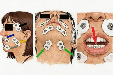
Schematic representation of electrode placement; from left to right: blue arrow, upper perioral; gray arrow, lower perioral; yellow arrow, masseter; black arrow, anterior temporalis; green arrows, submandibular muscle group; red arrow, magnet placement.
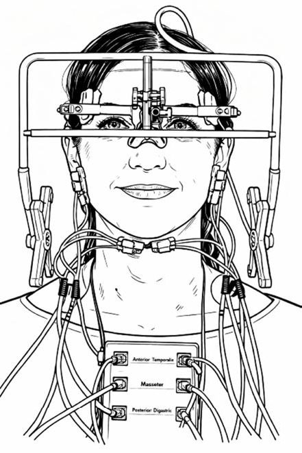
Frontal view of a participant wearing the K7 Evaluation System. A lightweight head frame supports the jaw-tracking sensor array positioned in front of the face, aligned relative to the nasion and lateral orbital regions. Bipolar surface electrodes are placed bilaterally on the anterior temporalis, masseter, and submental (anterior digastric) regions. This configuration enables synchronized three-dimensional recording of mandibular motion and multi-channel sEMG activity during standardized saliva-swallowing tasks.
2.6. Data Origin and Scope
The methodology presented in this article is based on retrospective review of anonymized sEMG and mandibular kinematic recordings acquired during routine neuromuscular functional assessments of the stomatognathic system in an outpatient dental setting. No additional procedures, patient visits, or data collection efforts were undertaken specifically for this methodological study; all recordings were originally obtained as part of the standard clinical evaluation of swallowing. The dataset comprises multiple synchronized sEMG-mandibular kinematic recordings from individuals who underwent routine functional assessment. A subset of this dataset was also used in a separate unpublished manuscript, currently under preparation, focusing on the qualitative characterization of voluntary and spontaneous saliva swallowing. In the present work, these recordings were reorganized and reviewed to illustrate the acquisition, synchronization, and interpretive phases of the integrated protocol. Quantitative summaries (such as the number of participants and scans) are reported only to characterize the scope of the available material and to exemplify the application of the protocol.
2.7. Swallowing Task
Participants were instructed to perform a Voluntary Swallow of Saliva (VSS). In addition, a Spontaneous Swallow of Saliva (SSS) was recorded during a passive observation period without swallowing-related cues; this approach was employed to minimize conscious modulation of the swallowing reflex [9, 10, 17]. All examinations were performed with the participant seated upright in a dental chair, feet supported, and the head in a natural, midline position. The backrest was positioned approximately vertically, and no headrest was provided to facilitate a natural head position. Participants were instructed to maintain a habitual mandibular rest position, with lips lightly closed and teeth not in contact, and to avoid deliberate head or trunk movements during acquisition. For the VSS task, participants were asked to accumulate saliva in their mouth and, upon a verbal cue (“swallow now”), to perform a deliberate swallow without additional bolus. A short practice trial was allowed if needed to ensure understanding. During the recording phase, individual VSS events were separated by rest periods of at least 20-30 seconds in the mandibular rest position to limit carry-over muscle activity and to re-establish a stable kinematic baseline. A minimum of two VSS events was recorded per participant. After each swallow event, the participant was instructed to close in centric occlusion by tapping lightly on the teeth (“tap-tap”). For the SSS task, spontaneous swallows were recorded during a passive observation period in which participants were asked to remain relaxed in mandibular rest position and were not informed that swallowing was the focus of recording. The operator refrained from providing cues or interacting with the participant, and swallows occurring within this interval were identified and marked in the synchronized sEMG-kinematic traces. A minimum of one SSS event was recorded per participant. After each SSS event, the participant was instructed to close into centric occlusion with the “tap-tap” command. In both tasks, the occlusal reference was defined as maximum intercuspation, which was recorded briefly at the end of each recording session to provide a stable positional reference for subsequent jaw-tracking trajectories. All swallows were performed without food or liquid bolus to avoid confounding effects of bolus volume or consistency and to maintain a standardized, non-invasive functional testing condition suitable for routine dental and neuromuscular evaluation. No pharmacological agents were administered during the recordings. sEMG and kinematic signals were acquired simultaneously to allow precise temporal synchronization between muscular activity and mandibular movement (Figs. 3 and 4).
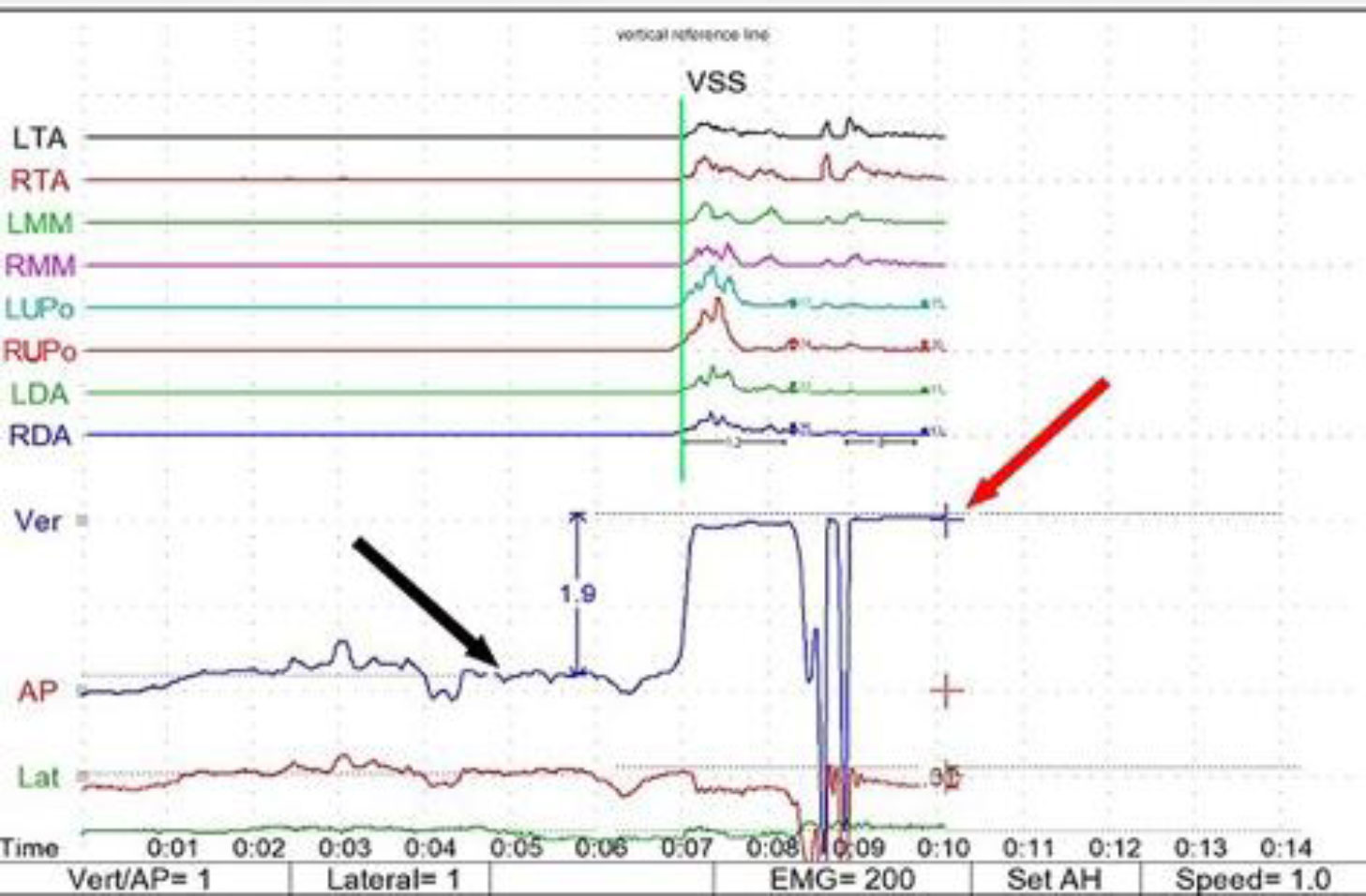
Temporal synchronization of sEMG and kinematic signals in sweep mode. Representative trace showing onset, peak, and offset of muscle activation relative to mandibular opening and closure during a single saliva-swallowing cycle (polygraphic scan, VSS, sweep mode). sEMG, surface electromyography; VSS, voluntary saliva swallow; LTA, left temporalis anterior; RTA, right temporalis anterior; LMM, left masseter muscle; RMM, right masseter muscle; LUPo, left upper peri-oral; RUPo, right upper peri-oral; AP, anteroposterior; Lat, lateral line; CO, centric occlusion; Ver, vertical. The green vertical reference line indicates the start of the VSS sequence; the black arrow indicates the mandibular rest position; the red arrow indicates the intercuspal vertical stop to mandibular closure. Example of a Voluntary Saliva Swallow (VSS). The figure illustrates the temporal scale typically displayed by the software interface during a swallowing recording.
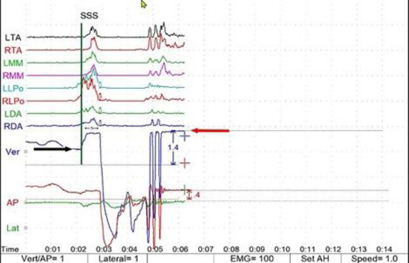
Combined sagittal and frontal trajectories of mandibular motion. The three-dimensional path of the incisal point is shown during standardized saliva swallowing, illustrating inferior, anterior, and lateral displacement phases relative to the reference position (polygraphic scan, SSS, sweep mode): SSS, spontaneous saliva swallow; LTA, left temporalis anterior; RTA, right temporalis anterior; LMM, left masseter muscle; RMM, right masseter muscle; LUPo, left upper peri-oral; RUPo, right upper peri-oral; AP, anteroposterior; Lat, lateral; CO, centric occlusion; Ver, vertical. The green vertical reference line indicates the start of the SSS sequence; the black arrow indicates the level of mandibular rest position; the red arrow indicates intercuspal vertical stop to mandibular closure. Example of a Spontaneous Saliva Swallow (SSS). The figure illustrates the temporal scale typically displayed by the software interface during a swallowing recording.
2.8. Signal Processing and Analytical Principles
Mandibular kinematic data were recorded concurrently and temporally synchronized with the sEMG signals in the same software environment. Within the clinical measurement space (10 mm × 10 mm × 8 mm in the vertical, anteroposterior, and lateral dimensions, respectively), the system's maximum measurement error was 0.5 mm in the vertical and anteroposterior directions and 0.7 mm in the lateral direction. Within the qualitative measurement space (50 mm × 50 mm × 30 mm), the maximum measurement error was 3.0 mm across all directions. The K7 evaluation system (US Food and Drug Administration clearance K003287) is approved for both sEMG and mandibular tracking applications. The analytical focus was on three descriptive aspects defined a priori: (1) temporal sequencing of muscle activation; (2) relative amplitude distribution among muscle groups; and (3) coordination between muscular activity and mandibular movement. In this methodological protocol, the analysis was descriptive and based on visual inspection of synchronized EMG and kinematic traces according to the predefined criteria described above [38]. Temporal sequencing was determined by identifying the order of onset, peak, and offset of rectified sEMG bursts across muscle channels relative to the initiation of mandibular opening in the kinematic trace. The relative amplitude distributions were assessed by comparing the amplitude envelope magnitudes of muscle groups during corresponding phases of mandibular displacement to observe general activation balance. Coordination between muscular activity and mandibular movement was evaluated by noting the temporal overlap between submental/perioral activation and mandibular opening-closing trajectories as well as by assessing the return to maximum intercuspation relative to jaw-closing muscle bursts. These criteria were applied consistently across all recordings to illustrate the descriptive application of the protocol. Because the purpose of this study was to describe and illustrate a methodological framework rather than to test hypotheses, compare groups, evaluate diagnostic performance, or assess treatment outcomes, no inferential statistical analysis was planned or performed. The recordings were used exclusively to demonstrate the acquisition, synchronization, visualization, and descriptive interpretation procedures that constitute the proposed protocol.
2.9. Integrated Electromyographic-kinematic Signal Acquisition and Visualization
The proposed methodological approach allows simultaneous acquisition and synchronization of sEMG and mandibular kinematic signals during swallowing. EMG recordings simultaneously monitor the activity of the selected orofacial and submental muscle groups, while mandibular tracking provides continuous three-dimensional movement data of the mandible throughout the swallowing task. Reliable temporal alignment between EMG-measured activity and mandibular movement enables clear visualization of muscle activation sequences in relation to mandibular motion. This is achieved using two primary visualization modalities, sweep mode and sagittal-frontal mode. Sweep mode is a continuous time-based sEMG acquisition mode in which raw or rectified muscle signals are displayed in a scrolling waveform over a fixed time base. Sagittal-frontal mode is a jaw-tracking display mode that simultaneously plots mandibular movement trajectories in the sagittal (anteroposterior/ vertical) and frontal (lateral/vertical) planes. These modalities can be displayed independently or combined within the same interface (Fig. 5), facilitating the identification of distinct phases of swallowing based on coordinated bioelectrical and kinematic events. In practice, signal preprocessing and standardized electrode placement aim to reduce motion artifacts and baseline noise, ensuring that swallowing-related activity and mandibular trajectories remain interpretable across repeated recordings. Verification of electrode contact, skin preparation, and jaw-tracking alignment was performed routinely both prior to and during recordings to minimize signal degradation and sensor drift.
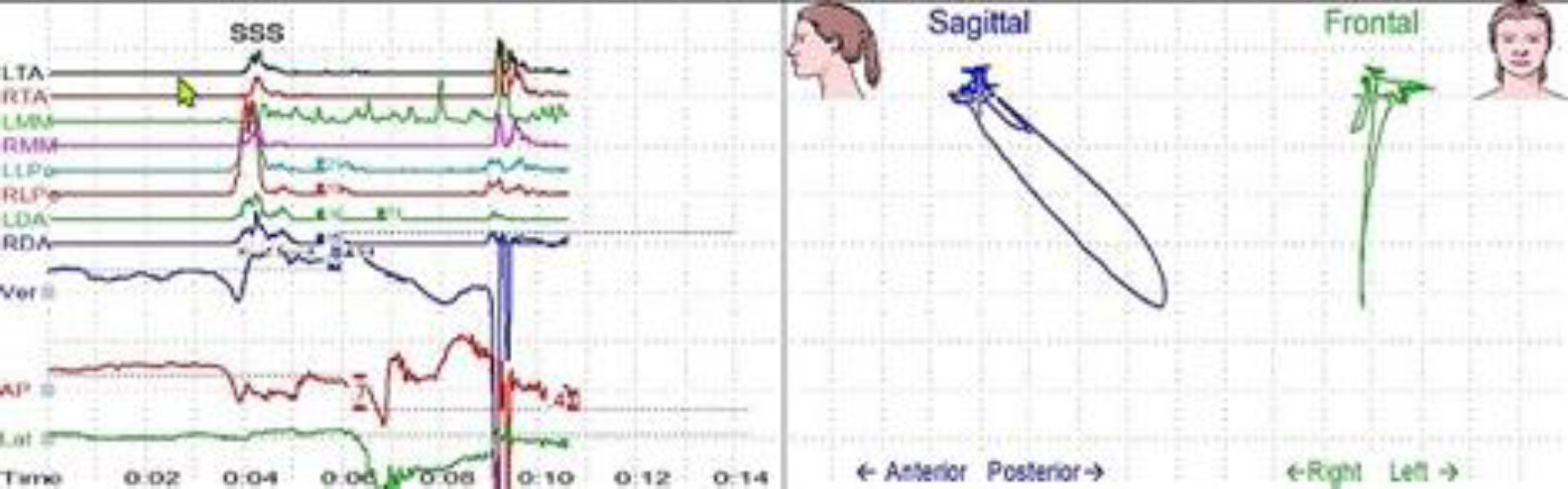
Synchronized display of EMG (sweep mode, left) and kinematic (sagittal-frontal mode, right) data during a swallowing event. EMG, electromyogram; LTA, left temporalis anterior; RTA, right temporalis anterior; LMM, left masseter muscle; RMM, right masseter Muscle; LLPo, left lower peri-oral; RLPo, right lower peri-oral; Ver, vertical; AP, anteroposterior; Lat, lateral.
3. RESULTS
Because the purpose of this article is methodological, the results section is limited to illustrating the application of the proposed acquisition, synchronization, and visualization workflow. The examples presented are intended to demonstrate the operational implementation of the protocol and the appearance of synchronized recordings under standardized conditions. They should not be interpreted as diagnostic findings, outcome measures, validation data, or quantitative study results.
3.1. Overview of Recordings
Previously acquired synchronized sEMG and mandibular kinematic recordings from 36 participants were reviewed to illustrate the methodological workflow. Each participant contributed multiple recordings during routine neuromuscular evaluation. The available dataset comprised 620 synchronized mandibular kinematic-sEMG recordings obtained during routine functional evaluations. These recordings served exclusively as source material for illustrating the acquisition, synchronization, visualization, and descriptive interpretation procedures of the proposed methodological framework. Although portions of the dataset have been used in a separate unpublished manuscript addressing the qualitative characterization of swallowing behavior, no statistical, diagnostic, validation, or outcome-based analyses were undertaken within the scope of the present methodological study. Routine quality verification steps, including checking electrode contact, baseline signal stability, and jaw-tracking alignment, were performed before each session to ensure signal integrity.
3.2. Integrated EMG-kinematic Data Visualization
The integrated acquisition protocol demonstrated clear temporal correspondence between sEMG activity and mandibular motion. In sweep mode, synchronized traces illustrated the relative timing of muscle activation and jaw displacement, allowing visual identification of onset, peak, and offset relationships across channels. The sagittal-frontal display simultaneously presented three-dimensional mandibular trajectories, showing typical “rest-opening-closure” paths toward maximum intercuspation. These visualizations serve as illustrative examples of how synchronized data can be inspected qualitatively to describe coordination patterns during swallowing. Mandibular movements are illustrated in the sweep through three colored lines indicating vertical, lateral, and anteroposterior displacement. In sagittal-frontal mode, mandibular trajectories typically showed a “rest-opening-closure” path toward maximum intercuspation, describing displacement in the vertical, anteroposterior, and lateral planes. The combined display allowed for qualitative identification of coordinated events, such as the relationship between submental and perioral activation and mandibular opening or the return to maximum intercuspation relative to jaw-closing muscle activity. Representative synchronized traces and trajectories in Fig. (5) illustrate how the protocol supports the descriptive analysis of swallowing.
3.3. Qualitative Characterization of Recordings
Selected examples (Figs. 3-5) illustrate the application of the descriptive approach described in the Materials and Methods section. They are presented solely to demonstrate the qualitative interpretive process applied to synchronized recordings without statistical quantification, grading, or diagnostic interpretation.
4. DISCUSSION
This work presents a standardized, non-invasive protocol for analyzing swallowing by synchronizing sEMG and mandibular kinematic data. Its primary contribution is methodological, providing a practical workflow for acquiring, aligning, and visually interpreting these signals during saliva swallowing in an outpatient dental/ neuromuscular clinic setting. Combining time-based sEMG traces with sagittal-frontal jaw trajectories allows synchronized visualization of the temporal relationship between orofacial and suprahyoid muscle activity and mandibular motion during swallowing [22, 30, 31]. Recent digital analyses have reported correlations between kinematics and EMG in masticatory cycles [31]. The protocol allows visualization of swallowing-related bursts alongside jaw opening and closure without reliance on imaging-based techniques, providing a structured graphical representation of synchronized muscular and mandibular events [22]. This is consistent with recent developments in computer-aided, wearable, and digital swallowing assessment systems that aim to provide objective, non-invasive, and potentially continuous functional monitoring outside traditional imaging settings [6, 39-41]. This methodological framework may facilitate standardized documentation of swallowing-related mandibular dynamics during routine dental functional evaluations. Recent work on standardized swallowing assessment protocols and multimodal wearable systems supports the need for reproducible acquisition pathways when translating such approaches into broader clinical use [42-44].
The descriptive dimensions of signal morphology, activation sequences, and kinematic patterns represent a framework to organize observations rather than diagnostic criteria. They offer a shared language for documenting recording characteristics and internal coherence, which may support clinical charting, within-patient follow-up, and future work aimed at defining quantitative indices.
Using routinely collected clinical recordings as examples demonstrated that this protocol can be integrated into everyday neuromuscular dental practice. The illustrations exemplify the application of the proposed protocol for documenting swallowing-related muscle activation and mandibular dynamics during standardized voluntary and spontaneous saliva swallowing under well-defined recording conditions.
5. METHODOLOGICAL LIMITATIONS
This protocol was developed using recordings from individuals in an outpatient dental/neuromuscular setting during saliva swallowing. It does not directly address the assessment of hospitalized patients with dysphagia, bolus swallowing, or acute pathological conditions. Consequently, its application in those contexts would require further adaptation and dedicated evaluation. The description herein is tailored to a specific hardware configuration (K7 system with Duotrode electrodes and magnet-sensor jaw tracking). Although the general principles of electrode placement, jaw tracking, and signal synchronization are transferable, other systems with different amplifier characteristics, sampling rates, or tracking technology will require independent alignment and verification procedures. Finally, only saliva swallows performed under standardized posture and occlusal conditions were considered. The behavior of integrated recordings during bolus ingestion, altered head positions [45], or more demanding functional tasks remains to be systematically explored before extending the protocol to a wider range of clinical scenarios.
6. FUTURE RESEARCH
Future work may extend the proposed acquisition and synchronization protocol to larger datasets, different recording systems, and patient populations with dysphagia or neuromuscular impairments. Recent reviews also suggest that future progress will depend on integrating multimodal sensors, standardized protocols, and digital health workflows for screening, monitoring, and rehabilitation [6, 39, 46]. Such studies may help refine quantitative descriptors and broaden the potential clinical applications of integrated sEMG-kinematic recordings. Recent developments also include portable and wearable systems for non-invasive swallowing assessment [7], and motion detection with highly sophisticated portable sensors [32, 39, 40, 43, 47].
CONCLUSION
This study presents a standardized, non-invasive protocol for the methodological analysis of swallowing based on synchronized sEMG and mandibular kinematic recordings. The approach specifies conditions for task execution, electrode placement, jaw tracking, signal preprocessing, and synchronized visualization. It illustrates their integration to describe swallowing-related muscle activity and mandibular motion in an outpatient dental/neuromuscular clinic setting. Rather than establishing normative values or diagnostic thresholds, this protocol is intended to be a methodological reference for clinicians and researchers who are interested in documenting swallowing function using integrated sEMG-kinematic recordings and serves as a foundation for future development of quantitative indices and clinical applications. Future investigations may use this framework as a basis for validation studies, quantitative analyses, and the development of clinically applicable diagnostic or monitoring tools. Such objectives, however, were beyond the scope of the present methodological report.
AUTHOR’S CONTRIBUTIONS
The author confirms his contributions to the paper as follows: F.S.: Conceived the study, collected, reviewed, and interpreted the recordings, and wrote and approved the final manuscript.
LIST OF ABBREVIATIONS
| EMG | = Electromyography |
| sEMG | = Surface Electromyography |
| VSS | = Voluntary Swallow of Saliva |
| SSS | = Spontaneous Swallow of Saliva |
ETHICS APPROVAL AND CONSENT TO PARTICIPATE
Ethical approval was granted by the Human Research Ethics Committee of Universitat Jaume I (CEISH), Castellón de la Plana, Spain (approval number CEISH/04/2025; approved on February 27, 2025). The approved protocol covered the acquisition and secondary analysis of anonymized surface electromyographic and mandibular kinematic data obtained during routine dental/orthodontic functional evaluation, including assessment of deglutition.
HUMAN AND ANIMAL RIGHTS
All human research procedures followed were in accordance with the ethical standards of the committee responsible for human experimentation (institutional and national), and with the Helsinki Declaration of 1975, as revised in 2013.
CONSENT FOR PUBLICATION
Written informed consent for publication of anonymized data and non-identifiable images was obtained from all participants and, when applicable, from the legal guardians of minors.
AVAILABILITY OF DATA AND MATERIALS
The anonymized data supporting the methodological framework described in this article are available from the corresponding author [F.S.] upon reasonable request.
ACKNOWLEDGEMENTS
Declared none.