All published articles of this journal are available on ScienceDirect.

Effect of Surface Conditioning on the Bond Strength Between 3D-printed PMMA Denture Bases and Resin-based Artificial Teeth

Abstract
Introduction
This study aimed to evaluate the effect of different surface conditioning protocols on the Shear Bond Strength (SBS) between SLA-based 3D-printed PMMA denture base and resin-based artificial teeth.
Methods
Forty denture base specimens were fabricated using stereolithography-based 3D printing and allocated into four groups (n = 10): control (unpolymerized resin-only), bonding agent, monomer + sandblasting, and monomer-only. The teeth were attached to the denture base specimens with the base resin monomer. SBS was measured using a universal testing machine. Failure modes were classified, and surface topographies were examined by Scanning Electron Microscopy (SEM). Statistical analyses were performed using Welch’s one-way ANOVA with Games-Howell post-hoc tests and Pearson’s chi-square test (α = 0.05).
Results
Significant differences in SBS were observed among the surface conditioning protocols (p < 0.001). The monomer-only group exhibited the highest SBS values, followed by the sandblasting + monomer group, the control group, and the bonding agent group. Mixed failures, defined as fractures occurring both at the tooth-denture base interface and within the material, were predominantly observed in the monomer-only and sandblasting + monomer groups, whereas adhesive failures were more frequent in the bonding agent group. SEM analysis revealed distinct qualitative surface morphologies associated with each conditioning protocol, including smoother surfaces in the control group and more porous and irregular surface structures after monomer application.
Discussion
The higher SBS values observed after monomer application may be associated with improved interaction between the denture base resin and artificial teeth. In addition, the qualitative SEM observations suggested that differences in surface morphologies may have contributed to variations in interfacial adhesion among the tested groups.
Conclusion
Monomer application demonstrated the highest bond strength values among the evaluated surface conditioning protocols for bonding resin-based artificial teeth to SLA-based 3D-printed PMMA denture base resin.
1. INTRODUCTION
Recent advances in computer-aided design and manufacturing (CAD/CAM) and additive manufacturing (3D printing) technologies have significantly transformed prosthodontics, particularly in the fabrication of complete dentures [1]. Among these technologies, Stereolithography (SLA)-based 3D printing has gained increasing popularity because of its accuracy and manufacturing efficiency [2]. However, most current dental 3D printing systems still require separate fabrication of denture bases and artificial teeth, making reliable bonding between these components a critical factor for prosthesis durability and clinical success [1, 3].
Consequently, prefabricated artificial teeth or digitally produced teeth must be bonded to the denture base in a secondary procedure [4]. For digital production, CAD/CAM PMMA discs or PMMA-based resins compatible with 3D printing are commonly used. Artificial teeth may be fabricated as a single arch or as individual units positioned into corresponding recesses in the base [3].
Bonding between denture teeth and acrylic resin bases has been extensively investigated because inadequate adhesion may result in tooth debonding, prosthesis repair, and reduced clinical longevity [5, 6]. Despite advances in digital denture fabrication and available bonding protocols, reliable adhesion between denture teeth and denture base materials remains a significant clinical challenge. Tooth debonding occurs particularly in the anterior region and accounts for approximately 22–30% of denture repairs [5, 6].
Various surface conditioning approaches have been proposed to improve adhesion between denture base materials and artificial teeth, including bonding agents, monomer application, and surface roughening procedures such as sandblasting [1, 7]. These methods aim to enhance chemical interaction and/or micromechanical retention at the tooth-denture base interface. In addition, because artificial teeth are prepolymerized, chemical copolymerization with denture base materials is limited, necessitating additional mechanical or chemical surface treatments to improve interfacial bond strength [8].
Although these approaches may improve adhesion, no standardized bonding protocol has yet been established for 3D-printed denture base materials. Previous studies have demonstrated variable bond strength outcomes depending on the denture base material, surface treatment method, and bonding protocol used [5, 8]. Furthermore, limited evidence is available regarding the effect of different surface conditioning protocols specifically on SLA-based 3D-printed PMMA denture bases bonded to resin-based artificial teeth [3, 8]. Direct comparisons of commonly used conditioning approaches under standardized experimental conditions also remain limited in the literature [8].
Therefore, this study aimed to evaluate the Shear Bond Strength (SBS) between SLA-based 3D-printed PMMA denture base resin and resin-based artificial teeth following different surface conditioning protocols, with the objective of identifying the most effective bonding strategy for clinical application. The null hypothesis of the study was that the bond strength between artificial teeth and the 3D-printed PMMA denture base resin does not differ according to the applied surface conditioning procedures.
2. MATERIALS AND METHODS
2.1. Sample Size Calculation
For the determination of the number of specimens to be included in the study, a sample size calculation was performed. For this purpose, a priori power analysis was conducted using the G*Power 3.1.9.7 (Franz Faul, Germany) software for one-way ANOVA comparisons. In the calculation, the effect size (Cohen’s f) was set at 0.769 based on previously published bond strength studies evaluating surface conditioning protocols [7, 9]. The significance level was set at 0.05 and statistical power at 95%. The analysis determined that a minimum of 36 specimens would be sufficient. Considering possible margins of error and specimen losses, it was decided to conduct the study on a total of 40 specimens.
2.2. Specimen Preparation
A total of 40 3D-printed PMMA denture base resin specimens (n = 10 per test group), each with a diameter of 25 mm and a height of 20 mm, were produced in accordance with the ISO/TS 19736:2017(E) standard [10]. PMMA-based resin (Allias Dental Denture, Dokuz Kimya, Istanbul, Turkey) was used as the base material, and the specimens were produced using 3D printing technology.
To ensure standardization of the artificial teeth, maxillary central incisors (Kaiplus, Huge Dental, Rizhao, China) compliant with the ISO 22112 standard [11], type 1 class, with a crown width of 8.5 mm and a crown height of 11 mm, were used. To ensure compatibility between the abutment and the artificial teeth, the base sections of the teeth that fit onto the base sample were scanned using a laboratory scanner (MEDIT T710, Medit Corp., Seoul, Korea), and the obtained digital data were modeled using Exocad CAM software (Exocad GmbH, Germany). The abutment resins were designed to cover the margins of the teeth by 1 mm and were prepared according to the manufacturer’s instructions. The created digital models were saved in STL format and used for production on a 3D printer.
The Saturn 4 Ultra 16K (Elegoo, Shenzhen, China) 3D printer, which operates using SLA (Stereolithography) technology, was selected for the production process of 3D-printed PMMA denture base resin specimens. The printer has a light source with a wavelength of 405 nm and a COB + Fresnel lens collimation system. It can print at 16K resolution, with a layer thickness ranging from 0.01 to 0.2 mm. Pre-print device calibration was performed according to the manufacturer’s recommendations. The photopolymer resin used was manually shaken for 5 minutes prior to printing to ensure a homogeneous mixture. The production parameters were set as follows: 0° print angle, 0.05 mm layer thickness, first-layer time 35 seconds, subsequent layer time 2 seconds, and 60% UV exposure.
After production was completed, the specimens were carefully removed from the platform and soaked for 15 minutes in an ultrasonic cleaning device (Soniret, Reitel Feinwerktechnik GmbH, Bad Essen, Germany) containing 99.9% pure isopropyl alcohol to remove any monomer residues from the surface. Then, following the manufacturer’s instructions, a post-polymerization process was performed for 10 minutes under light with a wavelength of 350–500 nm. This process was performed to ensure complete polymerization of the resin and to increase its mechanical strength. No additional oxygen inhibition layer removal procedure was performed beyond the post-polymerization and isopropyl alcohol cleaning procedures.
The glaze layer on the base surfaces of the artificial teeth was removed using a bur, and the specimens were then cleaned in an ultrasonic cleaning bath containing distilled water for 5 min and dried. The prepared specimens were stored in a light-free environment until the testing phase.
2.3. Surface Treatment Protocols
3D-printed PMMA denture base resin specimens were randomly divided into four test groups according to the surface conditioning protocol applied as follows: (1) Control group (unpolymerized resin-only), (2) Bonding agent, (3) monomer + sandblasting, and (4) monomer-only.
No additional surface conditioning procedure was applied beyond the use of unpolymerized resin during tooth fixation. After the 3D-printed PMMA denture base was fabricated, unpolymerized resin was dripped onto the base surface, and the artificial tooth was placed in the appropriate cavity. During tooth fixation, a force of 10 N was applied from the incisal edge, and excess material was cleaned with isopropyl alcohol and cotton. The specimens were polymerized for 10 minutes.
In the second test group, Ruby SE Bond (Inci Dental, İstanbul, Turkey), a universal self-etch adhesive containing functional methacrylate monomers, 2-Hydroxyethyl Methacrylate (HEMA), Bis-GMA, dimethacrylates, ethanol, water, silica-based fillers, and photoinitiator systems, was used as the bonding agent. The bonding agent was applied to the 3D-printed PMMA denture base resin surface for 10 seconds according to the manufacturer’s instructions, then air-dried for 5 seconds. After the teeth were placed, curing light was applied for 20 seconds using an LED light device (Woodpecker Light Cure I LED, Guilin Woodpecker, Guangxi, China) with a wavelength of 430-490 nm.
In the third test group, 3D-printed PMMA denture base resin specimens received sandblasting with 50 µm diameter Al2O3 particles from a distance of 10 mm, under 5 bar pressure for 10 seconds. After sandblasting, the specimens were cleaned with pressurized steam, then subjected to monomer (Probase Hot, Ivoclar Vivadent Inc., Liechtenstein) application for a total of 180 seconds in three cycles, each lasting 60 seconds. The liquid monomer component (ProBase Hot Liquid, Ivoclar Vivadent, Schaan, Liechtenstein) mainly consists of Methyl Methacrylate (MMA), ethylene glycol dimethacrylate as a cross-linking agent, and stabilizing additives. Following these procedures, unpolymerized resin dripped onto the base surface, and the teeth were placed under 10 N force to polymerize for 10 minutes.
In the fourth test group, the specimens were treated with monomer only (Probase Hot, Ivoclar Vivadent Inc., Liechtenstein) (without sandblasting). 3D-printed PMMA denture base resin specimen surface was exposed to monomer application for a total of 180 seconds in three cycles (each lasting 60 seconds) based on a previous study [9]. Subsequently, the base resin monomer was dripped onto the surface, the teeth were placed under 10 N force, and the specimens were polymerized after excess material was removed for 10 minutes.
All procedures and bond strength tests were performed at room temperature under dry laboratory conditions.
2.4. Shear Bond Strength Testing
For the assessment of the SBS of all test specimens, a universal testing machine (Lloyd-LRX, Lloyd Instruments, UK) compliant with the ISO/TS 19736:2017(E) standard [10] was used. The universal testing machine was calibrated according to the manufacturer’s instructions prior to testing. A 4 mm diameter shear pin and a 1000 N load cell were used in the tests. The bonded surface area (45.36 mm2) was calculated based on the designed tooth-base contact geometry obtained from the CAD model. The load was applied at an angle of approximately 90° to the palatal surface of the incisal edge along the long axis of the tooth. The crosshead speed was set to 1 mm/min, and loading was continued until bonding failure occurred. The maximum load value obtained was divided by the bonding surface area to calculate the SBS [10].
2.5. Fracture Type Analysis
Following the SBS tests, all specimens were examined using an optical stereomicroscope (SteREO Discovery.V20, ZEISS, Germany), and fracture types were classified as adhesive, cohesive, or mixed. Separations occurring at the interface between the artificial tooth and the base resin were defined as adhesive fractures, fractures occurring within the material were defined as cohesive fractures, and fractures occurring both within the material and at the interface were defined as mixed fractures. This classification was performed in accordance with the criteria specified in ISO/TS 19736:2017(E) [10] and the relevant literature [12] by a single calibrated examiner.
2.6. Evaluation of Surface Topographies
Surface topographies of the tested denture base specimens were analyzed by Scanning Electron Microscopy (SEM). One representative specimen from each group was selected for surface morphological evaluation. Prior to analysis, specimens were sputter-coated with a thin layer of gold using a sputter coater (Polaron SC 502, Fisons Instruments, UK). SEM imaging was then conducted (JSM 6060 LV, JEOL Ltd., Tokyo, Japan) to assess post-fracture surface characteristics of the tested denture base specimens.
2.7. Statistical Analysis
Statistical analysis was performed using the SPSS 31.0 package (IBM Corp., Chicago, Illinois, USA). The normality of continuous numerical variables was assessed using the Kolmogorov-Smirnov test, and the homogeneity of variances was examined using Bartlett’s Test. Statistical analysis of data showing normality was performed using Welch’s ANOVA test. Statistical analysis of the failure mode distribution among the groups was performed using the Pearson chi-square test. When expected cell frequencies were less than five, Fisher’s exact test was applied. Results were considered statistically significant at a p < 0.05 level of significance. Bonferroni correction was applied to control for Type I error in all possible multiple comparisons.
3. RESULTS
3.1. Shear Bond Strength Test Results
The SBS values showed normal distribution according to the Shapiro–Wilk test (p = 0.242). However, because variance homogeneity was not confirmed by Bartlett’s test (p = 0.028), Welch’s ANOVA was used for intergroup comparisons.
Statistical analysis demonstrated significant differences in SBS among the tested surface conditioning protocols (p < 0.001), indicating that the applied conditioning method had a considerable influence on bond strength performance. The statistical data table obtained from the pairwise comparisons of the test groups in terms of bond strength is presented in Table 1 and Fig. (1).
| Bonding Protocol | SBS (MPa) Mean ± SD | Min. | Max. |
|---|---|---|---|
| Unpolymerized resin-only | 0.717 ± 0.188 | 0.445 | 1.057 |
| Bonding agent | 0.303 ± 0.093 | 0.182 | 0.449 |
| Sandblasting + monomer | 0.880 ± 0.244 | 0.514 | 1.227 |
| Monomer | 1.139 ± 0.368 | 0.603 | 1.740 |
Note: The values are mean ± SD.
* Different lowercase letters indicate statistically significant differences between groups (Games-Howell, p<0.05).
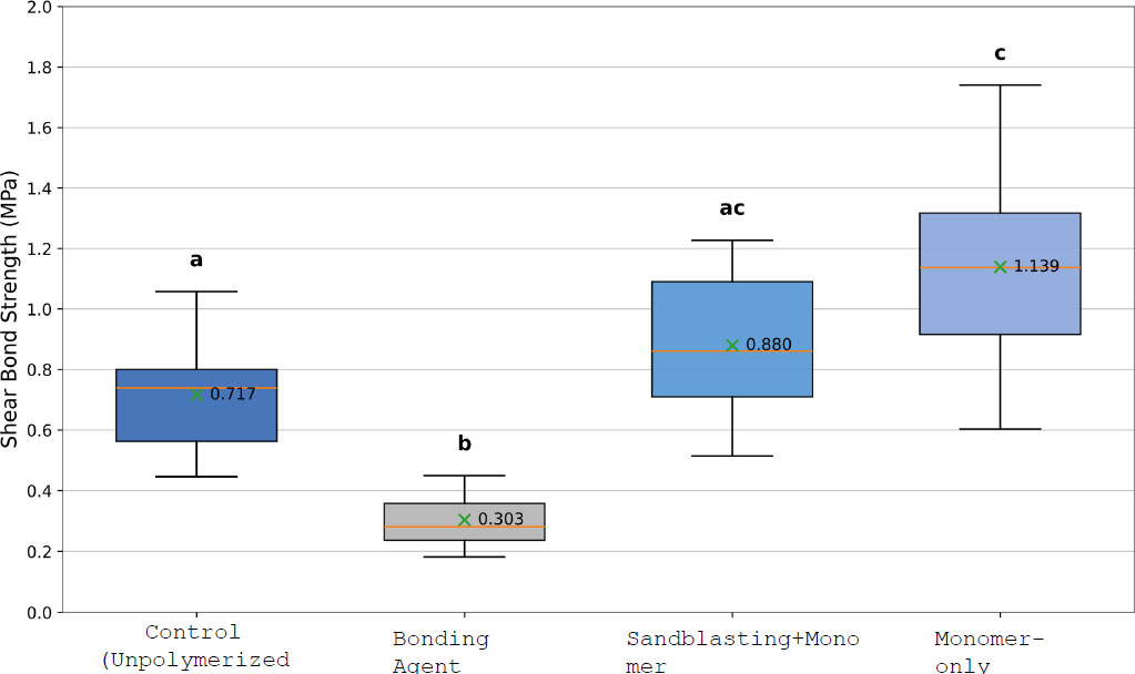
Distribution of Shear Bond Strength (SBS) values among the tested bonding protocols. Different letters indicate statistically significant differences between groups (Games-Howell, p < 0.05).
Pairwise comparisons demonstrated that the bonding agent group exhibited significantly lower SBS values than the control group, sandblasting + monomer, and monomer-only groups (p < 0.001). The monomer-only group showed significantly higher SBS values than the control group (p = 0.029) and the bonding agent group (p < 0.001). Although the sandblasting + monomer group demonstrated higher mean SBS values than the control group, this difference was not statistically significant (p = 0.369). Similarly, no statistically significant difference was observed between the sandblasting + monomer and monomer-only groups (p = 0.285).
Overall, the ranking of the test groups in terms of SBS was as follows: Monomer-only > Sandblasting + Monomer ≥ Control > Bonding agent.
3.2. Fracture Type Analysis Results
The distribution of fracture types among the test groups is presented in Fig. (2). In the control group, adhesive failures were predominant, accounting for 60% of the specimens, while the remaining 40% exhibited mixed failures. In contrast, the sandblasting + monomer and monomer-only groups demonstrated a markedly different failure pattern. In both groups, mixed failures were the most frequent, constituting about 80% of the total failures, whereas adhesive failures were observed in only 20% of the samples. No cohesive failures within the denture base or teeth were detected in any of the groups. The difference in fracture mode distribution among the tested groups was statistically significant (p = 0.003).
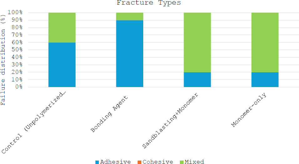
Distribution of fracture types (%) among the evaluated bonding protocols.
3.3. Topographic Evaluation Results
Figure 3 shows SEM images of tested 3D-printed resin base surfaces. Control group specimens typically showed a smooth regular surface (Fig. 3A). The bonding agent-treated specimen exhibited a surface morphology consisting of shallow pits and a micro-rough surface texture (Fig. 3B). Sandblasting + monomer application resulted in more pronounced surface irregularities than the control group (Fig. 3C). The monomer-only group exhibited a porous and protruding surface morphology oriented perpendicular to the resin base surface (Fig. 3D).
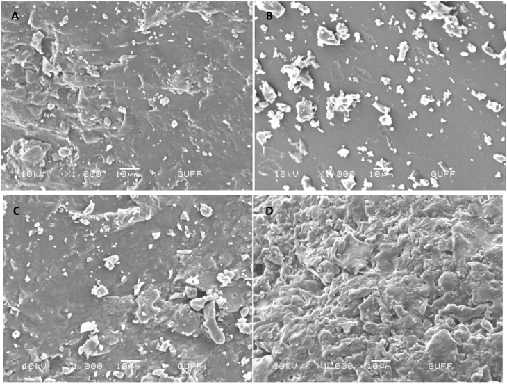
Scanning electron microscopy of tested 3D-printed resin base surfaces (10 kV, x1000 magnification) (A. control; B. bonding agent; C. sandblasting + monomer application; D. monomer application only).
4. DISCUSSION
The present study evaluated the effect of different surface conditioning protocols on the bond strength between SLA-based 3D-printed PMMA denture base resin and resin-based artificial teeth. Among the evaluated protocols, monomer application demonstrated the highest SBS values, whereas the bonding agent group exhibited the lowest bond strength values. The findings indicated that surface conditioning protocols significantly affected bond strength. Therefore, the null hypothesis was rejected.
Reliable bonding between denture teeth and denture base materials is essential for the long-term success of removable prostheses, particularly in digitally fabricated systems where denture bases and artificial teeth are produced separately and subsequently bonded [2, 13]. Tooth debonding remains one of the most common clinical complications associated with removable dentures and is frequently related to inadequate interfacial adhesion [6, 7]. Therefore, optimizing the bonding interface between 3D-printed denture base materials and artificial teeth remains clinically important.
In the present study, monomer application resulted in the highest bond strength values among the evaluated protocols. Previous studies have similarly reported that MMA-based surface treatments may enhance adhesion between denture teeth and denture base materials [6, 9]. This improvement has been associated with the solvent effect of MMA on PMMA surfaces, which may facilitate monomer diffusion and promote formation of an interpenetrating polymer network at the bonding interface [8]. The present findings are consistent with these observations and further support the potential effectiveness of monomer-based conditioning protocols for SLA-based 3D-printed denture base resins.
The sandblasting + monomer group demonstrated higher SBS values than the bonding agent group, although lower values were observed compared with the monomer-only group. Previous investigations have reported inconsistent findings regarding the effect of sandblasting on denture tooth adhesion [14, 15]. While surface roughening procedures may theoretically improve micromechanical retention, excessive roughness may adversely affect surface wettability and limit monomer diffusion [14]. This may explain why the combined sandblasting + monomer protocol did not surpass the bond strength achieved by monomer application alone in the present study.
The bonding agent group demonstrated the lowest SBS values among all evaluated protocols. This finding may suggest differences in interaction between the applied bonding agent and the SLA-based PMMA denture base resin under the tested experimental conditions. Although clinically acceptable bonding values have previously been reported using bonding agents in digitally fabricated denture systems [16], variations in adhesive composition, polymerization parameters, and resin chemistry may influence the effectiveness of these materials [5, 16].
Mixed failures were more frequently observed in the monomer-treated groups, whereas adhesive failures were more frequently observed in the control and bonding agent groups. Previous studies have also reported mixed fracture patterns as the dominant failure mode in 3D-printed denture base systems [8, 17]. In the present study, the predominance of mixed failures in the monomer-treated groups may indicate improved interfacial bonding compared with the bonding agent protocol.
SEM observations revealed distinct qualitative differences in surface morphology among the evaluated groups. Monomer-treated specimens exhibited more porous and irregular surface characteristics compared with the other protocols. However, because no quantitative surface roughness or interfacial analyses were performed, these observations should be interpreted descriptively and cautiously.
The experimental procedures used in this study were standardized to allow direct comparison among surface conditioning protocols under controlled laboratory conditions and should not be interpreted as modified clinical instructions for the evaluated commercial materials. Within the limitations of this study, monomer application demonstrated more favorable bond strength values compared with the other evaluated conditioning protocols.
5. STUDY LIMITATIONS
This study has several limitations. Because the experimental design was developed for standardized comparative testing, the procedures may not fully replicate manufacturer-recommended clinical workflows or intraoral conditions. In addition, the in vitro design cannot fully reproduce the complex mechanical, thermal, and chemical conditions of the oral environment. Only one type of denture base resin, artificial tooth material, and bonding system was evaluated, which may limit generalizability of the findings. Furthermore, long-term aging procedures such as thermocycling and fatigue loading were not performed.
Future studies should investigate different 3D printing technologies, commercially available resin systems, aging simulations, and combinations of chemical and mechanical surface treatments to better understand long-term bonding performance in digitally fabricated removable prostheses.
CONCLUSION
Within the limitations of this in vitro study, surface conditioning protocols significantly influenced the bond strength between SLA-based 3D-printed PMMA denture base resin and resin-based artificial teeth. Among the evaluated protocols, monomer application demonstrated the highest SBS values, whereas the bonding agent group showed the lowest bond strength values. The sandblasting + monomer and control groups exhibited intermediate bond strength values.
Fracture mode analysis revealed that mixed failures were more frequently observed in the monomer-treated groups, whereas adhesive failures were more common in the bonding agent group. Therefore, monomer-based surface conditioning protocols may provide more favorable bonding performance for digitally fabricated removable prostheses under the tested experimental conditions.
AUTHORS’ CONTRIBUTIONS
The authors confirm their contributions to the paper as follows: G.E., F.E.: Conceptualization; S.M.: Data curation; S.M., G.E., F.E.: Formal analysis; S.M.: Funding acquisition; S.M.: Investigation; G.E., F.E.: Methodology; G.E.: Project administration; S.M., G.E.: Resources; S.M., F.E.: Software; G.E., F.E.: Supervision; G.E., F.E.: Validation; G.E., F.E.: Visualization; S.M., F.E.: Writing - original draft; G.E., F.E.: Writing - review & editing.
LIST OF ABBREVIATIONS
| 3D | = 3 Dimensional |
| CAD/CAM | = Computer-Aided Design and Manufacturing |
| PMMA | = Polymethyl Methacrylate |
| SBS | = Shear Bond Strength |
AVAILABILITY OF DATA AND MATERIALS
The data supporting the findings of the article will be available from the corresponding author [G.E], upon reasonable request.
ACKNOWLEDGEMENTS
Declared none.