All published articles of this journal are available on ScienceDirect.

Assessment of Occlusal Relationships of Canines and Molars in Primary Dentition among Children Aged 2 to 6 Years: A Cross-sectional Study
Abstract
Background
The occlusion of deciduous teeth determines the natural occlusion of permanent teeth. The occlusion between deciduous and permanent teeth is critical in predicting malocclusion because the latter has a strong dependence on the former.
Objective
The study was intended to undertake baseline data related to the occlusion relationship between the primary canine and primary molar teeth among children aged between 2 to 6 years from public and private dental clinics in Madinah Al-Munawarah, Saudi Arabia.
Methods
A cross-sectional survey was designed and conducted to examine the occlusal relationship between the primary canines and molars among primary dentition from public and private dental clinics in Madinah Al-Munawarah, Saudi Arabia over the period of February, 2021 to March, 2022. A total of 357 primary school-going children (215 girls and 142 boys) aged between 2-6 years were enrolled through a convenient sampling technique.
Results
According to the results, primary canine occlusions (Class II: 64.7%, Class I: 71.1%) exhibited distinct molar associations. Consistent bilateral molar occlusion frequencies (flush terminal: 59.38%, distal step: 0.01, mesial step: 0.021) were noted. A prevailing ideal overjet (75%, mainly in females), increased overjet (17.9%), and reversed overjet (7%) were observed, with males showing a higher reversed overjet prevalence. Primate spacing clustered in the maxillary arch (42%), mandible (14%), and both (21%), with approximately 24% displaying no spacing.
Conclusion
The study indicated a relationship between the fitting of upper second molars together and the alignment of primary canine teeth in kids aged 2 to 6. Among this group, the most common occurrence of ideal front tooth overlap was observed more often in girls.
1. INTRODUCTION
The occlusion in primary dentition affects the natural alignment of the permanent dentition [1]. The occlusal relationship between the primary and permanent dentition is vital in predicting malocclusion because the latter has a strong dependency on the former. The characteristics of a normal occlusion that occurs in primary teeth in the early ages of a child include the gap between anterior teeth, ovoid arch form, flush terminal plane molar relation, primate spaces, and low overbite and overjet [2]. The diagnosis of malocclusion and treatment at an early stage is important to prevent further deterioration before the individual reaches the age of permanent dentition. Malocclusion is the third most frequent dentistry problem in children and young adults after dental caries and periodontal disease [3]. It influences an individual’s oral health as well as the level of self-confidence. The malocclusion relationship between the primary canine and primary molar teeth is not a disease but a morphological deformation that occurs without any pathological condition. The understanding of both primary and permanent occlusion is crucial in the treatment planning of pediatric dentistry [4]. Previously, few studies examined the prevalence of occlusion in children globally [5]. Following this stream, some studies in the context of Saudi Arabia, such as a study by Al-Emran et al. [6], found that malocclusion was observed in 62.4% of Saudi children, with 40% of these cases needing fixed orthodontic appliances. Asiry [7] revealed that about 60.11% of individuals in Saudi Arabia had Class I molar relationships, while 7.12% and 10.13% of the individuals had Class II and III molar relationships, respectively. In Riyadh, approximately 45.4% of Saudi adolescents experience dental crowding, 26.9% have spacing issues, and 16.4% exhibit increased overjet. This shows that the prevalence of occlusion relationships among children is common not only in Saudi Arabia but around the world. Baidas [8], in a descriptive correlational study, also confirmed the prevalence of malocclusion in pre-school children of 3 to 5 years old. Asiry [7], in a study involving Saudi school children aged 12-16, reported that malocclusion was most prevalent, followed by spacing issues in the primary dentition. These studies were conducted in different periods and regions of Saudi Arabia and noticed that the prevalence of malocclusion in primary dentition tends to increase over time. However, the evidence regarding the malocclusion relationship between primary canine and molar among children is lacking, specifically in Madinah Al-Munawarah, Saudi Arabia. Thus, this study was intended to undertake baseline data related to the occlusion relationship between the primary canine and primary molar teeth among children aged between 2 to 6 years from public and private dental clinics in Madinah Al-Munawarah, Saudi Arabia. In addition to this, the study also examined the distribution of overjet and spacing among primary dentition.
2. MATERIALS AND METHODS
A cross-sectional study design was adopted, focusing on individuals from public and private dental clinics in Madinah Al-Munawarah, Saudi Arabia over the period of February, 2021 to March, 2022. The study was initiated after approval from the institutional review board of Taibah University College of Dentistry Research Ethics Committee (IRB No: 0001037) and in accordance with the Helsinki Declaration of 1975, revised in 2000. The sample size for this study was calculated using “Raosoft” sample size calculator with a 95% confidence interval, 5% margin of error, and 50% response distribution. The recommended sample size on Raosoft came out to be 377. The study only included healthy children with no missing teeth, no developmental anomalies, no proximal restoration tooth, no stainless-steel crown in the posterior tooth, and no extensive dental carries. Based on this inclusion criteria, the study included a total of 357 primary school-going children (215 girls and 142 boys), aged between 2-6 years, who were enrolled through a convenient sampling technique. Two independent researchers communicated the study objective in detail to enrolled children's parents or guardians, and informed consent was obtained from parents. To ensure inter-examiner reliability, a calibration process was performed before data collection. Both researchers underwent joint training sessions to review the criteria for evaluating occlusion and malocclusion, including Foster and Hamilton criteria [9] and Kisling and Krebs criteria [10]. Periodic recalibration sessions were conducted throughout the study period to maintain consistency. The oral examination of enrolled children was performed carefully with properly sterile and complete instruments by a professionally trained dental examiner. The data on occlusion and malocclusion were recorded as described by the World Health Organization (WHO), the Oral Health Survey, and basic methods by using the Community Periodontal Index (CPI) probe [11]. The Foster and Hamilton criteria were applied to evaluate the relationship between the primary molars and primary canines when the teeth were in centric occlusion. The evaluation of primary canines and second molar was done in terms of occlusion relationships in centric occlusion. The flush terminal plane, distal step, and mesial step were noted to represent primary second molars occlusion relationships concerning the vertical plane passing the primary second molars mandibular and maxillary distal surface. The primary canine occlusion relationships were categorized as class I, class II, and class III, respectively, concerning the vertical plane passing the mandibular primary canine distal surface and primary maxillary canine cusp tip. The examiner used the “tell, show, and do” technique to attain better control over children's behavior during monitoring of centric occlusion, and the reliability of the dental exam was ensured through repetition of the process by the examiner in 15 cases within a week. Cohen's Kappa coefficient for categorical variables was computed to assess inter-examiner calibration agreement reliability. An excellent level of agreement was observed with a value of Cohen's Kappa to be greater than 0.75. Moreover, for continuous measurements, the intra-class correlation coefficient (ICC) was determined, which was calculated to be greater than 0.80, showing a good and excellent level of agreement. The reliability of the survey form was confirmed with a Cronbach's alpha of 0.81, indicating results within the standard range. The measurement of the overjet was carried out by using a millimeter gauge to measure the maximum distance between the incisal edges of the mandibular and maxillary incisors on the occlusive plane. If the positive overjet was equal to or less than 2 millimeters, it was recorded as ideal. If more than 2 millimeters, it was recorded as increased, and if found edge-to-edge relationship or anterior crossbite, it was referred to as reversed. Spacing conditions in the maxilla and mandible were recorded and categorized based on the Kisling and Krebs criteria, which include no contact or contact, spacing of 2 millimeters or more, and overlapping teeth. When there was uncertainty, dental floss was used to determine the presence or absence of contact between the teeth. Data was analyzed using SPSS version 23.0 (SPSS Inc., Chicago, USA). Median and range were computed for age, whereas frequency and percentages were used to calculate molar relationship, primary canine, and overjet. Chi-Square was used to compare categorical variables. p-value ≤ 0.05 was considered to be significant.
3. RESULTS
Our study examined the occlusion relationship between primary canine and molars among a total of 357 children (215 girls and 142 boys) with a median age of 4 (2-6) years. Boys and girls were categorized into five groups based on their age. Among boys, 28.9% of boys were 4 years old, and 24.2% of girls were 2 years old (Table 1).
| Age Group (Years) | Boys | Girls n (%) | Total n (%) |
|---|---|---|---|
| 2 | 20 (14.1) | 52 (24.2) | 72 (20.2) |
| 3 | 18 (12.7) | 41 (19.1) | 59 (16.5) |
| 4 | 41 (28.9) | 37 (17.2) | 78 (21.8) |
| 5 | 36 (25.4) | 43 (20.0) | 79 (22.1) |
| 6 | 27 (19.0) | 42 (19.5) | 69 (19.3) |
Our study compared relationships of molar occlusal (flush terminal, distal step, and medial step) with canine occlusal classes (I, II, and III) in (2-6 years) primary dentition during bilateral assessment. The results demonstrated a statistically significant difference (p-value: 0.019) between class II canine occlusions, as 64.7% of it intersects with flush terminal molar occlusions. Class I was also reported to be associated with the mesial step (71.1%) with a statistically significant difference (p-value: 0.001) (Table 2). The occlusion patterns of primary canine class II showed that the upper primary canine cusp tip was in line with the anterior, middle, and posterior of the one-third lower canine in 9 (2.5%), 33 (9.2%), and 146 (40.8%) participants, respectively.
The comparison of molar occlusion was also made in primary (2-6 years) dentition on both sides (right and left), and results showed no alteration in the molar occlusion frequency in most of the assessments. A statistically significant association was observed in the primary molar flush terminal on both sides (p-value: <0.001) with 59.38% coincidence. Significant associations were also reported for the distal step (p-value: 0.01) and mesial step (p-value: 0.021) (Table 3).
The results also revealed the distribution of overjet variation, and an ideal overjet was observed to be prevalent (75%) with female preponderance (75.7%), followed by increased overjet (17.9%), and then reversed overjet (7%). Male preponderance was observed in reversed overjet (Table 4).
| Molar Occlusion | - | Flush Terminal | Distal Step | Mesial Step | p-value |
|---|---|---|---|---|---|
| Canine Occlusion | Class I | 64 (17.9) | 39 (10.9) | 254 (71.1) | 0.001 |
| Class II | 231 (64.7) | 86 (24.1) | 40 (11.2) | 0.019 | |
| Class III | 141 (39.5) | 19 (5.3) | 197 (55.2) | 0.317 |
| Occlusion Pattern | Right Side | |||
|---|---|---|---|---|
| Flush Terminal | Distal Step | Mesial Step | ||
| Left Side | Flush Terminal | 212 (59.38) | 98 (27.5) | 97 (27.3) |
| Distal Step | 29 (8.12) | 232 (65) | 63 (17.6) | |
| Mesial Step | 116 (32.5) | 27 (7.5) | 197 (55.1) | |
| p-value | <0.001 | 0.01 | 0.021 | |
| Overjet | Boys | Girls | p-value |
|---|---|---|---|
| Ideal (n=268, 75%) | 65 (24.3) | 203 (75.7) | 0.003 |
| Increased (n=64, 17.9%) | 28 (43.7) | 36 (56.3) | 0.012 |
| Reversed (n=25, 7%) | 19 (76) | 6 (24) | 0.32 |
The most frequently observed primate spacing site was found in the maxillary arch (n=150, 42%), followed by the mandible (n=50, 14%) and in both maxilla and mandible (n=75, 21%), while spacing was absent in 82 (24%) children. The distribution of physiological spacing is shown in Fig. (1).
4. DISCUSSION
Our study examined the occlusion relationship between primary canine and molar dentition among the pediatric population aged between 2 to 6 years, and results demonstrated statistically significant differences (p-value: 0.019) between class II canine occlusion and molar occlusions. Class I was also reported to be associated with the mesial step (71.1%) with a statistically significant difference (p-value: 0.001). The comparison of molar occlusion showed no alteration in the molar occlusion frequency in most of the assessments. We also observed a distribution of overjet prevalence (75%) with female preponderance (75.7%). In another study, primary dentition occlusal is recognized between the ages of 3 and 6 years [12]. The association between primary dentition and permanent dentition transition predicts the final permanent occlusion [13].
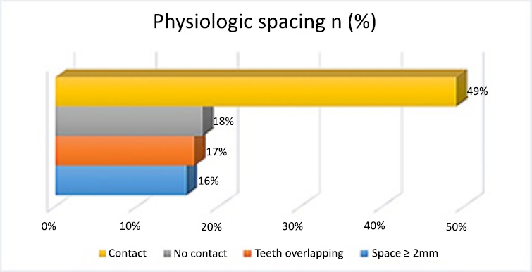
Percentage of physiological spacing in enrolled children.
In the present study, 42% of children had primate spaces in the maxilla, followed by 14% in the mandible. This finding was consistent with the findings reported by Pradhan et al. [14] and Abu Alhaija et al. [15], who also observed frequent primate spaces in the maxilla compared to the mandible, while another study by Shah et al. [16] reported that the most frequently observed molar relationship in all age groups was mesial, followed by the flush terminal and distal step. Primate spaces were observed in both mandible and maxilla among 67 (20.80%) participants. Kadiyala et al. [17] found primate spaces in the upper arch and lower arch, respectively. Our study result showed the most significant primate spacing observed in the maxillary arch (n=150, 42%).
Physiological spaces were commonly termed as the developmental or secondary spaces that appeared commonly between the incisors. In our study, 175 (49%) of the participants had contact, which was near to the findings observed by Sun et al. [18], where 55.7% had physiological spaces. The disproportion between the tooth size and the jaw in permanent dentition is reflected in the absence of physiological spaces among more than 50% of the studied participants. The authentic forecasting for the maxillary relationship in the permanent dentition is taken under consideration for both the molar and canine relationships. It was inferred that the flush terminal could have a preferable permanent molar relationship because it was the commonest molar relationship to be assumed. In our study, the flush terminal plane molar relationship was observed on both sides in 212 participants (59.38%), while the distal step relationship was found in 232 participants (65%). However, the flush terminal relationship was least prevalent in the Chinese study conducted by Zhou et al. [19]. In another study, Baral et al. [20] reported the prevalence of the mesial step molar relationship in the Nepalese population. In the Tunisian context, Maatouk et al. [21] reported that 15% of children with mesial steps exhibited the flush terminal molar relationship as compared to the findings of this study.
The study reported ideal overjet, class I canine, and flush terminal molar relation as the commonest in deciduous dentition. The prevalence of overjet in our study was 75%, with 75.7% female predominance. Another study by Sharma et al. [22] reported 88.4% ideal overjet presence and 10.3% with increased overjet. Another study reported an overjet extent of 2mm (49.1%) [23].
The current study has some limitations. For instance, gender was not evenly represented as more girls participated, which decreased the generalizability of the findings. Moreover, the cross-sectional design restricts the tracing of changes over time in occlusion, and focusing on a small age group from 2-6 years will not lead to any wider conclusion about occlusal development in later ages. Geographic and cultural factors also limit the generalizability of the results to other populations. Therefore, further studies in different communities and different regions of the Kingdom of Saudi Arabia can provide national epidemiological evidence to develop future strategies and guidelines to assure healthier dental health among children. Future studies can utilize these findings by undertaking longitudinal design for reporting occlusal relationships of permanent teeth.
CONCLUSION
The results of our study offer valuable insights into the occlusal relationships between primary canines and molars in children aged 2-6 years. A significant association was found between Class II canine occlusion and flush terminal molar occlusion, with most Class II cases intersecting with flush terminal molars. Similarly, Class I canine occlusion was predominantly associated with mesial step molar occlusion. The distribution of occlusal canine classes also indicates variation in the position of the upper primary canine cusp tip relative to the lower canine; a significant proportion of cases displayed alignment with the posterior third of the lower canine. Significant associations between the right and left sides were also observed at the molar occlusal level, particularly for flush terminal molar occlusion, suggesting a very high consistency across both sides of the dentition in such groups. Such findings are critical to understanding symmetry in primary dentition occlusal relations. Overjet variation showed that ideal overjet was the most common, especially in girls, and reversed overjet was more prevalent in boys. These results could indicate gender-related differences in occlusal development. The maxillary arch presented a higher frequency of primate spacing, thus indicating a natural spacing pattern in the primary dentition, although a minority of children did not present any spacing. These results contributed to a better understanding of occlusal relationships, overjet variation, and spacing in young children, providing valuable clinical insights into early orthodontic assessment and intervention.
AUTHOR'S CONTRIBUTION
The author confirms sole responsibility for the following: study conception and design, data collection, analysis and interpretation of results, and manuscript preparation.
LIST OF ABBREVIATIONS
| WHO | = World Health Organization |
| CPI | = Community Periodontal Index |
ETHICS APPROVAL AND CONSENT TO PARTICIPATE
The study was initiated after approval from the institutional review board of Taibah University, College of Dentistry Research Ethics Committee, Saudi Arabia (IRB No: 0001037).
HUMAN AND ANIMAL RIGHTS
All human research procedures followed were in accordance with the ethical standards of the committee responsible for human experimentation (institutional and national), and with the Helsinki Declaration of 1975, as revised in 2000.
AVAILABILITY OF DATA AND MATERIALS
The data will be available for review by the corresponding author [S.A.B] upon reasonable request.
ACKNOWLEDGEMENTS
The author is very thankful to all the associated personnel in any reference that contributed to this research.

