All published articles of this journal are available on ScienceDirect.

Influence of the Use of a Mixed Solution of Equal Amounts of Amyl Acetate, Acetone, and Ethanol on the Cleaning of Endodontic Sealer Residues on the Bond Strength of the Fiber Post Cementation System: A Laboratory Investigation
Authors Info & Affiliations
Abstract
Purpose
The study aimed to evaluate the effect of different chemical solutions (ET, 95% ethanol, and ES, experimental solution of amyl acetate, acetone, and 95% ethanol) on the removal of epoxy-based sealer residues (AH, AH Plus Jet, or AD, Adseal) from the adhesive interface between root dentin and universal adhesive in post space third, after 6 months control.
Methods
Eighty bovine roots were prepared and randomly divided according to the sealer and cleaning solution: AH Plus Jet + 95% ethanol (AH+ET), Adseal + 95% ethanol (AD+ET), AH Plus Jet + experimental solution (AH+ES), Adseal + experimental solution (AD+ES). Scanning electron microscopy was used to assess the incidence of residue in the post space, and the data obtained were subjected to Kruskal-Wallis/Dunn tests (α=0.05). Bond strength was evaluated in push-out tests, and the data were subjected to ANOVA/Tukey analysis. The adhesive failure pattern was assessed using stereomicroscopy, and the data have been evaluated through incidence frequency.
Results
AH+ES and AD+ES showed lower residues incidence and higher bond strength values (p<0.05). Type 4 and 3 failures were observed in the cervical and middle thirds in the groups AH+ET, AD+ET, and AH+ES, AD+ES, respectively.
Conclusion
The experimental solution positively influenced the bond strength of the post space.
1. INTRODUCTION
Endodontic residue sealer on the dentin surface of the root canal can negatively impact the clinical longevity of rehabilitative treatment with fiberglass posts [1]. Despite numerous research studies on the effectiveness of different solutions for cleaning endodontic sealer after obturation [2-9], few studies have addressed their relationship with the bond strength of the post space [7-9].
Furthermore, it is important to emphasize that, although epoxy-based sealer endodontics are commonly used for root canal obturation [10] due to their effectivity and cost-effectiveness, there is still no established ideal chemical solution, considered the gold standard, for their complete removal in intraradicular dentin after root canal cleaning in preparation for fiberglass post cementation. Additionally, considering the Etch-and-Rinse (ER) adhesive systems, which are commonly used by clinicians and have a mechanical effect on dentin, forming the hybrid layer [11], it can be speculated that this adhesive protocol may be even more effective on a dentin substrate free from any residual endodontic sealer [12].
In the face of literature review and the need to find viable and effective alternatives for the removal of endodontic sealer residues in post-obturation dentin, with a focus on improving the bond strength of the adhesive system to intraradicular dentin, it has been aimed to evaluate the efficacy of ethanol and an experimental solution (amyl acetate, ethanol, and acetone) using equal proportions to ensure a fair and balanced assessment of the effects of each component on cleaning post-obturation dentin. Additionally, by adopting equal proportions, it was made possible to achieve controlled conditions where each component contributes equitably to the properties of the test solution.
While ethanol is commonly used in the removal of sealer residues from coronal dentin [2], there are no reports on its use in the space prepared for the post. As for the experimental solution, little is known about its effectiveness in combining different chemical solutions. What is known is that acetone alone has been the subject of studies and is recognized for its effectiveness in removing epoxy resin sealer from endodontic instruments [3]. Meanwhile, amyl acetate is a solvent known for its ability to satisfactorily remove residues of epoxy resin-based sealants from dentin [3]. Therefore, by comparing the experimental solution with ethanol, a more in-depth assessment of its cleaning potential can be possible.
Therefore, this in vitro assay aimed to assess the efficacy of 95% ethanol and an experimental solution (amyl acetate, acetone, and 95% ethanol in a 1:1:1 ratio) in removing residues from two epoxy resin-based endodontic cements (AH Plus Jet or Adseal) and their effects on bond strength and adhesive failure pattern to intraradicular dentin in the space prepared for fiber post after 6 months of monitoring, using a conventional resin cement system (RelyX Ultimate) and a universal adhesive (Scotchbond Universal) in the etch-and-rinse strategy. The null hypothesis (H0) has proposed that the chemical solutions cannot be effective in removing residues of epoxy resin-based endodontic sealer from post-obturation dentin and cannot influence bond strength or adhesive failure in the thirds of the post space after 6 months of evaluation.
2. MATERIALS
2.1. Sample Size and Ethical Clearance
This study did not require ethical approval. The methodology followed the PRILE guidelines [13]. The sample size was calculated based on the pilot study results. Eighty samples were randomly divided into experimental subgroups (n = 20) based on the different study protocols.
2.2. Sample Selection and Specimen Preparation
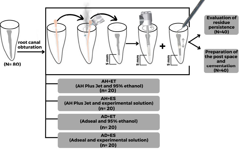
Eighty bovine incisors with similar root anatomy were selected and kept in a 0.1% thymol solution at 4°C until their use. Subsequently, a cutting machine (Isomet™ 1000; Buehler, Lake Bluff, IL, United States) was used to separate the crown, standardizing the specimens to a length of 17 mm from the root apex [14] (Fig. 1; PRILE flowchart).
2.3. Root Canal Preparation
Root canal preparations were performed following a previous study [15]. After achieving a glide path with the K#15 file (Angelus, Londrina, PR, Brazil), apical patency was standardized with a diameter equivalent to K#20 (Angelus, Londrina, PR, Brazil). Then, the root canals were prepared using the WaveOne Gold system to a length of 16 mm with the Large 45/.05 instrument (0.45/D0) (Maillefer, Ballaigues, Switzerland). Between each instrument change, the root canal was irrigated with 5 ml of 2.5% sodium hypochlorite solution (Rioquimica, São José do Rio Preto, São Paulo, SP, Brazil). Subsequently, the canals were flushed with 6 ml of distilled water, suctioned, and dried with absorbent paper points (Tanariman, Manaus, AM, Brazil).
2.4. Evaluated Protocols
After root canal preparation, the specimens were randomly allocated by numerical drawing from 1 to 80 into four groups (n=20), according to the sealer and residue cleaning solution (Fig. 2):
(1) AH+ET (AH Plus Jet and 95% ethanol): The resin-based epoxy root canal sealer (AH Plus Jet; Dentsply DeTrey, Konstanz, BW, Germany) was inserted into the root canal using an applicator attached to the automix tip. Subsequently, root canal obturation was performed with a Large gutta-percha cone (WaveOne Gold 45/.05; Dentsply Sirona, Pirassununga, São Paulo, SP, Brazil). Immediately afterward, the gutta-percha was sectioned 5 mm from the root apex using a special thermoplastic device (Fast Pack; MK Life, Porto Alegre, RS, Brazil), and vertical condensation was carried out using a #3 hand plugger (Easy; Belo Horizonte, MG, Brazil). Then, the dentin surface of the cervical and middle thirds of the root canal was cleaned using 95% ethanol (Rinse-N-Dry; Vista Dental, Racine, WI, United States). For this, the solvent was introduced into the canal with an irrigation syringe, and a rotary brush adapted to an electric motor (X-Smart Plus; Dentsply Sirona, Ballaigues, Switzerland), in continuous rotation (950 rpm), was used to agitate the solvent. Then, a cotton pellet soaked in the solution was used to rub against the canal walls.
(2) AH+ES (AH Plus and experimental solution): Similar to the description for AH+ET, an experimental solution composed of amyl acetate, acetone, and ethanol (1:1:1) was used to clean the cervical and middle thirds of the root canal [16]. For this purpose, the solution was introduced into the canal with an irrigation syringe and agitated first using a rotary brush adapted to an electric motor (X-Smart Plus; Dentsply Sirona, Ballaigues, Switzerland), in continuous rotation (950 rpm), followed by a cotton pellet.
(3) AD+ET (Adseal and ethanol 95%): Similar to AH+ET, the resin-based endodontic sealer has been found to have a different chemical composition (Adseal; Meta Biomed, Chungcheongbuk-do, Republic of Korea).
(4) AD+ES (Adseal and experimental solution): Similar to the method described for AD+ET, the experimental solution described in AH+ES was used for the removal of endodontic cement residues.
After the different cleaning protocols, the canal was irrigated with 5 ml of distilled water to remove the solvents (95% ethanol or experimental solution) and aspirated.
2.5. Post-space Preparation
After the cleaning protocols, all specimens were then used for post-space preparation using a Largo #1 drill (MK Life, Porto Alegre, RS, Brazil) without irrigation at a length of 11 mm. The post space was then standardized using White Post DC FIT #0.4 (FGM, Joinville, SC, Brazil). The post space was rinsed again with 5 ml of distilled water, aspirated using endodontic aspiration tips (Capillary tips; Ultradent, South Jordan, UT, United States), and dried with absorbent paper points [15].
Forty specimens (n=10) were separated for residue persistence analysis using Scanning Electron Microscopy (SEM) and forty specimens (n=10) were cemented with a post and subjected to push-out bond strength and adhesive failure pattern analysis. Table 1 shows the dental products used in the present study, including their materials, origin, and chemical composition.
2.6. Fiber Post Cementation
Forty specimens (n = 10) were used, and a #0.4 fiberglass post (White Post DC FIT; FGM, Joinville, SC, Brazil) was selected for each specimen. The external surface of the fiberglass post was cleaned with 95% ethanol (Sigma-Aldrich, Oakville, Canada), and imme diately afterward, two layers of universal adhesive (Scotchbond Universal; 3M ESPE, St. Louis, MO, United States) were vigorously applied and light-cured using an LED Valo (Ultradent, South Jordan, UT, United States) for 20 seconds [15].
The post-space dentin was conditioned with 37% phosphoric acid (Scotchbond Phosphoric Etchant; 3M ESPE, St. Paul, MO, United States) for 15 seconds, rinsed with distilled water for twice the time, and gently dried with absorbent paper points. Next, two layers of universal adhesive (Scotchbond Universal; 3M ESPE, St. Louis, MO, United States) were actively applied to the dentin surface, and the excess adhesive system was removed with absorbent paper tips. Subsequently, the resin cement RelyX Ultimate (St. Paul, MO, United States) was inserted into the post space using a syringe (Maquira, Maringá, PR, Brazil), and the fiber post was positioned. The assembly was photoactivated with an LED curing device (Valo; Ultradent, South Jordan, UT, United States) at a power of 1.000 mW/cm2 for 40 seconds on each side of the specimen, 1 mm away from the cervical face of the specimen. Finally, the specimens were stored in mineral oil (Nujol; Mantecorp, São Paulo, SP, Brazil) for 6 months for bond strength evaluation and adhesive failure pattern assessment [15].
| Material | Manufacturer | Composition |
|---|---|---|
| AH Plus Jet | Dentsply | Paste A: Epoxy bisphenol-A resin and epoxy bisphenol-F, calcium tungstate (CaWO4), zirconium oxide (ZrO2), silica, and iron oxide. Paste B: Dibenzyl-diamine-aminoadamantane, CaWO4, ZrO2, silic, and silicone. |
| Adseal | Meta | Paste A: Epoxy oligomer resin, ethylene glycol salicylate, calcium phosphate, bismuth subcarbonate, zirconium oxide. Paste B: Poly-aminobenzoate, triethanolamine, calcium phosphate, bismuth subcarbonate, zirconium oxide, and calcium oxide. |
| RelyX Ultimate |
3M ESPE | Base paste: Methacrylate monomers, initiator components, radiopaque, silanated fillers, stabilizers, and rheological additives. Catalyst paste: Methacrylate monomers, radiopaque alkaline (basic) fillers, initiator components, stabilizers, pigments, rheological additives, fluorescence dye, dark cure activator for Scotchbond universal adhesive. |
| Scotchbond Universal | 3M ESPE | 10-methacryloxydecyl dihydrogen phosphate, phosphate monomer, dimethacrylate resins, 2-hydroxyethyl methacrylate, methacrylate-modified polyalkenoic acid copolymer, filler, ethanol, water, initiators, silane. |
2.7. Evaluation of Residue Incidence
After the obturation and dentin surface cleaning protocol, according to the evaluation groups (AH+ET, AH+ES, AD+ET, AD+ES) and post-space preparation, the analysis of residue incidence on dentin surface was performed in forty specimens (n=10). For this purpose, two longitudinal grooves were made, one on the buccal and one on the lingual radicular face, using a double-sided diamond disc (7020, KG Sorensen, São Paulo, SP, Brazil) at low speed. Then, the root was cleaved with a chisel, and the distal section was used for microscopic analysis. The specimens were mounted on metal stubs and placed in a dehydration chamber containing silica gel for 24 hours.
Residue incidence was evaluated using SEM (DSM 940; Carl Zeiss, Oberkochen, BW, Germany). Four different images of the root surface from the cervical, middle, and apical post-space thirds were obtained at a magnification of 500× [17]. The images were consistently captured by the same operator. One representative image from each third was selected for microscopic analysis. Two independent and properly calibrated examiners (Kappa=0.93) classified the residue incidence on the dentin surface, adapted from the classification proposed by Belizário et al. (2022) [18], with score 0 implying no residues on the dentin surface, score 1 indicating between 75% and 100% of the intertubular dentin area without residue, score 2 pointing to between 50% and 75% of the intertubular dentin area without residue, score 3 indicating between 25% and 50% of the intertubular dentin area without residues, and score 4 referring to from 0% to 25% of the intertubular dentin area without residue.
2.8. Bond Strength
After 6 months, the specimens were vertically positioned within a PVC matrix (21.3 mm diameter x 20.0 mm length) and embedded in vinyl resin (Maxi Rubber, São Paulo, SP, Brazil). After 24h, the specimens were removed from the molds and sectioned perpendicular to their longitudinal axis using an Isomet 2000 cutting machine (Buehler Ltd, Lake Bluff, IL, United States) with water cooling.
Three sections, each with a thickness of 2.0 mm ± 0.1 mm, were obtained from the apical, middle, and cervical thirds of the post space. The cervical, middle, and apical sections were obtained, respectively, from 1.0 mm, 5.0 mm, and 9.0 mm apical to the root cervical face. To ensure smoothness, any irregularities on the sections were eliminated using 1200-grit silicon carbide sandpaper (Norton, São Paulo, SP, Brazil) with water cooling.
The specimens underwent push-out bond strength testing using an electromechanical testing machine (EMIC, São José dos Pinhais, PR, Brazil). The apical, middle, and cervical thirds were marked, and a 5 kN load cell, with a crosshead speed of 0.5 mm/min, was applied until complete displacement of the fiber post and/or cementation system occurred. Crosshead diameters of 1.2 mm, 0.9 mm, and 0.5 mm were used for the cervical, middle, and apical thirds of the post space, respectively. The Force (F) required for specimen displacement was measured in Newtons (N) and converted to bond strength (MPa) [14, 19].
2.9. Adhesive Failure Pattern
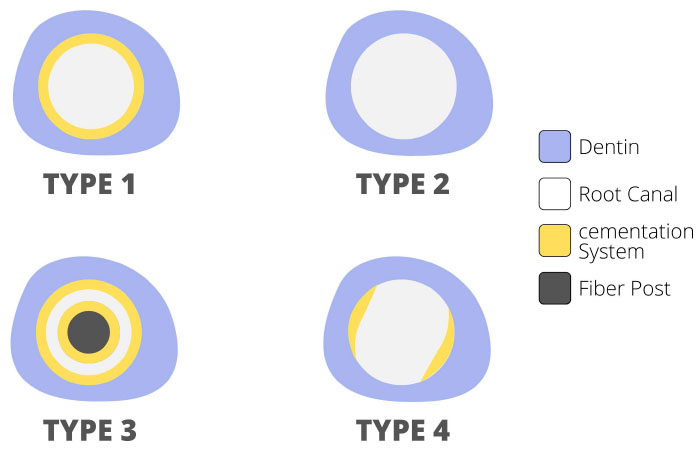
After the push-out test, the slices were analyzed using stereomicroscope Leica DFC295 attached to a Leica S8 APO (Leica Microsystems, Wetzlar, Hesse, Germany), with 10× magnification, to evaluate the adhesive failure pattern. The failure pattern was classified as follows [17, 15]: type 1 (adhesive 1), between the post and the cement; type 2 (adhesive 2), between dentin and the cement; type 3 (cohesive), within the cement; type 4 (mixed), a combination of all failure types. Fig. (3) illustrates the adhesive failure patterns.
2.10. Statistical Analysis
Statistical analysis was performed using the computer program BioEstat 5.0 (Civil Society, Mamirauá, PA, Brazil). The data obtained from the incidence of endodontic sealer residues were subjected to the Kruskal Wallis test, followed by the Dunn test (α=0.05). The data obtained from the bond strength at 6 months were initially evaluated by the Shapiro-Wilk test in order to verify the homoscedasticity of the distribution of the results. Once the normality of the distribution of the results was verified, the data were subjected to two-way ANOVA, followed by Tukey post-hoc tests (α=0.05). Each third of the post space was described in terms of the frequency of incidence of the adhesive failure pattern.
3. RESULTS
3.1. Residue Incidence
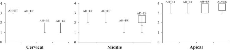
In the cervical and middle thirds post space, AH+ES and AD+ES showed a lower incidence of endodontic sealer residues (score 1, Fig. 4) compared to AH+ET and AD+ET (score 3, Fig. 4; P < 0.05). There was no difference between AH+ES and AD+ES or between AH+ET and AD+ET (P > 0.05). In the apical third, the protocols showed similarity in the incidence of residues in the post space (score 3, Fig. 4; P > 0.05).
Fig. (5) shows the median, maximum, and minimum values, and the first and third quartiles of the scores of the residues incidence on the dentin surface, in cervical, middle, and apical thirds of the post space. In Fig. (5), residues incidence can be observed according to the epoxy resin-based endodontic sealer and the post-space cleaning protocol.
3.2. Bond Strength
In the cervical and middle thirds of the post space, AH+ES and AD+ES provided higher bond strength values compared to AH+ET and AD+ET (P < 0.05). There was no difference between AH+ES and AD+ES or between AH+ET and AD+ET (P > 0.05). In the apical third, there was no difference in bond strength values between the cementation system and the dentin post space provided by the different protocols evaluated in the present study (P > 0.05).
Table 2 shows the arithmetic mean and standard deviation of the bond strength values (in MPa) at the adhesive interface, after endodontic sealer residues removal and fiber post space preparation in the thirds of the post space, after 6 months of evaluation.
3.3. Adhesive Failure Pattern
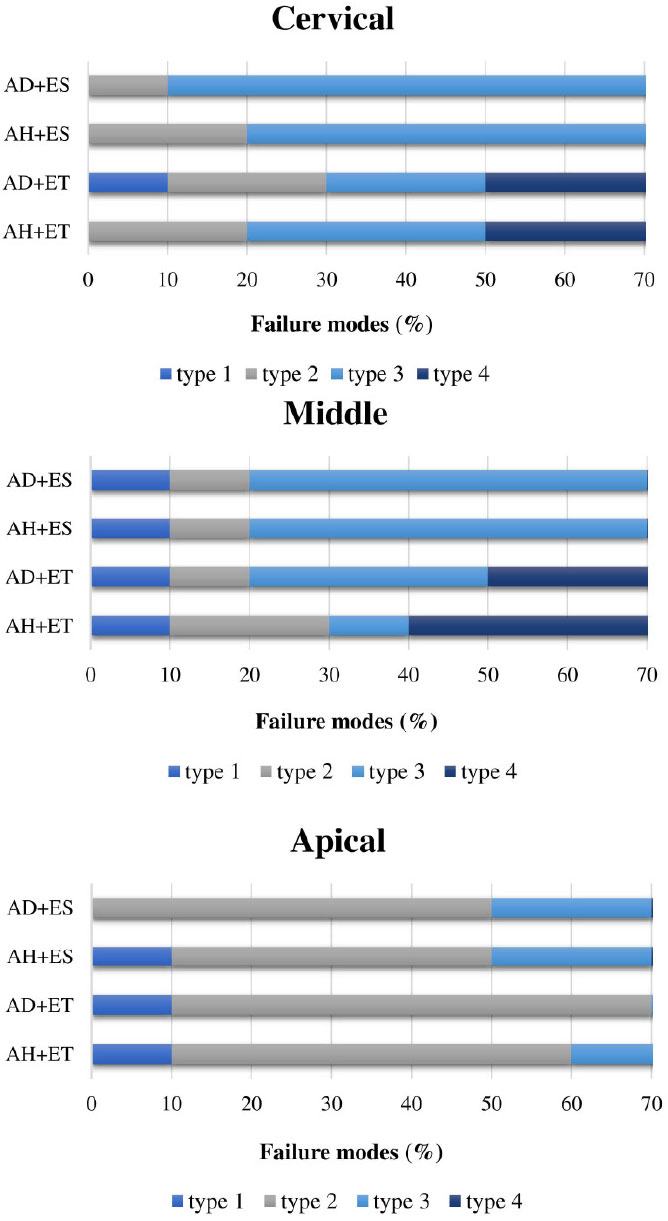
In the cervical and middle thirds, AH+ET and AD+ET showed a higher incidence of type 4 failures (mixed). On the other hand, AH+ES and AD+ES showed a higher incidence of type 3 failures (cohesive). In the apical third, the incidence of type 2 failures (adhesive) was higher in all evaluated protocols.
Fig. (6) shows the failure patterns (%) observed in the cervical, middle, and apical post space thirds, at 6 months control, in the different evaluation groups.

| - | AH+ET | AD+ET | AH+ES | AD+ES |
|---|---|---|---|---|
| C | 7.96 (0.56) b | 7.89 (0.78) b | 9.97 (0.68) a | 9.71 (0.87) a |
| M | 7.77 (0.51) b | 7.67 (0.83) b | 9.39 (0.71) a | 9.22 (0.57) a |
| A | 7.33 (0.51) a | 7.24 (0.67) a | 7.75 (0.33) a | 7.72 (0.68) a |
4. DISCUSSION
The experimental solution composed of amyl acetate, acetone, and ethanol resulted in a lower incidence of epoxy-based resin endodontic sealer residues (AH Plus Jet and Adseal) in the root dentin and higher bond strength values in the cervical and middle thirds of the post space, on adhesive interface between universal adhesive (Scotchbond Universal), in etch-ad-rinse strategy, and radicular dentin, after 6 months control. Therefore, the null hypothesis was rejected.
The literature presents three widely recommended mechanisms for removing dentin contaminated with epoxy-based resin endodontic sealer: mechanical removal, physical dissolution, and surface modification followed by removal. Among these methods, physical dissolution using an organic solvent is widely employed [20]. It is known that the solubility of a substance is intrinsically linked to its molecular structure, particularly the polarity of the bonds present [21]. Amyl acetate is an organic solvent recommended as an alternative for removing epoxy resin-based endodontic sealer residues due to its apolar chemical nature, as well as acetone [3]. On the other hand, 95% ethanol, despite being commonly used for surface cleaning due to its widespread availability, has limited efficacy due to its high polarity [22].
The results of this study demonstrated that the experimental solution (amyl acetate, acetone, and ethanol) promoted more effective removal of obturating sealer residues in dentin, regardless of the type of sealer used, when compared to 95% ethanol. It can be presumed that ethanol alone is immiscible or only partially miscible with the epoxy resin component of the sealer [2], but the combination of solutions may have generated a synergistic effect, contributing to a more effective removal of residues in intraradicular dentin, as shown in Fig. (5).
When evaluating bond strength at 6 months, AH+ES and AD+ES demonstrated higher values in the cervical and middle thirds of the post space. The etch-and-rinse strategy using universal adhesive systems requires that dentin walls be properly prepared for hybridization, which involves the complete removal of gutta-percha and endodontic sealer residues, which is essential to promote mechanical interlocking of resin monomers with intraradicular dentin, ensuring effective adhesion through the formation of resin tags [23, 24]. Subsequently, with the polymerization of monomers within the collagen fiber network, a hybrid layer is created, providing mechanical retention to the substrate [25]. Therefore, the presence of epoxy-based resin sealer residues on the dentin surface in the groups treated with 95% ethanol likely limited the action of phosphoric acid and its ability to demineralize dentin in depth, thus reducing subsequent penetration of monomers into the dental structure and decreasing bond strength. This may not have occurred in the groups treated with the experimental solution, possibly due to the reduced presence of obturating sealer residues on the dentin surface, due to possible cleaning of the dentinal substrate to a greater depth [6].
Adhesive failure type 4 (mixed) was predominantly found in the cervical and middle thirds in the AH+ET and AD+ET groups. It is known that hydrolysis of exposed and non-hybridized collagen fibrils breaks the covalent bonds with the resin polymers, resulting in adhesive failure [26]. In this sense, it is possible that endodontic sealer residues on the dentin surface acted as a physical barrier between the adhesive universal and dentin, impairing their interaction with the substrate [27]. On the other hand, type 3 failures (cohesive) were the most frequent in the AH+ES and AD+ES groups. It is reported that cohesive failures are generally caused by low polymerization of the resin cement [28] due to difficulties in light transmission inside the canal [29-32].
Basic science is important for investigating unique hypotheses that can contribute to a deeper understanding of complex processes. This study has introduced significant results to restorative dentistry, demonstrating that the experimental solution was effective in removing residues from the epoxy resin-based endodontic sealer, with a positive effect on the bond strength of conventional cement to intraradicular dentin. This distinguishes it from previous literature [16] that has not assessed the effects of this material on dentin in the post space. However, the development of new investigations comparing different adhesive strategies, as well as different resin cements (conventional vs. self-adhesive), is crucial for an enhanced understanding of the effects of the evaluated solutions on the adhesive interface between the universal adhesive, in the acid etching and rinsing strategy, and the dentin of the post space.
CONCLUSION
In summary, the experimental solution composed of amyl acetate, acetone, and ethanol has been found effective in removing residues of epoxy resin-based endodontic sealer (AH Plus Jet or Adseal) in the post space and provided higher bond strength values in the cementation system with universal adhesive in the etch-and-rinse strategy.
LIST OF ABBREVIATIONS
| CaWO4 | = Calcium tungstate |
| ZrO2 | = Zirconium oxide |
| EMIC | = Electromechanical testing machine |
| SEM | = Scanning electron microscopy |
| AD+ES | = Adseal and experimental solution |
| AD+ET | = Adseal and ethanol |
ETHICAL STATEMENT
This study did not require ethical approval. This study employed samples of bovine teeth. According to the ethical committee regulations in the country in which the study was conducted, obtaining approval for research involving animal-derived teeth is not required.
STANDARDS OF REPORTING
All the procedures involving animals were performed according to the PRILE Guideline.
AVAILABILITY OF DATA AND MATERIALS
The data that support the findings of this study are available from the corresponding author, [A.P.O.B.], on special request.

