All published articles of this journal are available on ScienceDirect.
Mineralized Plasmatic Matrix for Horizontal Ridge Augmentation in Anterior Maxilla with and without a Covering Collagen Membrane
We would like to apologize for the errors that occurred in the online version of the article. Duplicate paragraphs in abstract and incorrect figures 1 and 2 have been published in the article entitled “Mineralized Plasmatic Matrix for Horizontal Ridge Augmentation in Anterior Maxilla with and without a Covering Collagen Membrane” in “The Open Dentistry Journal”, 2020 Dec 31;14(1) [ 1].
The original article can be found online at https://opendentistryjournal.com/VOLUME/14/PAGE/743/FULLTEXT/
Original:
Materials and Methods:
Sixteen edentulous spaces were randomly divided into 2 equal groups. MPM was used for horizontal ridge augmentation with and without a covering collagen membrane (group 1 and 2, respectively). Cone Beam CT images were obtained preoperatively as well as 1 week and 4 months postoperatively to evaluate alveolar ridge and the resorption of the grafting material at 3 predetermined points along the site where the future dental implant will be placed.
Student’s t-test (Unpaired) was used for comparing two different groups with quantitative parametric data and student’s t-test (Paired) was used for comparing two related groups with quantitative parametric data while repeated measures ANOVA (Analysis of variance) followed by post-hoc Bonferroni was used for comparing more than two related groups with quantitative parametric data.
Student’s t-test (Unpaired) was used for comparing two different groups with quantitative parametric data and student’s t-test (Paired) was used for comparing two related groups with quantitative parametric data while repeated measures ANOVA (Analysis of variance) followed by post-hoc Bonferroni was used for comparing more than two related groups with quantitative parametric data.
Corrected:
Materials and Methods:
Sixteen edentulous spaces were randomly divided into 2 equal groups. MPM was used for horizontal ridge augmentation with and without a covering collagen membrane (group 1 and 2, respectively). Cone Beam CT images were obtained preoperatively as well as 1 week and 4 months postoperatively to evaluate alveolar ridge and the resorption of the grafting material at 3 predetermined points along the site where the future dental implant will be placed.
Student’s t-test (Unpaired) was used for comparing two different groups with quantitative parametric data and student’s t-test (Paired) was used for comparing two related groups with quantitative parametric data while repeated measures ANOVA (Analysis of variance) followed by post-hoc Bonferroni was used for comparing more than two related groups with quantitative parametric data.
Original:


Corrected:
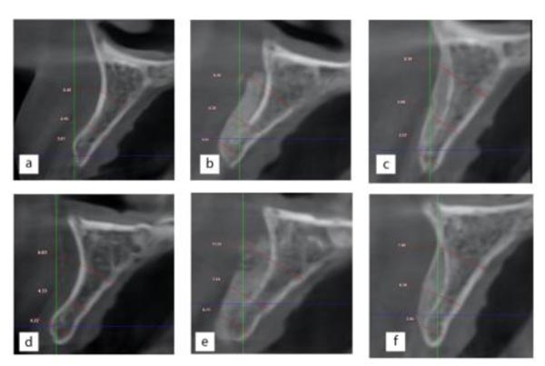
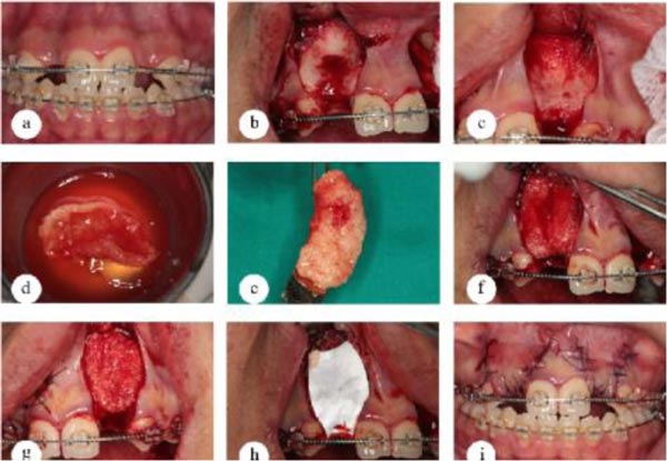
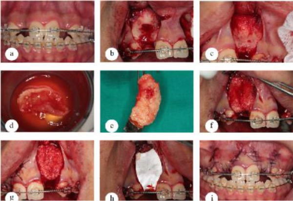
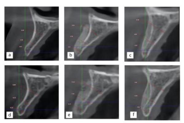
Figs. 1 and 2 have been revised as:


