All published articles of this journal are available on ScienceDirect.

Evaluation of Sexual Dimorphism with Mandibular Parameters by Digital Panoramic Radiography
Authors Info & Affiliations
Abstract
Introduction:
Sex determination is the first step of personal identification in the field of forensics and is essential for reconstructive profiling. The skull appears to be the most reliable part of the skeleton, apart from the pelvis, in sex determination. Tooth and bone are used as a key tool for personal identification due to their high resistance to decomposition and degradation. The present study aimed to evaluate the sexual dimorphism with mandibular parameters by digital panoramic radiography.
Materials and Methods:
In this analytical-descriptive study, the mandibular parameters in 315 females and 217 males, including the ramus height, the coronoid height, the mental height, the mandible body height, the minimum width of the ramus, the distance between the right and left gonial angle, the distance between the right and left condyle and the distance between the right and left coronoid, were measured in the panoramic radiography via SCANORA software. Discriminant function and canonical regression methods were used to determine the predictability of mandibular parameters in sexual dimorphism. P < 0.05 was considered to be statistically significant.
Results:
All parameters were statistically significant between genders (P<0.05). The mean of all parameters, except the gonial angle, in males, was higher than that of the female. Percentage of correctly classified in discriminant function based on the central and right side and left side dimorphic parameters of the mandible is 82.5% and 82.9%, respectively.
Conclusion:
According to the present study, panoramic radiography can be considered as a valuable tool in sex determination (with an accuracy of 82.5%), and all parameters of mandible had sexual dimorphism and showed that they are reliable parameters with a total accuracy of 82.5% in the sexual dimorphism.
1. INTRODUCTION
The main issue in forensic anthropology is human identification, most often of human remains [1]. Sex determination is the first step of personal identification in the field of forensics and is essential for reconstructive profiling. In general, the sex of an unidentified person can be determined based on the anatomical characteristics [2]. Tooth and bone are used as a key tool for personal identification due to their high resistance to decomposition and degradation. Different bones of the body are used in sex determination [3]. The pelvic bone is the most reliable source for sex determination, but when the pelvic bone is not available, the skull is used for this matter; and among skull bones, mandible plays an important role; since it is the strongest, largest and the most dimorphic bone of the skull [4].
The relative position of the upper and lower teeth in jaws determines occlusal stability, which is related to muscular performance. Subjects with high forces exhibit different kinematics of the mandible during mastication, resulting in an increased frequency of chewing cycles [5].
Mandibular dimorphism is affected by the size and shape of masticatory muscles, since the chewing force varies for men and women, and this causes the men’s bones to be generally larger and stronger than the women’s bones [4]. Age, sex, race, and occlusion status affect the morphological characteristics of the mandible. Studies have shown that remodeling of the mandibular bone occurs with aging [1].
Radiographic images are an essential tool for use in forensic anthropology. Panoramic is the most available and widely used extra-oral radiography, and its lower cost and wide area coverage and ease of preparation have made it an excellent choice for examination of many structures [6].
Many studies showed that linear and angular cephalometric measurements of the face and cranial base differed between girls and boys, and changed with age [7].
Among the craniometric measurements, head length and head width are considered most important, as they are used to derive the cranial size expressed as a cephalic index [8]. Several studies have explored and proved the effectiveness of these parameters in racial or ethnic differentiation amongst various populations [9, 10]. However, there is little information regarding their sex discrimination potential.
Recently, studies have been conducted on the ability of radiographic images to determine sex in different societies [1, 11, 12]. Considering the importance of the subject and the lack of research in the Iranian population, this study aimed to assess sexual dimorphism with mandibular parameters such as including the ramus height, the coronoid height, the mental height, the mandible body height, by using panoramic radiographs in Rasht-Iran population.
2. MATERIALS AND METHODS
This study was performed using 541 panoramic radiographs of both genders, referring to a private clinic. The inclusion criteria were the proper quality of radiography in patients with complete dentitions and the age between 20 to 55 years. Radiographies of patients who had lost more than one tooth in each quadrant, patients with bony lesions, fracture, history of surgery, deformed mandible, erosion, malocclusion, anomalies of TMJ (temporomandibular joint), as well as radiographs with positioning errors were excluded from this study. This study was approved by the ethics committee of School of Dentistry, Guilan University of Medical Sciences,Rasht,Iran by ethical code (2909606042).
Radiographs were acquired using the CRANEX D panoramic system (Soredex, Tuusula, Finland) with the kvp=66 , mA=10, t=14.3 s, and the mandibular parameters were measured using a 12.6-inch LED monitor in SCANORA ver. 5.2.1 software (Soredex, Tuusula, Finland).
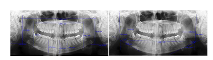
These variables are coronoid height (the distance between the coronion and the lower wall of the bone) [4], mandible body height (direct distance from the alveolar process to the lower border of mandible), chin height (the distance between the menton and the alveolar crest), ramus height (the distance between the highest and the lowest posterior point of ramus), the minimum width of ramus (the lowest posterior-anterior distance of ramus) [12], the distance between the gonial of both sides (the horizontal distance from the right gonion to the left gonion) [1], the distance between the condyle of both sides (the direct distance between the most lateral points on both condyles), the distance between the coronoid of both sides (the direct distance between the most interior points on coronoid) [12] and the gonial angle (a line is tangential to the most inferior points in gonial angle and lower border of mandible body, and another line is tangential to the posterior borders of the ramus and condyle. The angle between these lines forms the gonial angle) [1].
The variables were measured, as shown in Fig. (1) and the information for each measurement was entered into the checklist. All images were evaluated by two oral and maxillofacial radiologists on a 17-inch 32-bit monitor (SyncMaster 740 N, Samsung, Korea) with 1280×1024 pixels resolution in a semi-dark room.
Data were entered into SPSS ver. 21 software. The independent T-test in a single-variable analysis was used to compare the parameters of mandible based on sex. Then, the canonical correlation coefficient was used to determine the relationship between each variable with the sex variable. Also, statistical methods of discriminant function and regression canonical were used for determining the predictability of mandiblar parameters for sex determination.
After determining the proper cutoff points according to the ROC curve, the diagnostic indices(sensitivity, specificity, LR +, NPP, PPV, LR-) were calculated. The significance level of the tests in this study was considered as 0.05 (P<0.05) (Fig. 2).
3. RESULTS
In this study, 532 panoramic radiographs 59.2% (315 radiographs) were for women and 40.8% (217 radiographs) were for men. Mean and standard deviation of the age of samples was 34.4 ± 9.9 years. The inter-observer and intra-observer agreement between the first and the second observer were 0.80 and 0.92, respectively. On both sides, the results were similar and the differences were very small. Due to the high rate of Eigenvalue (expressing the differentiation power of the model in detecting not detecting) on the right side, the right side was considered for the evaluation of parameters.
All parameters were statistically significant between the two genders (P=0.0001). The mean of all parameters, except the gonial angle, in males, was higher than that of the female. (Table 1).
| - | - | Parameter | Gender | Number | Mean(mm) |
|---|---|---|---|---|---|
| Standard Deviation | p-value | Left Coronoid Height | Female | 315 | 63.83 |
| 4.52 | 0.0001 | Male | 217 | ||
| 69.01 | 4.82 | left mandibular body height | Female | 315 | 31.73 |
| 2.70 | 0.0001 | Male | 217 | ||
| 35.08 | 2.88 | chin height | Female | 315 | 31.42 |
| 2.47 | 0.0001 | Male | 217 | ||
| 34.55 | 2.74 | left ramus height | Female | 315 | 64.60 |
| 4.15 | 0.0001 | Male | 217 | ||
| 70.35 | 4.62 | minimum width of left ramus | Female | 315 | 26.86 |
| 2.38 | 0.0001 | Male | 217 | ||
| 27.98 | 2.67 | inter gonial distance | Female | 315 | 163.35 |
| 8.63 | 0.0001 | Male | 217 | ||
| 171.34 | 10.18 | inter condylar distance | Female | 315 | 173.17 |
| 9.04 | 0.0001 | Male | 217 | ||
| 183.34 | 11.17 | inter coronoid distance | Female | 315 | 105.29 |
| 7.07 | 0.0001 | Male | 217 | ||
| 111.36 | 8.11 | left gonial angle | Female | 315 | 122.15 ᵒ |
| 6.54 | 0.0265 | Male | 217 | ||
| 121.48 ᵒ | 7.12 | right coronoid height | Female | 315 | 63.68 |
| 4.47 | 0.0001 | Male | 217 | ||
| 68.87 | 4.84 | right mandibular body height | Female | 315 | 31.82 |
| 2.73 | 0.0001 | Male | 217 | ||
| 35.12 | 2.83 | right ramus height | Female | 315 | 64.48 |
| 3.70 | 0.0001 | Male | 217 | ||
| 69.98 | 4.66 | minimum width of right ramus | Female | 315 | 26.46 |
| 2.44 | 0.0001 | Male | 217 | ||
| 27.97 | 2.58 | right gonial angle | Female | 315 | 123.16 ᵒ |
| 6.65 | 0.0012 | Male | 217 |
Among the central and right side dimorphic parameters of the mandible, the height of right ramus (λ=0.698, P=0.0001), chin height (λ=0.614, P=0.0001), the distance between condyles of both sides (λ=0.578, P=0.0001) and height of right coronoid (λ=0.558, P=0.0001) are considered as diagnostic (discriminative) parameters of sex, respectively (Wilks ‘lambda (λ)).
Table 2 shows the Canonical discriminant function coefficients of these parameters. Based on acquired data, sex discriminant function is equal to the following equation:
Z= -23.746 + 0.111 X 1 + 0.128 X2 + 0.042 X 3 + 0.070 X 4
X 1 = the height of right ramus
X 2 = chin height
X 3 = the distance between condyles of both sides
X 4 = height of right coronoid
Statistically, this discriminant function is significant.
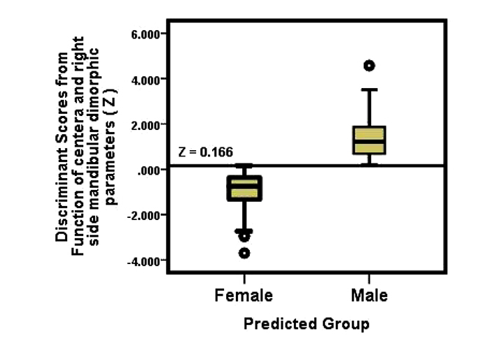
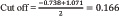
Centroids mean in women group is -0.738 and in men group is 1.071. Therefore, the discriminant function, based on(x1-x4) values for less than 0.166 represents woman sex and for more than 0.166 represents the man sex. (Diagram 1)
 |
(1) |
Based on the classification results (Table 3), the percentage of correctly classified in discriminant function based on the central and right side and left side dimorphic parameters of the mandible is 82.5% and 82.9%, respectively. Thus based on these four factors, the sex determination will be done correctly in 82.5% of cases.
4. DISCUSSION
Determining the identity of human skeletal remains is of fundamental importance in forensic medical science. The skull and pelvic bones are known to elicit up to 100% success in sex classification, and more recently teeth have also been considered a useful adjunct in this context [8, 13]. Mandible bone is the most dimorphic, large and strong bone of skull and it is resistant to decomposition and disintegration. So it can be a good tool for determination of sex [4]. Additionally, the anatomical landmarks on them are standardized, well distinct, and easy to locate [8]. Among the cranial anthropometric measurements, jaw length and width are considered most important as they are used in determination of the cranial size expressed as cephalic index [14]. Several studies explore the effectiveness of these parameters in racial differentiation among various populations [13]. However, their sex discrimination potential is not extensively studied.
In this study, all parameters had a statistically significant difference between both sex, but in the meantime, four parameters of ramus height, chin height, the distance between both condyles and coronoid height showed the highest sexual dimorphism, respectively.
In the present study, the ramus height showed the highest sexual dimorphism, which was similar to the results of Damera and Sairam [1, 6]. Also, in the study by Indira, ramus height showed a significant sexual dimorphism, but the minimum width of ramus was a more effective index than ramus height. In the study by Sambhana, the coronoid height followed by ramus height also had the highest sexual dimorphism. Differences in study population, race, and method of measuring can also be effective in these differences [4, 12].
The chin height in the present study was the second parameter with sexual dimorphism, in which the study by Hazari had also presented this parameter as a dimorphic index [15].
The distance between the condyles of both sides was identified as the predictor of sex in the present study and studies by Hazari and Kharoshah. In the study by Kharoshah, this parameter was 108.9 ± 7.7 in men and 99.6 ± 6.4 in women. According to the study by Kharoshah, CT accuracy in sex determination was calculated as 83.9%, while panoramic accuracy in the present study was obtained as 82.5%. However, this difference in measurements can be due to differences in race or the points considered [15, 16].
In the present study, the height of coronoid, as in the studies by Sairam and Damera, had sexual dimorphism, and the ramus height in these three studies showed the highest sexual dimorphism. However, in the Sambhana study, the height of the coronoid was the most significant parameter with sexual dimorphism [1, 6, 12].
Sambhana also provided an equation with an accuracy of 75.8% by examination of 10 parameters [12]. Sairam by examining 7 parameters and Indira by examining 5 parameters also obtained equations with an accuracy of 79.5% and 76%, respectively [4, 6]. The accuracy of all of these mentioned equations is lower than that of the present study (82.5%).
| Canonical Discriminant Function Coefficients | Wilks' Lambda | Chi-square | df | P. | Eigenvalue | Canonical Corelation | |
|---|---|---|---|---|---|---|---|
| Discriminative Parameters | Unstandardized Coefficients | 0.558 | 308.218 | 4 | 0.0001 | 0.793 | 0.665 |
| Ramus Height | 0.698 | ||||||
| Chin Height | 0.614 | ||||||
| Distance between Condyles | 0.578 | ||||||
| Coronoid Height | 0.558 | ||||||
| - | - | Sex | Predicted Group Membership | Total | |
|---|---|---|---|---|---|
| Female | Male | ||||
| Original | Right side | Female | 271 (86%) | 44 (14%) | 315 (100%) |
| Male | 49 (22.6%) | 168 (77.4%) | 217 (100%) | ||
| Left side | Female | 269 (85.4%) | 46 (14.6%) | 315 (100%) | |
| Male | 45 (20.7%) | 172 (79.3%) | 217 (100%) | ||
Predictability percent in Left side:82.9%.
On the other hand, Damera presented an equation with an accuracy of 83.8% by studying 7 parameters, which is slightly higher than the present study. Ramus height and coronoid height were common in both equations (the present study and the Damera study), but chin height and the distance between two condyles did not exist in the Damera equation. Out of parameters of the Damera equation that was not examined in the present study was the maximum width of the ramus. However, despite the similarity of other parameters in two studies, according to statistical analysis, their significant arrangement was different in both genders [1].
In all of these equations, more parameters were used in comparison to the present study; therefore, the equation obtained in our research can be considered the easiest and the most practical one.
Several studies have used CT images involving other bony structures to analyze sexual prediction. Uthmann et al., using measurements of the maxillary sinus on CT images, reported an accuracy rate of 73.9% and showed viable results for sex estimation [17]. In Ghamba study, using different mandibular measurements made on 3D CBCT images, revealed a high accuracy rate (95.1%) for sexual prediction, suggesting that such images can be used for effective and accurate anthropometric measurements [18].
conventional CT has shortcomings such as high cost and relatively high patient radiation dose. CBCT was introduced for dentistry in 1988, and it is currently an ideal imaging modality for many dental applications. This modality has superiority over conventional CT mainly owing to having a lower patient radiation dose, lower cost and higher spatial resolution.CBCT also enables reconstruction of two-dimensional (2D) views from three-dimensional (3D) images for conventional cephalometric analysis [19].
Traditionally it has been assumed that sexual dimorphism emerges primarily at puberty, resulting from an influx of sex hormones [20, 21]. Contrary to this view, a few studies have reported sex-differences in facial measurements in pre-pubescent children [22, 23].
CONCLUSION
According to the present study, panoramic radiography can be considered as a valuable tool in sex determination (with an accuracy of 82.5%), and all parameters of mandible had sexual dimorphism and showed that they are reliable parameters with a total accuracy of 82.5% in the sexual dimorphism.
ETHICS APPROVAL AND CONSENT TO PARTICIPATE
This study was approved by the ethics committee Guilan University of Medical Sciences, Iran, with approval no 2909606042.
HUMAN AND ANIMAL RIGHTS
No animals were used in this research. All human research procedures followed were in accordance with the ethical standards of the committee responsible for human experimentation (institutional and national), and with the Helsinki Declaration of 1975, as revised in 2013.
CONSENT FOR PUBLICATION
Not applicable.
AVAILABILITY OF DATA AND MATERIALS
The authors confirm that the data supporting the findings of this study are available within the article.
FUNDING
None.
CONFLICT OF INTEREST
The authors declare no conflict of interest, financial or otherwise.
ACKNOWLEDGEMENTS
This manuscript was a part of doctorate thesis in dentistry (thesis number 2909606042) .We would like to appreciate the Vice-Chancellor for Research and Technology of the Guilan University of Medical Sciences for approval of this work.

