All published articles of this journal are available on ScienceDirect.

Evaluating Tooth–denture Base Bonding Performance in Conventional and CAD/CAM Denture Bases with Different Surface Treatments: An In Vitro Study
 iD
iD
Authors Info & Affiliations
Abstract
Introduction/ Background
The integrity of the bond between acrylic denture teeth and denture base resin is essential for the longevity and clinical success of complete dentures. However, limited evidence exists regarding the influence of CAD/CAM fabrication methods and surface treatments on the performance of this interface.
Materials and Methods
A total of 42 acrylic canine denture teeth (21 left and 21 right) were fabricated using either heat-cured acrylic resin or the CAD/CAM production method. Each group of 21 specimens was further divided into three subgroups (n = 7), which received one of the following surface treatments: no treatment, creation of a diatoric recess on the ridge lap surface, or creation of a diatoric recess combined with the application of a bonding agent (methyl methacrylate). Maximum load and mode of failure were evaluated using a universal testing machine at a crosshead speed of 1 mm/min. Data were analyzed using two-way Analysis of Variance (ANOVA) followed by Tukey post hoc tests at a significance level of p < 0.05.
Results
The Diatoric + Monomer Heat-Cure group exhibited the highest mean maximum load, whereas the CAD/CAM groups showed lower values. Two-way ANOVA revealed significant effects of production method (p < 0.001) and surface treatment (p = 0.036), while no significant interaction was observed (p = 0.066). Tukey post hoc analysis showed no significant pairwise differences between treatment groups (p > 0.05).
Discussion
The findings indicate that the maximum load sustained at the tooth–denture base interface is influenced by both fabrication method and surface treatment. Conventional heat-cured specimens generally demonstrated higher maximum load values than CAD/CAM specimens, while the combination of diatoric preparation and methyl methacrylate application produced the most favorable interface performance.
Conclusion
Conventional denture bases demonstrated higher maximum load values than CAD/CAM-milled bases. Surface treatment showed a significant overall effect; however, no significant pairwise differences were found between treatment groups. Further studies with larger sample sizes are needed.
1. INTRODUCTION
Replacement of a single tooth or multiple teeth can be achieved using a fixed prosthesis or a dental implant. However, these treatment options may be costly for some patients or unsuitable for others [1]. As an alternative, removable dentures remain an effective option for restoring esthetics, function, and speech [2].
The use of acrylic resin polymers in denture base fabrication was first introduced in 1937, and since then they have become the most widely used materials for this purpose [3]. Among these materials, Polymethyl Methacrylate (PMMA) remains popular despite its limitations, including low fatigue strength, polymerization shrinkage, and relatively low flexural strength [4].
Conventional denture fabrication typically involves injecting or pouring acrylic resin around prefabricated denture teeth [5]. However, polymerization shrinkage may adversely affect denture adaptation and durability [6]. Although compression molding remains the most commonly used technique, it is time-consuming and may produce inconsistent results [7]. Injection molding offers improved accuracy but also requires more extensive laboratory procedures [7].
A critical factor in denture performance is the bonding performance of the interface between acrylic denture teeth and the denture base. This bond can be influenced by bonding methods, resin dough stage, processing conditions, and the surface characteristics of the teeth [7]. Previous studies have highlighted the importance of selecting appropriate materials and processing parameters to optimize this bond [8]. Accordingly, numerous investigations have evaluated chemical and mechanical surface treatments to improve bonding [9, 10]. Other studies have examined the effects of bonding agents, surface grinding, and retentive grooves, with mixed results [11, 12].
More recently, Computer-Aided Design/Computer-Aided Manufacturing (CAD/CAM) systems have transformed dental prosthetics [12, 13]. These systems, which incorporate scanners, three-dimensional design software, and manufacturing devices, use either additive (3D printing) or subtractive (milling) methods to fabricate restorations [14]. CAD/CAM dentures offer several advantages over conventional techniques, including fewer clinical appointments, digital data storage, and improved material stability through the use of prepolymerized acrylic blocks [14, 15]. Subtractive manufacturing, in particular, has gained popularity because of its speed, precision, and reproducibility [16]. In addition, denture bases can be milled directly from acrylic resin blocks using Computer Numerical Control (CNC) machining, potentially overcoming some limitations of conventional fabrication techniques [17].
Unlike conventionally processed denture base resins, CAD/CAM denture bases are typically fabricated from prepolymerized PMMA blocks manufactured under controlled industrial conditions [14, 18]. These materials exhibit higher density, a greater degree of polymerization, lower residual monomer content, and reduced polymerization shrinkage [14, 19]. While these characteristics may improve dimensional stability and mechanical properties, they may also alter the bonding mechanism between the denture base and acrylic teeth [14, 20]. The highly polymerized and cross-linked structure of milled PMMA may limit monomer diffusion and chemical interaction at the bonding interface, potentially reducing the effectiveness of conventional bonding protocols. Recent studies and reviews have highlighted the need for optimized mechanical and chemical surface treatments to improve bonding to CAD/CAM denture materials and have emphasized the limited evidence currently available regarding tooth–denture base adhesion in digitally fabricated dentures [18-20].
Despite these advancements, the bonding interface between acrylic teeth and digitally milled denture bases remains underexplored. Given the potential for bond failure, it is essential to evaluate the mechanical performance and nature of this interface using standardized mechanical testing methods. To date, few studies have specifically assessed the maximum load sustained at the interface between acrylic teeth and denture bases fabricated using CAD/CAM milling:
- Compare the maximum load sustained at the tooth–denture base interface for acrylic teeth produced using conventional and digital fabrication methods following the application of different surface treatments.
- Record the failure modes (adhesive, cohesive, or mixed) occurring at the interface between denture teeth and denture base materials following surface treatments.
The null hypothesis was that there would be no statistically significant difference in maximum load between denture teeth and denture base materials fabricated using the conventional heat-polymerization method and the CAD/CAM milling process.
2. MATERIALS AND METHODS
2.1. Specimen Categories and Grouping
Anterior teeth were selected in accordance with ISO 22112:2006, using Enigma Cosmetic canine teeth [21]. A total of 42 teeth (21 left and 21 right), identical in size and shape, were obtained from the manufacturer’s plates to evaluate the maximum load sustained at the tooth–denture base interface. Residual wax was removed from the ridge lap surface of each tooth. Specimens were divided into two categories: (1) heat-cured acrylic resin specimens and (2) CAD/CAM-milled specimens. Each category was further divided into three groups (n = 7) according to the surface treatment protocol. For clarity, Groups A–C represented the conventional heat-cured specimens (Normal, Diatoric, and Diatoric + Monomer, respectively), whereas Groups D–F represented the corresponding CAD/CAM specimens (Normal, Diatoric, and Diatoric + Monomer, respectively).
A post hoc power analysis was performed to evaluate the adequacy of the sample size for detecting statistically significant differences. Based on the observed effect size from the two-way ANOVA (η2 = 0.437), the corresponding Cohen’s f was calculated as 0.88, indicating a large effect size. With a total sample size of 42 specimens and an alpha level of 0.05, the study was adequately powered (>0.90) to detect large main effects. However, the statistical power to detect smaller differences among treatment groups and their interaction effects was comparatively lower.
2.2. Specimen Preparation
The master specimen used to duplicate the acrylic teeth consisted of a wax block measuring 20 × 20 mm in cross-section and 15 mm in height and was designed to fit the Lloyd LRX universal testing machine (Lloyd Instruments Ltd., Fareham, UK). Each test specimen consisted of a block with a 45° taper on one side, bonded to the ridge lap surface of the tooth (Fig. 1A), in accordance with the Japanese standard JIS T6506 for bond testing of anterior maxillary acrylic teeth. The master specimen was secured in a rectangular plastic flask, and a reference mark was made 1 mm above the canine neck according to ISO 19736 [21] (Fig. 1B). A silicone mold (Dreve Dublisil 15) was fabricated over the master specimen and allowed to set. Identical molds were prepared for the left and right canines to ensure consistent angulation and height.
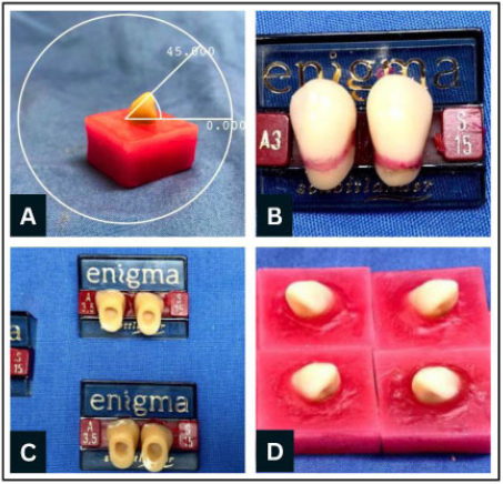
(A) Test specimen with 45° angulated taper. (B) Specimen embedded in silicon 1 mm over the neck. (C) Groove in ridge lap surface created for sample preparation. (D) Prepared samples.
After polymerization of the silicone mold, the master specimen was removed, resulting in standardized molds. Groups B and C underwent ridge lap surface preparation using a carbide bur, whereas Group A remained untreated (Fig. 1C). A standardized carbide bur was used for all preparations, and all procedures were performed by a single operator under controlled conditions to ensure consistency. The bur was inspected regularly and replaced when necessary to minimize variability associated with bur wear.
For all groups, an acrylic tooth was positioned within the mold, and molten wax (Metrowax) was poured into the mold cavity. A total of 21 specimens were prepared (Fig. 1D).
2.3. Preparation of Conventional Heat-cured Specimens
For category 1 specimens, flasking, packing, and finishing procedures were performed to bond the teeth to heat-cured acrylic resin using conventional denture-processing techniques [22]. A thin layer of separating agent (Candulor ISO-K) was applied to the inner surface of the flask and allowed to dry. Gypsum powder (Formula Saint-Gobain) was mixed with water and poured into the lower half of the flask using a vibrator (Krapp Medizintechnik, Germany) to eliminate air bubbles. The waxed teeth were embedded in the plaster before setting, ensuring that only the denture base portion and not the teeth were enclosed within the mold.
After the plaster had set, a separating agent was reapplied to the plaster surface without contacting the teeth. The upper half of the flask was assembled, filled with plaster, and allowed to set for 30 minutes. The flasks were then boiled for 15 minutes (Dreve Machine, Germany) to remove the wax, followed by a 10-minute rinse with hot water. A separating agent was reapplied before packing. Groups A and B received no surface treatment, whereas Group C was treated with methyl methacrylate (ProBase Hot).
The heat-cured monomer and polymer were mixed according to the manufacturer’s instructions and allowed to reach a dough-like consistency. The material was packed into the molds, the flasks were closed, and polymerization was performed at 100°C for 45 minutes (Eclipse Dental Manufacturing Ltd., England). The specimens were then finished and polished.
2.4. CAD/CAM samples
CAD/CAM denture bases were fabricated using a subtractive manufacturing technique (milling). Prepolymerized PMMA blocks (IvoBase CAD, Ivoclar Digital) were used as the starting material to ensure high material density and minimal polymerization shrinkage.
After fabrication of the heat-cured specimens, a standardized mold was created to reproduce identical specimens for digital fabrication. Left and right canine specimens were prepared using the same mold to ensure consistency in shape and dimensions. A cold-cure acrylic index (ProBase Cold, Ivoclar Vivadent) was used instead of wax to minimize dimensional changes and facilitate specimen preparation. To facilitate removal of the teeth from the acrylic index and prevent unintended adhesion, a thin layer of Vaseline was applied as a separating medium. The acrylic resin was polymerized in a pressure pot (Kulzer and Co GmbH, Germany) at 3 bar (0.3 MPa) for 10 minutes. This index was used solely for positioning and did not interfere with the bonding surface. Following removal from the acrylic index, the teeth were thoroughly cleaned and rinsed to remove any residual separating medium before the bonding procedures were performed.
The prepared specimens were scanned using a laboratory scanner, and the resulting data were processed using EXOCAD software to generate STL files (Fig. 2A). These files were used to digitally design the denture bases, which were subsequently milled from prepolymerized PMMA disks using a Roland DWX-50 milling machine (Roland DG Corporation, Hamamatsu, Japan) (Fig. 2B–D). The completed CAD/CAM specimens following the milling and specimen preparation procedures are shown in Fig. 2E and Fig. 2F. A total of 21 CAD/CAM specimens were fabricated [23].
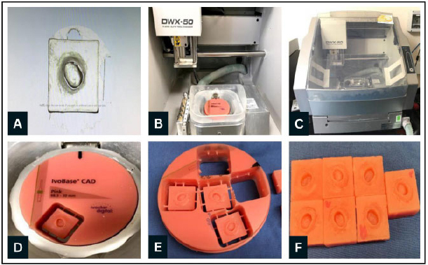
(A) Conversion of the scanned specimen into an STL file. (B–D) Digital design and milling of the specimens using the Roland DWX-50 milling machine. (E, F) Final CAD/CAM specimens after completion of the specimen preparation process.
The milled denture bases were bonded to prefabricated acrylic teeth. Bonding was performed using a conventional acrylic resin mixture (powder and monomer), with or without surface treatment depending on the experimental group. In the designated groups, a bonding agent (IvoBase CAD Bond; Ivoclar Vivadent AG, Schaan, Liechtenstein) containing methyl methacrylate was applied according to the manufacturer’s recommended protocol and previously reported methods for bonding CAD/CAM denture base materials [24].
The specimens were subsequently polymerized in a pressure pot for 20 minutes before mechanical testing.
2.5. Maximum Fracture Load and Mode of Failure
Testing was performed using a Lloyd LRX universal testing machine (Lloyd Instruments Ltd., Fareham, UK) equipped with a 2.5-kN load cell. Each specimen was securely mounted on a stainless-steel jig and positioned within the testing machine, with a polycarbonate shield used for safety. The jig was aligned such that a vertically oriented cylindrical pin applied force to the palatal surface of the acrylic tooth at approximately 90°.
A continuous compressive load was applied at a crosshead speed of 1 mm/min in accordance with ISO 19736 until the tooth fractured from the denture base. The maximum load (N) and deflection (mm) were recorded using a connected computer. Following testing, the fracture surfaces were examined visually and under an optical stereomicroscope (Stereo Discovery V8; Carl Zeiss AG, Oberkochen, Germany).
Fractures were classified as adhesive (fracture at the bond interface, with no denture base resin remaining on the tooth surface), cohesive (fracture occurring within the tooth or denture base material), or mixed (a combination of adhesive and cohesive features) (Fig. 3).
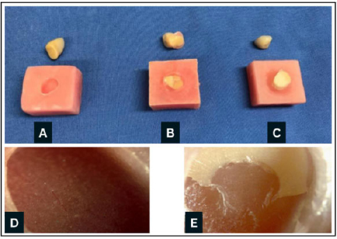
(A) Adhesive, (B) mixed, (C) cohesive fracture types; (D) adhesive fracture under the microscope; (E) mixed fracture under the microscope.
2.6. Data Analysis
Data analysis was conducted using SPSS version 25 (IBM Corp., Armonk, NY, USA). Descriptive statistics, including frequencies, percentages, means, and standard deviations, were used to summarize the data. Normality was assessed using the Kolmogorov–Smirnov and Shapiro–Wilk tests together with graphical inspection of histograms.
For the treatment groups, the Kolmogorov–Smirnov test statistics (D, df = 7) were 0.182 (Normal), 0.262 (Diatoric), and 0.447 (Diatoric + Monomer), with corresponding p-values of 0.200, 0.112, and 0.001, respectively. The corresponding Shapiro–Wilk statistics (W, df = 7) were 0.964, 0.890, and 0.706, with p-values of 0.851, 0.336, and 0.008, respectively.
For the fabrication methods, the Kolmogorov–Smirnov statistics (D, df = 21) were 0.181 (Conventional) and 0.239 (CAD/CAM), with p-values of 0.200 and 0.130, respectively. The corresponding Shapiro–Wilk statistics (W, df = 21) were 0.946 and 0.955, with p-values of 0.476 and 0.569, respectively.
Although the Diatoric + Monomer subgroup showed some deviation from normality, the balanced factorial design, equal sample sizes across groups, and graphical inspection of the data supported the use of two-way ANOVA. Tukey post hoc tests were subsequently performed for pairwise comparisons. Statistical significance was set at p ≤ 0.05.
3. RESULTS
3.1. Descriptive Statistics
As shown in Fig. (4), the Diatoric + Monomer Heat-Cure group exhibited the highest mean maximum load (369.4 ± 42.7 N), followed by the Diatoric Heat-Cure group (341.9 ± 36.4 N). The Normal Heat-Cure group demonstrated a mean maximum load of 256.0 ± 52.4 N. In contrast, the CAD/CAM groups exhibited lower and relatively similar mean maximum load values, with the Diatoric + Monomer CAD/CAM group recording 255.8 ± 29.7 N, the Normal CAD/CAM group 250.9 ± 31.8 N, and the Diatoric CAD/CAM group 247.5 ± 28.6 N.
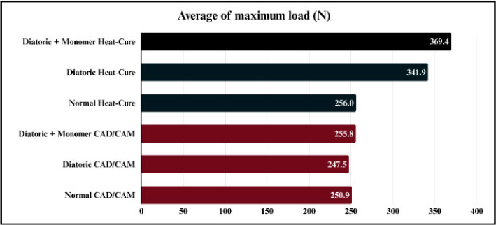
Mean maximum load (N) values for conventional heat-cured and CAD/CAM-milled specimens subjected to different surface treatments (Normal, Diatoric, and Diatoric + Monomer). The Diatoric + Monomer Heat-Cure group exhibited the highest mean maximum load, whereas the CAD/CAM groups showed comparatively lower values.
Descriptive analysis revealed that most conventional groups exhibited mixed failure. Diatoric Heat-Cure showed 85.7% mixed and 14.3% cohesive failure, while Diatoric + Monomer Heat-Cure had 71.4% mixed and 28.6% cohesive failure. Neither showed adhesive failure. Normal Heat-Cure had 42.9% adhesive and 57.1% mixed failure (Fig. 5).
In CAD/CAM groups, mixed failure dominated. Diatoric CAD/CAM and Diatoric + Monomer CAD/CAM showed 100% mixed failure, while Normal CAD/CAM had 85.7% mixed and 14.3% adhesive failure (Fig. 5).
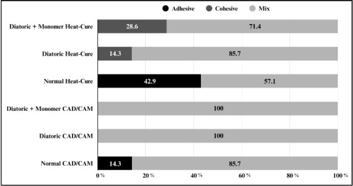
Distribution of failure modes (adhesive, cohesive, and mixed) according to fabrication method and surface treatment. Mixed failures predominated across most groups, whereas adhesive failures were mainly observed in untreated specimens.
3.2. TWO-WAY ANOVA
A two-way ANOVA was conducted to evaluate the effects of fabrication method and surface treatment on maximum load values. Significant effects were observed for fabrication method (F = 14.741, p < 0.001) and surface treatment (F = 3.640, p = 0.036). The interaction between fabrication method and surface treatment was not statistically significant (F = 2.939, p = 0.066) (Table 1).
| Source | Type III Sum of | Df | Mean Square | F | Sig. |
|---|---|---|---|---|---|
| Squares | |||||
| Corrected Model a | 123667.352a | 5 | 24733.470 | 5.580 | .001 |
| Intercept | 3553375.246 | 1 | 3553375.246 | 801.674 | .000 |
| Condition | 65338.988 | 1 | 65338.988 | 14.741 | .000 |
| Treatment | 32272.564 | 2 | 16136.282 | 3.640 | .036 |
| Condition * Treatment | 26055.801 | 2 | 13027.900 | 2.939 | .066 |
| Error | 159568.013 | 36 | 4432.445 | - | - |
| Total | 3836610.610 | 42 | - | - | - |
| Corrected Total | 283235.365 | 41 | - | - | - |
The plot of estimated marginal means (Fig. 6) showed relatively similar maximum load values across the CAD/CAM groups. In contrast, greater variation was observed among the conventional groups, particularly for the untreated specimens.
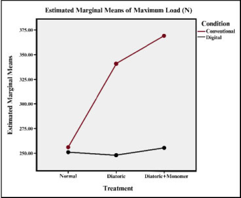
Plot for estimated marginal means of maximum loads.
Tukey post hoc analysis revealed no statistically significant pairwise differences between individual treatment groups (p > 0.05; Table 2). Therefore, although the ANOVA indicated a significant overall effect of surface treatment, the differences between individual treatment groups were not statistically significant.
| (I) Treatment | (J) Treatment | Mean Difference (I-J) |
Std. Error | Sig. | 95% Confidence Interval | |
|---|---|---|---|---|---|---|
| Lower Bound | Upper Bound | |||||
| Normal | Diatoric | 56.5252 | 25.16359 | .077 | -4.9821 | 118.0325 |
| - | Diatoric + Monomer | -4.3174 | 25.16359 | .984 | -65.8247 | 57.1899 |
| Diatoric | Normal | -56.5252 | 25.16359 | .077 | -118.0325 | 4.9821 |
| - | Diatoric + Monomer | -60.8425 | 25.16359 | .053 | -122.3498 | .6648 |
| Diatoric + Monomer | Normal | 4.3174 | 25.16359 | .984 | -57.1899 | 65.8247 |
| - | Diatoric | 60.8425 | 25.16359 | .053 | -.6648 | 122.3498 |
4. DISCUSSION
This study aimed to investigate the effects of denture fabrication method (conventional heat-cured and CAD/CAM) and surface treatment on the bonding performance between acrylic denture teeth and denture base resin, as assessed by maximum fracture load. The results demonstrated that conventionally fabricated specimens exhibited significantly higher maximum load values than CAD/CAM specimens. Therefore, the null hypothesis was rejected, and the alternative hypothesis was accepted, indicating a statistically significant relationship between maximum load, fabrication method, and surface treatment.
The findings indicate that the maximum load sustained by the tooth–denture base interface is influenced by the fabrication method, with the conventional heat-polymerization technique demonstrating superior performance. This observation is consistent with previous studies reporting that the bonding performance between acrylic denture teeth and denture base resin is affected by multiple factors, including the polymerization method, thermocycling procedures, denture tooth composition, laboratory processing techniques, monomer-to-polymer ratio, adhesive materials, and compatibility between denture base resins and acrylic teeth [10, 11, 25, 26].
There were statistically significant effects of fabrication method and surface treatment on the average maximum load. These findings confirm that both variables significantly influence the performance of the tooth–denture base interface. They are also consistent with previously published studies emphasizing the importance of selecting appropriate fabrication methods and surface treatments to improve bonding performance [27, 28]. Although the two-way ANOVA indicated a statistically significant overall effect of surface treatment, the Tukey post hoc analysis did not demonstrate statistically significant pairwise differences among individual treatment groups. This may be attributed to the relatively small sample size (n = 7), which limits statistical power, as well as the conservative nature of the Tukey test. The borderline significance observed in some comparisons (e.g., p = 0.053) suggests a trend toward differences between treatments; however, these differences were not strong enough to reach statistical significance at the pairwise level.
Although the interaction between fabrication method and surface treatment did not reach statistical significance (p = 0.066), the result approached the conventional threshold for significance. Examination of the group means suggested that the effect of surface treatment may vary with the fabrication method. Surface treatments appeared to produce greater increases in maximum load among conventionally processed specimens, whereas only limited changes were observed in the CAD/CAM groups. Clinically, this may indicate that the effectiveness of mechanical and chemical surface modifications is influenced by the material characteristics and fabrication process of the denture base. Because of the relatively small sample size used in the present study, the possibility of a Type II error cannot be excluded. Therefore, future studies with larger sample sizes are recommended to further investigate the interaction between fabrication method and surface treatment and determine whether treatment effectiveness differs between conventional and CAD/CAM denture systems.
The two-way ANOVA model yielded an adjusted R2 value of 0.358, indicating that approximately 35.8% of the variation in maximum load was explained by the factors included in the model. This suggests that additional variables not evaluated in the present study may also influence the mechanical performance of the tooth–denture base interface. Potential contributors include minor variations in tooth morphology, differences in the bonding interface, specimen preparation variability, and material-related factors. Although efforts were made to standardize specimen preparation and testing procedures, some degree of unexplained variability is expected in in vitro investigations involving acrylic denture teeth and denture base materials. Future studies should consider evaluating additional variables that may contribute to variability in maximum load and overall bonding performance.
The average maximum load was significantly higher in conventionally produced specimens than in digitally produced specimens. This difference was confirmed to be statistically significant. Across all surface treatments (Diatoric + Monomer, Diatoric, and Normal), conventional specimens consistently exhibited higher maximum load values than digitally produced specimens. Given the lack of studies directly comparing CAD/CAM and conventional methods in this context, the findings offer new insights rather than a synthesis of prior evidence.
Despite the superior performance observed in the conventionally fabricated specimens in the present study, previous studies have reported favorable outcomes for CAD/CAM denture systems with respect to tooth–denture base adhesion and other mechanical properties. These differences may be attributable to variations in CAD/CAM systems, material formulations, specimen preparation methods, and bonding protocols. CAD/CAM materials are manufactured under highly controlled industrial conditions and exhibit a high degree of polymerization, reduced residual monomer content, and increased cross-link density. While these characteristics may improve certain mechanical properties, they may also reduce the potential for chemical interaction at the tooth–denture base interface. Consequently, bonding performance may depend not only on the fabrication method itself but also on the specific materials, surface treatments, and bonding agents used. These methodological and material differences may help explain the conflicting findings reported in the literature.
The trend toward lower maximum load values observed in the CAD/CAM diatoric group may be explained by differences in material properties and bonding mechanisms. Unlike conventional heat-cured acrylic, CAD/CAM materials are pre-polymerized under controlled industrial conditions, resulting in a highly cross-linked structure with limited availability of free monomer. This reduces the potential for chemical bonding between the denture tooth and the denture base. In this context, the introduction of diatoric recesses may not provide the expected mechanical advantage. Instead, these recesses may act as stress concentration points, particularly if resin penetration into the recess is incomplete or insufficient. Additionally, the smooth and dense surface of CAD/CAM materials may limit mechanical interlocking, thereby reducing the effectiveness of diatoric modifications. This could explain the trend toward lower maximum load values observed in the CAD/CAM specimens compared with the untreated group. These findings suggest that mechanical retention alone may not be sufficient to improve bonding performance in CAD/CAM systems and that chemical surface treatments may play a more critical role. Further studies are recommended to optimize bonding strategies specific to digitally fabricated denture materials.
The observed variation may stem from differences in resin penetration. While diatoric recesses can increase mechanical retention by enlarging the contact area, poor resin penetration, possibly due to inconsistent recess diameters, may reduce the maximum load sustained by the tooth–denture base interface, as observed in the digitally produced specimens in the present study.
Additionally, the milling process used in CAD/CAM fabrication may contribute to the formation of a superficial stressed or altered layer on the PMMA surface. This layer, combined with the inherently high degree of polymerization and cross-linking of pre-polymerized PMMA blocks, may reduce the material’s ability to undergo plastic deformation and limit effective mechanical interlocking. Further surface modification, such as grinding to create diatoric recesses, may exacerbate this effect by introducing microcracks or stress concentrations that can act as weak points under load.
Moreover, the increased hardness and density of CAD/CAM materials may hinder resin penetration into mechanically prepared features, reducing the effectiveness of diatoric retention compared to conventional acrylic systems. This suggests that, in CAD/CAM materials, bonding may rely more heavily on chemical interaction rather than mechanical retention, highlighting the importance of surface treatment protocols that enhance chemical adhesion.
In recent years, improved bonding agents have been developed specifically for CAD/CAM denture systems to enhance adhesion to pre-polymerized PMMA materials. These materials exhibit reduced free monomer content and higher cross-link density, which can limit conventional chemical bonding. To address this, manufacturers have introduced surface primers and bonding agents containing methyl methacrylate and other functional monomers designed to promote surface swelling and enhance chemical interactions.
Previous studies have shown that the use of dedicated bonding agents can significantly improve the bond strength between CAD/CAM denture bases and acrylic teeth compared to untreated surfaces. These agents enhance surface wettability and promote diffusion at the bonding interface, compensating for the limited reactivity of highly polymerized materials. This may explain why, in the present study, the application of monomer in combination with mechanical treatment produced improved bonding outcomes compared to mechanical retention alone. These findings highlight the importance of combining appropriate surface treatment with chemical bonding strategies when working with CAD/CAM denture materials. Future developments in adhesive systems may further improve bonding performance and should be explored in subsequent studies.
The secondary objective of the study was to understand the mode of bonding failure, whether adhesive, cohesive, or mixed, depending on the mode of production. Notable differences regarding the mode of bonding failure have been observed among the different methods of production and surface treatments. In conventionally prepared specimens, bonding failures were predominantly mixed, whereas surface treatments prevented bonding failures. This agrees with earlier observations that the application of monomer prevents bonding failure.13 Studies have validated the application of monomer for at least 180 seconds to improve bonding performance and prevent bonding failures [29].
In addition, a previous study has suggested that adhesive failures may result from contamination of the bonding surface as well as incompatibility and inadequate copolymerization between denture teeth and denture base materials [30]. In our study, it was found that without treatment, the digital group had fewer cases of adhesive failure when compared to the conventional group due to the effect of CAD/CAM in reducing contamination.
The maximum load was measured using a universal testing machine with a cross-head speed of 1 mm/min. This speed has been found to be quite reliable and precise. This was supported by previous research [31, 32].
This finding may have important clinical implications. Although the CAD/CAM groups generally demonstrated lower maximum load values than the conventional groups, the reduced frequency of adhesive failures in untreated CAD/CAM specimens suggests a potentially more consistent tooth–denture base interface. A possible explanation is that the digital workflow minimizes several laboratory procedures associated with conventional denture fabrication, thereby reducing the likelihood of contamination from wax residues, separating media, or processing errors. As adhesive failure is often associated with weaknesses at the tooth–denture base interface, a lower occurrence of this failure type may indicate improved interface quality in digitally fabricated specimens. Further studies are required to determine whether this observation contributes to improved long-term clinical performance of CAD/CAM dentures.
Recent advances in CAD/CAM denture fabrication have stimulated the development of bonding strategies aimed at improving adhesion between denture teeth and pre-polymerized PMMA denture bases. Due to their high degree of polymerization and cross-link density, CAD/CAM materials may exhibit reduced chemical reactivity compared with conventional heat-polymerized acrylic resins. Consequently, both chemical and mechanical surface treatments have been proposed to enhance bonding, although their effectiveness remains variable depending on the materials used and the treatment protocol employed [18, 20].
Previous studies have reported that material incompatibility and insufficient copolymerization are major causes of bond failure between denture teeth and denture base resins [18, 20]. This may explain why, in the present study, the combination of monomer application and mechanical retention produced higher maximum load values than mechanical treatment alone. The findings also suggest that chemical surface treatment may play a greater role than mechanical modification in improving adhesion to CAD/CAM denture bases. Further research is needed to identify the most effective bonding protocols for digitally fabricated dentures.
From a clinical perspective, the findings suggest that the selection of appropriate surface treatment protocols is particularly important when working with CAD/CAM denture systems. Although CAD/CAM specimens demonstrated lower maximum load values than conventionally processed specimens, the combination of mechanical modification and monomer application produced the highest mean maximum load among the CAD/CAM groups. Therefore, clinicians and dental technicians should consider incorporating both mechanical and chemical surface treatments when bonding acrylic teeth to CAD/CAM denture bases to optimize adhesion and potentially reduce the risk of tooth debonding.
Several limitations should be considered when interpreting the findings of this study. Failure modes were assessed using stereomicroscopy without Scanning Electron Microscopy (SEM), limiting detailed characterization of the fracture surfaces. The primary outcome was reported as maximum load (N) because the bonded surface area was not independently measured during specimen preparation. Consequently, conversion to stress values (MPa) was not possible, which may limit direct comparison with studies reporting bond strength in MPa. In addition, different bonding protocols were required for the conventional and CAD/CAM groups because of the materials and fabrication systems used. Consequently, the observed differences may reflect the combined effects of fabrication method and bonding chemistry rather than fabrication method alone. Furthermore, the CAD/CAM workflow required the use of a cold-cure acrylic index to reproduce the tooth socket geometry, representing a procedural difference from the conventional wax-based technique. Although the material was not incorporated into the final bonded specimens, a potential indirect influence on specimen preparation cannot be completely excluded. Although specimen preparation and testing were standardized, formal randomization and operator blinding were not implemented. Furthermore, the relatively small sample size may have limited the ability to detect subtle differences between treatment groups and interactions.
The study was limited to maxillary canine teeth and did not include thermocycling or artificial aging; therefore, the findings reflect initial bond performance under controlled laboratory conditions rather than long-term clinical behavior. Furthermore, the CAD/CAM workflow required the use of a cold-cure acrylic index during specimen preparation, representing a procedural difference from the conventional technique. Future studies should address these factors to further validate and expand upon the present findings.
CONCLUSION
The following conclusions can be drawn:
- Conventionally processed specimens demonstrated significantly higher maximum load values than digitally produced specimens.
- A statistically significant overall effect of surface treatment on maximum load was identified by two-way ANOVA. However, no statistically significant pairwise differences were detected between individual treatment groups in the post hoc analysis. Although the Diatoric + Monomer groups demonstrated the highest mean maximum load values, these findings should be interpreted with caution.
- Surface treatments appeared to yield greater improvements in maximum load among conventionally processed specimens than among digitally produced specimens; however, these trends require confirmation in studies with larger sample sizes.
- The application of surface treatments eliminated adhesive failures in all treated groups, whereas no cohesive failures were observed in digitally produced specimens.
- The observed differences between conventional and CAD/CAM groups should be interpreted with caution, as the two fabrication systems employed different bonding procedures. Therefore, the effects of denture base fabrication method cannot be completely separated from the effects of bonding chemistry. Further studies using standardized bonding protocols are required to clarify the individual contribution of each factor.
AUTHORS’ CONTRIBUTIONS
The authors confirm contribution to the paper as follows: M.A., M.A.: Study conception and design; M.A, M.A.: Data collection; M.A., Y.E., R.S.: Analysis and interpretation of results; M.A., M.A., R.S.: Draft manuscript. All authors reviewed the results and approved the final version of the manuscript.
LIST OF ABBREVIATIONS
| CAD/CAM | = Computer-Aided Design and Computer-Aided Manufacturing. |
| PMMA | = Polymethyl Methacrylate. |
ETHICAL STATEMENT
This in vitro laboratory study did not involve human participants, patient data, human tissues, or animal subjects. Therefore, formal ethical approval and informed consent were not required.
AVAILABILITY OF DATA AND MATERIALS
All the data and supporting information is provided within the article.
ACKNOWLEDGEMENTS
Declared none.