All published articles of this journal are available on ScienceDirect.
Fixed Prosthodontic Rehabilitation of a Patient with Cleidocranial Dysplasia: A Case Report
Abstract
Introduction
Cleidocranial Dysplasia (CCD) is a rare congenital disease characterized by skeletal and dental anomalies. Clinical findings of CCD patients include low facial height, pseudoprognathism, unerupted teeth, an excessive deep bite, chewing difficulties, and unsatisfied dentofacial appearance. These patients’ dental treatments present a substantial challenge.
Case Representation
This case report describes the prosthodontic treatment of a 29-year-old male CCD patient using porcelain-fused-to-metal restorations. The avoidance of a surgical procedure serves to minimize the potential for complications and expedites the attainment of outcomes with greater celerity. Throughout the follow-up period of 1 year, the patient maintained good periodontal health. The restoration of masticatory function and enhancement of facial esthetics were successfully achieved and the patient expressed a high degree of satisfaction with the outcome of the treatment.
Conclusion
The use of fixed prostheses in CCD patients is a treatment modality that resolves many of the issues caused by the surgical approach.
1. INTRODUCTION
Cleidocranial Dysplasia (CCD) is a rare, autosomal dominant genetic disorder that prevents the development of bones and teeth, and has no correlation with gender or ethnic origin [1]. Sainton and Marie initially documented this phenomenon in 1898 [2], and its prevalence is estimated to occur in approximately 1 in 1,000,000 individuals globally [3]. This dysplasia affects the entire skeletal system, giving rise to multiple abnormalities that pose significant threats to the patient’s life. Skeletal symptoms of the disease include clavicular aplasia or hypoplasia and a conical thorax with short ribs. Within the cephalic region, reported symptoms involve frontal protrusion, hypertelorism, a collapsed bridge, and a widened nasal base, accompanied by the presence of fontanelles and delayed closure of sutures [4]. Dental anomalies are considered major characteristics of CCD and are the primary basis for patient complaints. Retention of deciduous dentition and the presence of supernumerary teeth that prevent the eruption of permanent teeth are common findings in patients with CCD [5]. Another disruption can be described as the underdevelopment of maxilla, resulting in skeletal class III malocclusion tendency and pseudo-prognathism [6]. Despite the existence of such problems, patients may remain asymptomatic, with an absence of pain, swelling problems, and oral function challenges, particularly during the time that deciduous teeth are still in the mouth. Dental disability begins at a subsequent stage with the progressive damage of deciduous dentition. Oral degeneration undergoes accelerated progression in a few years, resulting in an edentulous and aged facial appearance [7]. Treatment planning has critical importance for patients with CCD. The treatment aims of these patients should include restoration of the Vertical Dimension of Occlusion (VDO) and masticatory function, improvement of the patient's dentofacial aesthetics, and advancement of the psychosocial situation [8, 9]. Several therapeutic approaches have been reported in the literature, including surgical, orthodontic, and prosthodontic procedures. In the literature, it has been noted that a combination of surgical and orthodontic treatment is usually recommended for patients at an early age [8, 10, 11]. Eruption of well-developed impacted teeth can be accomplished through the extraction of deciduous and supernumerary teeth, removal of obstructing osseous tissue, orthodontic traction, and their subsequent alignment [8, 12]. It has been reported that decreased lower facial height and pseudo prognathism in CCD patients do not improve despite surgical-orthodontic treatment [6, 13]. Therefore, orthodontic treatment of the permanent teeth and subsequent orthognathic LeFort I surgery may be required to create the appropriate vertical dimension and correct the skeletal defects, which are the original cause of dental anomalies [6, 8, 14]. Implant surgery is another invasive treatment in CCD patients. Following the osseointegration, a fixed or removable prosthesis can be fabricated [7]. However, surgical procedures are not always possible for patients with CCD and prosthetic treatments may remain the only viable option [15]. Teeth exhibiting a favorable prognosis and positioned appropriately for prosthetic applications can serve as abutments for either fixed or removable dentures [1]. While a range of therapeutic options exist, there is no consensus on the treatment strategy [7]. The aim of this case report was to present the treatment procedure for various dental and skeletal anomalies due to CCD, using fixed prostheses.
2. MATERIALS AND METHODS
2.1. Case Presentation
A 29-year-old male patient with CCD presented at the prosthodontics department for an examination. The patient’s chief complaint was difficulty chewing and lack of aesthetics, and he has not undergone any dental interventions before. There were no diagnostic challenges observed.
2.2. Extraoral Evaluation
Extraoral examination revealed an enlarged skull, clavicular hyperelasticity, and protruding frontal region. The Vertical Dimension of Occlusion (VDO) was found to be significantly decreased. The tip of the nose and the chin were converged as older individuals with complete edentulism. The lower lip was outwardly deviated, the sublabial groove was deepened, and the dentofacial composition appeared disproportionately unfavorable considering the age of the patient.
2.3. Intraoral Evaluation
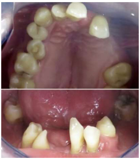
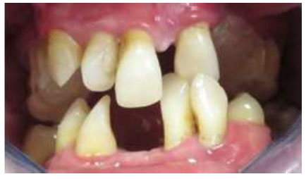
The intraoral examination showed a significant number of permanent teeth that failed to attain complete eruption. Supernumerary teeth were present and 12 mm overbite existed due to supereruption of the anterior teeth (Fig. 1). Teeth 21 and 46 were extracted before the patient was referred to the department of prosthodontics (Fig. 2).

Intraoral photograph showing an anterior overbite.

Occlusal view of maxillary and mandibular arch.
2.4. Radiographic Evaluation
Radiological examination showed teeth 18, 35, 38, 45, and 48 to be impacted and their position was not suitable for eruption (Fig. 3).
2.5. Dental Treatment
The planned treatment involved the rehabilitation of edentulous spaces through fixed prostheses. The use of implants was eliminated because there was a potential risk of bone fragility due to a high number of supernumerary and unerupted teeth to be extracted. The aim of the prosthetic rehabilitation was to create a functional occlusal relationship, correct the excessive overbite, improve the chewing quality, and increase the VDO and aesthetic of the dentofacial composition. After patient consent, teeth 25 and 36 were extracted. Also, unerupted teeth 22, 24, 31, 41, 42, and 45 were extracted to prevent any possible eruption in the future. The remaining unerupted teeth were not extracted due to their location in the basis mandibulae, their horizontal orientation, and their anatomical proximity to the mandibular canal.

Panoramic radiograph before treatment.
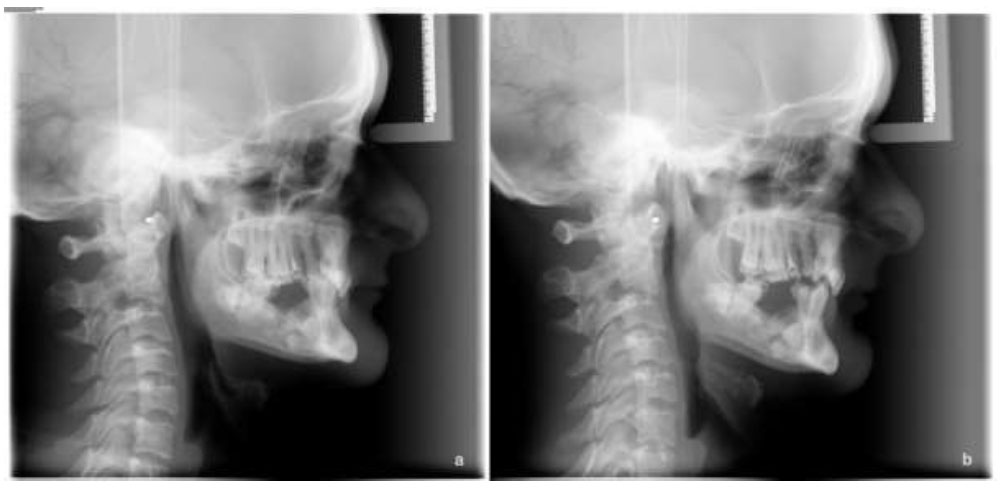
The treatment was delineated into two phases. The first stage was the fabrication of a maxillary splint to establish the appropriate VDO. The optimal VDO was estimated with bite registration (Cavex Modelling Wax, The Netherlands, Holland) by evaluating the height of the upper lip, swallowing, phonetics, and smile line [17]. An auto-polymerizing transparent acrylic splint (Temdent Classic, Schütz Dental Group, Rosbach, Germany) was prepared according to the determined VDO. As a result, the VDO was increased by 9 mm and overbite was reduced to 3 mm (Fig. 4). The patient was informed to use a splint continuously, both day and night, except at meal times, for 8 weeks. It was determined that the patient successfully adapted to the increased VDO and encountered no functional problems related to the neuromuscular components of the stomatognathic system. Furthermore, it was noted that the increase in lower facial height yielded favorable outcomes in terms of facial aesthetics (Fig. 5). The assessment of the increased VDO was evaluated through a comparative analysis of cephalometric radiographs (Fig. 6).

Increase in VDO with the acrylic splint.
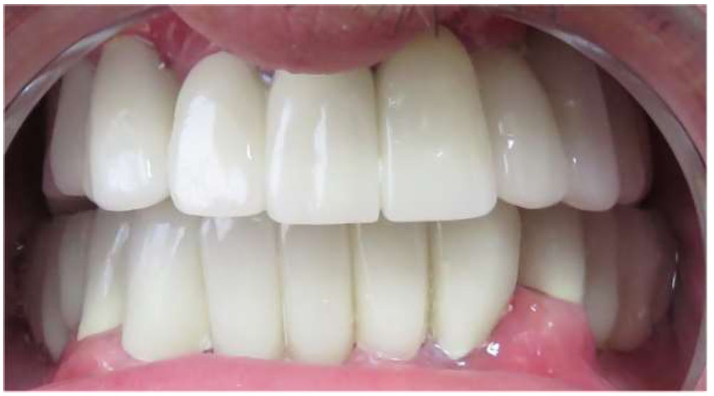
For the second stage of the treatment, porcelain-fused-to-metal restorations were planned. Preparation was completed via 360o chamfer margin (G&Z Instrumente, Lustenau, Austria). Following the gingival displacement with an aluminum chloride-absorbed retraction cord (ViscoStat Clear, Ultradent, Utah, USA; Ultrapak, Ultradent, Utah, USA), impressions were made with condensation silicone elastomeric material (Optosil/ Xantopren, Heraeus-Kulzer, Wasserburg, Germany) with stock trays. A facebow transfer (Dentatus Facebow, Stockholm, Sweden) and maxillomandibular relationship were recorded. The VDO with the splint was transferred to the definitive restoration and the casts were articulated to a semiadjustable articulator (Dentatus ARO Articulator, Stockholm, Sweden). After confirming the fit of the metal frameworks (S&S Scheftner GmbH, Mainz, Germany) intraorally, a veneering porcelain material (Noritake Dental Supply Co., Nagoya, Japan) was built and the occlusion was adjusted to be unilaterally balanced. Porcelain-fused-to-metal restorations were luted with zinc polycarboxylate cement (Poly-F Plus, Dentsply Sirona, Konstanz, Germany) (Figs. 7, 8). The patient expressed a high degree of satisfaction with the outcome of the treatment. The patient had bi-monthly consultations to assess the potential eruption of the impacted teeth. Radiographic examination is a fundamental method for diagnosing and locating impacted teeth [7]. In case of a potential eruption, an intervention could be carried out by removing the fixed prostheses. After 1-year follow-up period, there were no adverse or unanticipated outcomes detected.

Extraoral evaluation. (a) Extraoral frontal view before acrylic splint; (b) extraoral frontal view after acrylic splint.

Cephalometric evaluation. (a) Cephalometric radiograph before acrylic splint; (b) cephalometric radiograph after acrylic splint.
3. DISCUSSION
This case report has described the fixed prosthodontic treatment of a patient who showed all of the dental symptoms of CCD. The diagnostic and therapeutic aspects of managing CCD patients pose numerous challenges. Considerations related to supernumerary teeth, retention or extraction of remaining deciduous teeth, and the optimal timing for intervention can be decisive in the treatment choice [16, 17]. A variety of treatment approaches have been reported in the reviewed literature, including orthodontics, surgery, or prosthodontics [7]. The surgical exposure of the impacted permanent teeth, their orthodontic traction, and subsequent alignment at an early age are the preferred solutions [18]. This approach can preserve the patient’s natural dentition. According to Kargul et al. [19], surgically exposing unerupted teeth has the potential to induce cementum formation and facilitate the eruption of a dentition characterized by normal root formation at an early age. However, this approach is accompanied by some disadvantages, including the prolonged duration of treatment and the requirement of multiple surgical procedures, which may be expensive for the patient and pose challenges for the clinician [14]. Limitations of this method include the inability to address certain dental issues, such as shape abnormalities. Additionally, the literature highlights occurrences of calcification, tooth loss due to caries subsequent to orthodontic treatment, and the potential for failures in the orthodontic treatment.

Definitive prostheses (intraoral view).

Panoramic radiograph after the fitting of definitive prostheses.
Implant surgery has been described as an alternative surgical procedure [18]. This alternative has appeared to be a compelling option yielding favorable outcomes within a brief timeframe [7]. The literature presents a contentious discourse on the use of implants for CCD patients. According to Butterworth et al. [4], the use of implants is typically contraindicated due to the presence of numerous unerupted teeth, which diminishes the available bone volume. It has also been reported that this genetic disease can negatively affect osteoblastic activity around the implant [7]. However, some reports have documented bone formation after orthodontically erupting teeth in patients with CCD, demonstrating that implant therapy can be an option [20]. Some reports have demonstrated that once osseointegration is achieved, prosthetic treatment can be completed with fixed or removable prostheses, as prescribed in the pre-implant treatment plan [21]. Despite reports indicating a high incidence of surgical complications in the treatment of CCD [22, 23], implant-supported fixed dental prostheses may be beneficial in terms of preventing bone loss and providing comfort through fixed prostheses [24]. A long-term follow-up period is necessary to confirm the efficacy of this treatment and assess the osseointegration quality of these implants in relation to bone defects. Implant surgery usually requires the removal of primary and unerupted teeth that obstruct implant placement [18]. Extraction of the unerupted teeth may traumatize the jaw continuity and neuromuscular bundle, and cause anatomic structure damage [25]. Considering this factor, prosthodontic treatment is another option for the rehabilitation of CCD patients. Chang et al. [26] reported that occlusion and esthetics cannot be improved without prosthetic treatment.
Removable prostheses stand out as expedient solutions for restoring both esthetic and function. The potential use of deciduous teeth as abutments for such dentures raises concerns about their capacity to endure functional forces or whether premature resorption may appear [4]. Notably, the extraction of these teeth does not constantly prompt the eruption of permanent teeth [27]; thus, they could be preserved where removable prostheses are recommended. Trimble et al. [28] advocated the application of removable prostheses after the extraction of both primary and permanent teeth by acknowledging the associated risk of alveolar hypoplasia. Their findings indicated an adverse impact on the retention of the removable prostheses in cases where alveolar hypoplasia was present. Rushton [29] and Pusey [30] recommended the exclusive use of erupted teeth in removable prostheses to reduce alveolar bone loss. However, they pointed out the potential drawbacks, emphasizing that the ongoing eruption of teeth might result in mucosal ulcerations and cyst formation in persistent teeth. Surgical removal of the follicle surrounding an unerupted tooth establishes direct contact with the enamel [31]. Literature indicates that the atrophy or loss of the tooth follicle can induce replacement resorption of the crowns of impacted teeth over time. Consequently, the removal of the follicle surrounding an impacted tooth, without actively promoting its eruption, may cause a reaction similar to replacement resorption of the crown of the tooth [31, 32]. Some authors have remarked on the utilization of removable prostheses as a therapeutic approach for addressing stomatognathic dysfunction in patients with CCD. This treatment option keeps primary, permanent, and supernumerary teeth unless pathologic changes occur. Removable prosthetic treatments have been documented as beneficial for pediatric patients, aiding in their early integration into society. Additionally, this approach is deemed suitable for elderly patients who may not be available for orthodontic or surgical interventions. D’Alessandro reported that the majority of cases treated with removable prostheses pertained to pediatric patients [18]. With respect to the present case, the patient was a 29-year-old adult who expressed a preference for a fixed prosthesis. Some authors have suggested the use of spontaneously erupted permanent teeth as abutments for fixed prosthodontic treatment [21]. Nonetheless, it has been noted that the occurrence of such spontaneously erupted permanent teeth is less prevalent in patients with CCD. Consequently, the combined use of fixed and removable prostheses may be recommended [21]. Due to the increased VDO in CCD patients, the crown-to-root ratio was compromised for the fixed prostheses. However, erupted permanent teeth were found to be suitable for a fixed prosthesis in the present case. The limitation of this approach is the requirement for teeth with an ideal prognosis and appropriate position to serve as abutments. Throughout the follow-up period of 1 year, the patient maintained good periodontal health. Symptoms associated with temporomandibular joint mechanical complications were not observed despite the intentional increase in VDO. Furthermore, the restoration of masticatory function and enhancement of facial esthetics were successfully achieved.
CONCLUSION
The management of patients with CCD is complex. Selection of the most suitable treatment option based on factors, such as the patient’s age, requirements, dental status, and compliance with the treatment, is crucial. Teeth-supported fixed restorations offer a good result if the permanent erupted teeth are suitable. During 1 year of follow-up, there were no technical and biological complications reported in the present case. Teeth-supported fixed prostheses can be considered a rapid and non-surgical alternative treatment option for adult CCD patients.
AUTHORS’ CONTRIBUTION
Merve Karakaya collected the data, wrote the paper, performed data analysis or interpretation, and contributed to the study concept or design. All authors have reviewed the results and approved the final version of the manuscript.
LIST OF ABBREVIATIONS
| CCD | = Cleidocranial Dysplasia |
| VDO | = Vertical Dimension of Occlusion |
CONSENT FOR PUBLICATION
Informed consent was given by the patient for inclusion before participation in the study and informed consent form with HHD.RB.19 code was signed.


