All published articles of this journal are available on ScienceDirect.
Efficacy of Various Heat-treated Retreatment File Systems on the Apical Deformity and Canal Centering Ability in a Single-rooted Teeth using Nano CT
Abstract
Aim
To analyze the efficacy of various heat-treated retreatment file systems on the apical deformity and canal centering ability in single-rooted teeth using nano CT.
Materials and Methods
A total of 45 single-rooted teeth were decoronated to 18mm and obturated. Three retreatment file systems were used, such as: Group 1- Solite RS3 Retreatment, Group 2- Solite RS3 Black Retreatment, and Group 3 - Solite RE Black Retreatment. All these procedures were carried out by a single operator. Nano-Computed tomography (CT) scans, pre- and post-operative, were obtained and superimposed for analysis. One-way ANOVA and post hoc tests were done to test the significance between the three groups.
Results
Based on the results, it was inferred that the Solite RE Black file system had better canal centering ratio and less apical deformity during the retreatment compared to Solite RS3 and Solite RS3 Black (p<0.05).
Conclusion
Solite RE Black file systems had superior centering ability in all apical thirds and less transportation in the middle and coronal thirds when compared to the other two retreatment file systems.
1. INTRODUCTION
Failure of root canal therapy is typically caused by the persistence of microbes inside the intricate structures of the root canal [1]. A root canal system's chemical-mechanical preparation, followed by hermetic obturation and coronal sealing, is essential for the successful removal of pathogens during endodontic treatment [2].
The process of endodontic retreatment entails re-instrumenting the root canal after the filling material has been removed using either mechanical or rotary instruments [3]. Because of its efficacy and capacity to safeguard dental structures, non-surgical endodontic retreatment is generally the primary treatment option advised for teeth previously treated with endodontics that show persistent apical periodontitis, Retreatment was found to have a 77%–80% effectiveness rate in certain studies [2]. H-files, GP solvent, Gates-Glidden (GG) drills, pluggers, ultrasonic technology, and lasers are some of the techniques that can be used to remove GP [4].
Superplastic nickel-titanium (Ni-Ti) alloys, after under- going thermomechanical processing and maintaining stability in the martensitic phase during clinical use, are employed in the production of endodontic files. When compared to conventional Ni-Ti alloys, these alloys yield conically shaped root canal preparations and reduced failures due to their enhanced flexibility and resistance to cyclic fatigue. Studies suggest that Ni-Ti files achieve a more centralized shape with minimal deviations from the central axis of the root canal during the creation of conical root canal preparations [5]. Several factors contribute to vertical root fractures. These include extensive biomechanical preparation, excessive dentin removal during and after preparation, the amount of coronal structure that remains, different types of parafunction, and using excessive force when filling the root canal [6].
Retreatment files such as the Solite RS3 system, the Solite RS3 Black system, and the Solite RE Black system have been introduced. The three-file system and heat-treated technology is called Solite RS3. The Solite RE Black has a two-file system that utilizes c-wire technology, while the Solite RE black system combines c-wire technology with a variable taper design. The present study aims to analyze which of the three systems has a more centered preparation and less apical deformation during the endodontic retreatment.
The noninvasive method known as micro-computed tomography, or micro-CT, enables the three-dimensional study of different endodontic outcomes [7]. Although nano-CT offers better resolutions than micro-CT in terms of quantification [8] having a higher spatial resolution of up to 400 nm-it has been used in numerous studies that assessed the effectiveness of endodontic instruments in the preparation of flat-oval root canals and the removal of the filling material. These studies allow for non-destructive volumetric quantitative and qualitative assessments with precise comparisons of defects in the same specimen before and after endodontic procedures [9]. The main aim of the study is to analyze the efficacy of various heat-treated retreatment file systems on the apical deformity and canal centering ability in single-rooted teeth using nano CT. The null hypothesis states that there is no significant difference in the canal centering ratio and apical deformity between the three file systems assessed.
2. MATERIALS AND METHODS
2.1. Sample Size Calculation
The study received approval by the Scientific Review Board of Institutional human ethics committee from Saveetha Dental College (SRB/SDC/ENDO-2106/23/077). The research was conducted on humans by the Helsinki declaration of 1975. The sample size for the current study was calculated from the previous study by our colleagues [10]. Based on the assessment, a total sample size of 45 was achieved at a power of 95% (1- β = 95%, α= 0.05). Based on the experimentation, the specimens were randomly divided into three groups (n=15).
2.2. Specimen Selection
Forty-five freshly extracted teeth for periodontal reasons were collected. The collected teeth were evaluated radiographically and teeth having additional canals, fractures, severe canal curvatures, calcifications, internal and external resorption were excluded from the study. Only teeth with intact root apices, fully formed apices, and patent canals with minimal curvatures were selected for the study. For this investigation, teeth that had been removed due to periodontal disease but showed no symptoms of caries were included. After using a scaler to remove any remaining tissue and debris, the extracted teeth were disinfected for an hour in a 5.25% sodium hypochlorite solution (Acquafarma; Niteroi, RJ, Brazil). Prior to the experiment, the teeth were preserved in a saline solution.
2.3. Root Canal Preparation
Digital calipers were used to confirm that all of the samples were standardized to 18mm, which was achieved via decoronation of the samples. The access aperture was finished with an Endo Access bur of Size 2, 21mm (Dentsply, Maillefer). The patency was then attained using a #10 hand K-file Dentsply Maillefer, Switzerland). As directed by the manufacturer, the coronal and apical parts are enlarged using a P0 rotary file until the taper reaches PF2 6% till the working length. During the preliminary stage, all the groups utilized a 15% ethylene-diamine-tetra-acetic acid (RC Help, Prime Dental Products Pvt Ltd, Thane, India) for the initial negotiation. 5.25 percent sodium hypochlorite irrigant (Acquafarma; Niteroi, RJ, Brazil) was used as an intermittent irrigant. The Profit S3 rotary file system (Profit Dental, India) was employed precisely as instructed by the manufacturer. During canal preparation, irrigation was carried out following each intensification in file size. Final rinse of 5 ml of EDTA solution, and 5ml of 5.25% NaOCl was done. Final flush was carried out using a 5 ml of saline and the canals were paper dried. 6% taper size 25 Gutta Percha (DIADENT, India) was used for a matched taper single cone obturation, using an AH plus sealer (Dentsply Maillefer, Switzerland). In order to confirm the quality of the obturation, the teeth were radiographically examined. Radiographs were collected buccolingually and mesio- distally to assess if the obturation was sufficient.
2.4. Nano CT Scanning
The samples “were scanned employing the Bruker Skyscan 2211 Nano-CT imaging system (Bruker micro-CT, Kontich, Belgium). The images were obtained utilizing an 80kV accelerating voltage, 170μA current, a 0.25mm aluminum filter in front of the camera, and a final isotropic resolution of 450nm per voxel”. The samples underwent a 360° rotation with a step size of 0.43° around the vertical axis, and each projection was exposed for 1700ms. Two frames were captured on average, for a total of 3400 ms for each projection. A total of one hour was spent on scanning each sample. Software called InstaNRecon was used to recreate the images [11].
2.5. Retreatment Procedures
There was only one operator that carried out the retreatment process. The forty five teeth were divided into three groups at random using an electronic technique (http://www.random.org), with fifteen specimens allotted to each group. Group I: Solite RS3, Group II: Solite RS3 black, and Group III: Solite RE black Retreatment file system were the three different retrieval systems that were used.
2.5.1. Group 1 - Solite RS3
The instruments were made to rotate continuously in a clockwise direction by carefully pecking in and out at a torque of 2.6 N/cm and a speed of 350 rpm. The root canal filling removal was carried out using instrument RS1 - 30/.08 (15mm) in the coronal third, RS2 - 25/.07 (18mm) in the middle third, and instrument RS3 - 20.06 (23mm) at the WL. The entire procedure was carried out without the use of a gutta percha solvent. During the entire course of gutta percha removal and instrumentation, sequential irrigation of 5.25% NaOCl irrigation was carried out. A total of 10 ml of sodium hypochlorite irrigating solution was used in total. The entire course of irrigation was only carried out using a disposable syringes with a 30-gauge side vented needle (Profit Dental, India) kept 1mm short of the working length, with continuous oscillations. No activation devices were employed. Final rinse of 5 ml of EDTA solution, and 5ml of 5.25% NaOCl was done. Final flush was carried out using a 5 ml of saline and the canals were paper dried.
2.5.2. Group 2- Solite RS3 Black
The instruments were made to rotate continuously in a clockwise direction by carefully pecking in and out at a torque of 2.6N/cm and a speed of 350 rpm. Instrument RS1 Black - 30/.08 (15mm) was used to remove filling in the coronal third; instrument RS2 Black - 25/.07 (18mm) was used to fill the middle third, which is made up of c wire technology; and instrument RS3 Black - 20.06 (23mm) was used to fill the WL. The entire procedure was carried out without the use of a gutta percha solvent. During the entire course of gutta percha removal and instrumentation, sequential irrigation of 5.25% NaOCl irrigation was carried out. A total of 10 ml of sodium hypochlorite irrigating solution was used in total. The entire course of irrigation was only carried out using a Using disposable syringes with a 30-gauge side vented needle (Profit Dental, India) kept 1mm short of the working length, with continuous oscillations. No activation devices were employed. Final rinse of 5 ml of EDTA solution, and 5ml of 5.25% NaOCl was done. Final flush was carried out using a 5 ml of saline and the canals were paper dried.
2.5.3. Group 3 - Solite RE Black
The two file systems in this tapering design have different levels of flexibility. Using a torque of 2.6 N/cm and a gentle in-and-out pecking motion, the instruments were continually rotated in a clockwise orientation. The coronal and middle third of the canal is treated with instrument RE1, which has a tip size of 0.30 and a variable taper. The middle, as well as apical thirds of the canal, are treated with instrument RE2, which has a tip size of 0.20 and a variable taper composed of c-wire technology. The entire procedure was carried out without the use of a gutta percha solvent. During the entire course of gutta percha removal and instrumentation, sequential irrigation of 5.25% NaOCl irrigation was carried out. A total of 10 ml of sodium hypochlorite irrigating solution was used in total. The entire course of irrigation was only carried out using a Using disposable syringes with a 30-gauge side vented needle (Profit Dental, India) kept 1mm short of the working length, with continuous oscillations. No activation devices were employed. Final rinse of 5 ml of EDTA solution, and 5ml of 5.25% NaOCl was done. Final flush was carried out using a 5 ml of saline and the canals were paper dried.
The removal of filling material was deemed complete without the use of a solvent [12, 13]. Following the retreatment process, the scans are processed in the same way as previously mentioned.
2.6. Imaging Reconstruction and Processing
NRecon (version 2.1.0.2, SkyScan, Kontich, Belgium) was utilized to reconstruct the images. Employing an innovative approach, the software produced axial, two-dimensional images with a resolution of 1000 pixels. Parameters for ring rectification and artifact reduction were kept at 0 to preserve the integrity of the original image data during reconstruction. The resulting scans delivered a precise and detailed 3D depiction of the root canal structure.
Following image reconstruction, 3D volumetric imaging was conducted, utilizing the CTAn Application (version 1.21.2.0, Skyscan, Aartselaar, Belgium), to analyze the root canals. This application facilitated both examination and measurement of canal volumes, enabling a comprehensive assessment of changes in canal morphology resulting from root canal preparation tech- niques. The integration of NRecon and CTAn software provides a detailed and thorough 3D visualization and analysis of root canal structure.
2.7. Apical Transportation
Cross-sectional images of the roots were used to measure the apical transportation both before and after instrumentation. Gambill et al. proposed a formula to determine root canal transportation [14]: (X1–X2) − (Y1–Y2). Before and after instrumentation, the shortest distances from the outer to the inner curvature of the root were denoted as X1 and Y1, respectively. The shortest distance from the outer to the inner curvature of the root was represented by Y2 and X2, while X2 represented the shortest distance from the outer to the inner curvature of the root. Ten cross-sectional images were taken in the final 2mm of each root's apex, as indicated by the arithmetical mean value.
2.8. Centering Ability
Additionally, the centering ability of the instruments was determined for the 1mm, 3mm, 5mm, & 7mm of the apical third using the formula given by Gambill et al. (14). The results were contrasted with the values attained throughout the transportation examination. CA = X1–X2/Y1–Y2 or CA = Y1–Y2/X1–X2. The instrument's ability to sustain centralization in the root canal was shown to be worse when the values were close to 0 (zero) than when they were close to 1 (one).
One examiner, who had already undergone calibration, worked in blind mode to analyze the images and compute the apical transportation and centering abilities.
2.9. Statistical Analysis
The “statistical software SPSS (version 23, SPSS Inc., Chicago, IL, USA) was employed to carry out the analysis. The statistical significance for both within-group and between-group differences was ascertained utilizing one-way ANOVA (analysis of variance) and post-hoc examination. A statistically significant difference between the groups or” circumstances under comparison was indicated by a p-value of < 0.05.
3. RESULTS
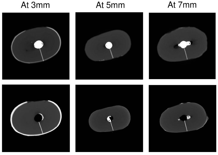
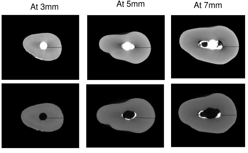
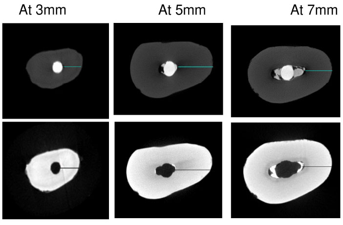
Nano-CT revealed (Figs. 1-3) represents canal centering ability between groups, and Tables 1 and 2 depicting the mean canal centering ability and apical transportation between Solite RS3, Solite RS3 Black, and Solite RE Black Retreatment files at 3mm, 5mm & 7mm on the mesial and distal sides. *Indicates a significant diff- erence (P < 0.05). After retreatment, there was a statistically significant difference (p-value<0.05) in the apical deformity and canal centering ability between the groups (Tables 1 and 2). Solite RE black showed a statistically significant difference when compared to the other two groups (p<0.05). No significant differences were observed between Solite RS3 and Solite RS3 Black for canal centering ability and apical transportation respectively (p>0.05). Thus, the study's null hypothesis is disproved.

Represents the measurement of the shortest distance between the canal wall and root pre and post retreatment using Solite RS3 system.

Represents the measurement of the shortest distance between the canal wall and root pre and post retreatment using Solite RS3 Black system.

Represents the measurement of the shortest distance between the canal wall and root pre and post retreatment using Solite RE Black system.
| Groups | Levels | ||
|---|---|---|---|
| At 3mm | At 5mm | At 7mm | |
| Solite RE Black | 0.59±0.071A | 0.59±0.05 A | 0.71±0.05 B |
| Solite RS3 Black | 0.62±0.051 A | 0.67±0.05 A | 0.75±0.03 B |
| Solite RS3 | 0.64±0.022 A | 0.73±.003 A | 0.83±0.05 B |
| Oneway -ANOVA (p-value) |
0.008* | 0.000* | 0.000* |
| Groups | Levels | ||
|---|---|---|---|
| At 3mm | At 5mm | At 7mm | |
| Solite RE Black | .022±.015 A | .0360±.015 A | .0450±.017 A |
| Solite RS3 Black | .039±.016 B | .0640±.012 B | .0850±.017 B |
| Solite RS3 | .0430±.01 B | .0800±.030 B | .0870±.029 B |
| Oneway -ANOVA (p-value) |
0.000* | 0.000* | 0.000* |
4. DISCUSSION
A preparation that maintains strictly the original canal curvature and avoids procedural errors is the aim of both the main and retreatment procedures according to Gogulnath [15] In this study, the three distinct retreatment file systems were compared and evaluated for their canal transportation and apical deformity. The noninvasive gold standard for evaluating canal geometry, nano-CT, was used to obtain the images. This imaging technique allowed for a comparison of the root canal's anatomical structure before and after instrumentation.
The transition between the periodontal and pulpal tissues is represented by the CDJ, which is the apical termination of the canal [16]. The transportation may suddenly alter its course based on the evaluation of the apical location. The root's radius and angle of curvature are intimately related to the apical transportation in various directions within the same root canal [3]. For all methods of canal preparation, reports of apical extrusion of debris during initial root canal therapy exist. One of the theories for the worst results of root canal therapy is the release of irrigants and infected tissue into the periapical area [17].
The taper of the instruments is another aspect that can affect the outcomes. Greater taper instruments are better at removing fillings and encourage larger root canal enlargement, but they also run the risk of damaging the root from excessive dentin removal [2]. The study employed progressive taper retreatment files, which preserve dentin thickness as well as canal centering ability. Canal centering ability and apical deformity did not differ statistically significantly between rotary and reciprocating devices [18]. Currently, multiple studies are available comparing the efficiency of the Solite RS3 system to other systems like ProTaper Universal Retreatment, Hyflex Remover, etc. When comparing the ability to remove gutta-percha between Solite RS3 and ProTaper Universal Retreatment, there seems to be no significant difference between the two systems, however, with respect to dentin removal the latter has a greater taper, thereby resulting in significantly more dentin removal as compared to the former. Consequently, due to its lesser taper, Solite RS3 is expected to conserve more dentin. [10]
The present study compares the Solite RS3 system to the Solite RS3 Black system and Solite RE Black systems, which are relatively new and hence have scarce literature. The results of the present study show that Solite RE Black system and solite RS3 Black system show more centered canal preparations. This can be attributed to the differences in metallurgy between the three systems with the RE2 file in the Solite RE system being machined using the C wire technology, thus being more resistant to cyclic fatigue while maintaining canal curvature in comparison to Solite RS3 system which is heat treated.
After instrumentation, alterations in the central axis of the root canal were taken into consideration when evaluating canal transportation [5]. Usually, the mesiodistal and buccolingual features of the curvature were used to assess canal transportation. However, transportation could not be measured since the teeth did not exhibit their greatest curvature in those planes. Therefore, in the apical, middle, and coronal portions of the root canals, linear measurements of the canal transportation were carried out in eight distinct directions. According to the study [19], this implemen- tation made it possible to get precise information regarding the location and direction of the canal transportation. As per Sairaman et al., Solite RS3 resulted in minimal canal transportation and created a more centrally positioned preparation compared to the ProTaper retreatment system. This is attributed to its heat treatment and reduced taper, which aid in gutta-percha removal while preserving dentin [20, 21].
The increased risk of vertical root fracture in teeth treated with endodontic therapy is amply demonstrated by endodontic literature. Therefore, it is imperative to stop more dentin loss from occurring as a result of over-instrumentation. The tooth will thereafter have more structural durability as a result of this [22]. However, the current study has the following limitations: it only tests the file systems on straight root canal systems, it only uses one obturation approach, and it does not use solvents or PUI to augment the removal of filling materials. To determine which file system is actually superior, additional research taking into account all of these extra criteria is required.
The outcomes of the current investigation were utilized in drawing the conclusion that the Solite RE Black file systems had superior centering ability in all apical thirds and less transportation in the middle and coronal thirds when compared to the other two retreatment file systems. This is because the Solite RS3 file has alternating cutting edges, which makes it easier to remove the obturating material, thus rejecting the null hypothesis. Furthermore, their exceptional flexibility serves as a safeguard against the excessive removal of dentin [23]. The transit and focus capabilities of several retreat file systems with differing technological as well as compositional components have not, as far as we are aware, been studied.
CONCLUSION
According to the study's findings, using Solite RE Black files during retreatment procedures may result in canal centering ratio and apical deformity, which is something that should be taken into account at all three levels. However, it is crucial to emphasize the need for additional research to look into other factors and characteristics that could affect retreatment's efficacy. These include employing solvents, trying out different retreatment instruments, and doing root canal fillings in a different way. Future research should examine a larger variety of parameters and think about doing clinical trials to confirm the results of laboratory testing in order to come to more conclusive results.
AUTHORS' CONTRIBUTION
It is hereby acknowledged that all authors have accepted responsibility for the manuscript's content and consented to its submission. They have meticulously reviewed all results and unanimously approved the final version of the manuscript.
ETHICS APPROVAL AND CONSENT TO PARTICIPATE
The Study received approval by the Scientific Review Board of Institutional human ethics committee from Saveetha Dental College, India (SRB/SDC/ENDO-2106/23 /077).
HUMAN AND ANIMAL RIGHTS
All human research procedures followed were in accordance with the ethical standards of the committee responsible for human experimentation (institutional and national), and with the Helsinki Declaration of 1975, as revised in 2013.


