All published articles of this journal are available on ScienceDirect.
Gingival Retraction with 980-nm Diode Laser Compared to Double-Cord Technique: An In vitro Study
Abstract
Background
In fixed prosthodontics, the need for gingival retraction prior to impression plays a fundamental role in ensuring the reproduction of anatomical details in the preparation using traditional impression materials or Intraoral Scanner (IOS) light beams. The recent introduction of laser in dentistry has opened new therapeutic options in the displacement of the gingival margin.
Objective
This study aimed to compare the efficacy of a 980 nm diode laser with the double gingival retraction cord technique to obtain a longitudinal and vertical displacement of the gingival margin.
Methods
Four bovine mandibles were used as the experimental model (n=32 teeth). Sixteen teeth were randomly assigned to the laser group (group A), and the other 16 to the retraction cord group (group B). For each group, the initial status was considered the corresponding control group. After tooth preparation, in both groups, a conventional impression was taken with Polyvinyl Siloxane (PVS). Successively, the gingival margins were displaced by using a 980nm-diode laser for group A, and by means of two retraction cords for group B. After the retraction process, a new impression was taken with the same polyvinyl siloxane. The amount of horizontal and vertical gingival displacement (HD, VD) was measured by an image analyzer. Tukey’s, Student’s t-test, and One-way ANOVA test were employed.
Results
There was a statistically significant difference (P<0.001) found in both methods compared to the corresponding control groups. A statistically significant difference (P<0.05) between the means of laser vertical displacement and the cord vertical displacement was observed. Conversely, no statistically significant difference was observed when the laser horizontal displacement and cord longitudinal displacement were compared (P>0.05).
Conclusion
The gingival displacement obtained by means of a 980 nm diode laser has been found to be similar to the displacement obtained with the double-cord technique. However, the double-cord technique provided a better vertical displacement. The findings suggest that the 980 nm diode laser may represent a valuable alternative in the displacement of the gingival margin.
1. INTRODUCTION
In fixed prosthodontics, it is not always possible to work with supragingival preparations. Juxta-gingival or intrasulcular finishing margin indications are related to high aesthetic requirements and increased restoration retention or they address dental pathological conditions, such as deep cervical caries [1]. Furthermore, in clinical cases of replacement of previous prosthetic crowns or of dyschromic abutments in the anterior area, it is inevitable that the preparation margin should be placed juxta-gingivally, that is to say, preparation at the same level as the gingival margin or intrasulcularly, namely subgingival, apical to the gingival margin [2].
For gingival retraction prior to the impression, there is a need to record all the anatomical details of the preparation, including the preparation margins, the finishing line, and the untouched areas. Given that one of the most relevant factors for fixed dental restorations with natural teeth is marginal adaptation, the more accurate it is, the lower the probability of developing a secondary disease [2]. Alternatively, in the absence of a readable margin, the dental technician may find it difficult to design a restoration that seals and adapts perfectly to the existing prosthetic preparation. This can result in the fabrication of an incongruous prosthetic restoration, which presents non-optimal marginal adaptation, such as horizontal or vertical discrepancy [3]. This inaccuracy may lead to a series of negative side effects, such as greater plaque retention and, consequently, gingival inflammation that can subsequently develop into periodontal pocket formation [4]. In the long term, complications, such as secondary caries, and in some susceptible gingival phenotypes, gingival recession could occur [5]. For these reasons, the accuracy of a restoration does not exclusively represent the search for an optimal transition between the natural element and the prosthesis, but it is key to the longevity of the restoration itself and periodontal health [6].
Gingival retraction, also referred to as gingival displacement, is the procedure of reversible deflection or removal of the inner surface of the gingival sulcus. The purpose is to create a dry environment to allow the impression material to penetrate into the sulcus in order to read beyond the preparation margins or increase the abutment surface visible to the Intraoral Scanner (IOS) light beam to correctly capture the margins of prosthetic preparations in natural teeth.
Gingival displacement methods can be categorized into two main groups [6] as follows:
a) Conventional mechanical, chemical, and mechano-chemical methods: These approaches involve techniques that use physical instruments, such as retraction cords; chemical agents, such as astringent pastes, or a combination of both, as well as retraction pastes containing specific compounds, like aluminum chloride and kaolin, or expanding polyvinyl siloxane material to displace the gingival tissues for impression taking.
b) Surgical methods: This group includes surgical techniques, such as rotary curettage with a suitably shaped diamond or ceramic bur, electro-surgical, and laser gingival tissue displacement.
The latest advances in laser in dentistry have opened new therapeutic options in the displacement of the gingival margin as an adjunct to traditional and digital impression techniques in fixed prosthodontics. Various dental lasers, such as Neodymium-doped Yttrium-aluminum-garnet (Nd:YAG), CO2, Erbium-doped Yttrium-aluminum-garnet (Er:YAG), Erbium, Chromium-doped Yttrium-scandium-gallium-garnet (Er, Cr:YSGG), and diode lasers have been described as a valuable surgical gingival retraction method [7, 8].
Diode lasers are solid-state Aluminum Gallium Arsenide (AlGaAs) semiconductor lasers that efficiently convert electrical energy into coherent light energy. The diode laser has wavelengths ranging between 800 and 980 nm. This wavelength range is well absorbed by pigmented tissue and hemoglobin, promoting water evaporation, which leads to ablation. This laser is used for cutting and coagulating gingiva and mucosa, and is, therefore, a soft-tissue laser.
Recently, a few studies have claimed that the 980-nm diode laser is also suitable for gingival retraction [3, 9, 10]. The potential advantage of this new technique could be a more simplified soft tissue removal inside the gingival sulcus, hemostasis, and a lower risk of gingival recession [11]. There are studies indicating that gingival tissue displacement with laser is less painful and can be used without anesthesia in selected cases for greater patient comfort [12].
Few studies have been carried out to assess the amount of gingival retraction achieved quantitatively by using diode lasers [7, 9, 13]. To the authors’ knowledge, no study has compared the degree of both the horizontal and vertical gingival displacement obtained with the diode laser surgical technique to the conventional double-cord mechanical method. Therefore, the purpose of this study was to analyze in vitro the horizontal and vertical gingival displacement measured at the juxta-gingival level by means of both a double retraction cord and the 980-nm diode laser. The null hypotheses tested were the following: (1) there is no statistically significant difference between the two techniques in terms of vertical gingival displacement; (2) there is no statistically significant difference between the two techniques in terms of horizontal gingival displacement.
2. MATERIALS AND METHODS
2.1. Samples
After review and approval by the local ethical committee (protocol number: 2402/2018), 4 bovine jaws were selected with standard age and dimensions. Each anatomical piece presented 8 teeth. Sixteen elements were randomly assigned to group A (gingival retraction by means of a diode laser), and the other 16 to group B (gingival retraction with two retraction cords), accounting for a total of 32 teeth. The randomization was performed by putting the letters in a sealed and opaque envelope, pointing out which teeth would be used for each of the selected gingival retraction techniques. The envelope was opened by an independent operator just before the restorative procedure began. All clinical procedures were performed by a single experienced operator with the aid of 4.3x400 surgical head-worn loupes (KS, Carl Zeiss Vision, Jena, Germany). Each tooth was prepared to provide a conical chamfer finishing margin at the juxta-gingival level. The reduction of tooth substance was achieved using water spray with a dental turbine (Kavo GmbH, Biberach, Germany) at 400,000 rpm and diamond burs with successive decreasing grain sizes 2886K, 2979K green, and 8979K (Komet Dental Brasseler GmbH, Lemgo, Germany), following gross tooth reduction using a 859K (Komet Dental Brasseler GmbH, Lemgo, Germany) bur. Following tooth preparation, a traditional impression was taken using a single-step double-mix impression technique with Polyvinyl Siloxane (PVS) according to the manufacturer's instructions at a controlled temperature (22 ±1°C).
The regular body impression material (Honigum Mono, DMG, Hamburg, Germany) was placed on a standard perforated steel tray, immediately followed by light body material (Honigum Light, DMG, Hamburg, Germany) spread around the dental elements involved in the preparation, ensuring that no bubbles are formed. The impressions were marked for each group and stored at room temperature for 2 hours to allow elastic recovery of the elastomeric materials and release of hydrogen from the polyvinyl siloxane.
2.2. Gingival Retraction
Gingival retraction was undertaken using two different methods: a 980 nm diode laser for group A and a double retraction cord for group B.
In Group A, a diode laser (Wiser - Doctor smile - Lambda SpA, Brendola, Vicenza, Italy) with a wavelength of 980nm was used. In accordance with the laser pre-select “surgery gingivectomy” program, 1.0 Watts of power in continuous wave mode and with a 300μm diameter optic fiber was applied. The optic fiber was inserted into the sulcus at a depth of 1 mm, and with a circular movement around the dental element for about 15 seconds, the retraction was completed.
For Group B, first, a non-impregnated 000-retraction cord (ULTRAPAK, Ultradent Products Inc., South Jordan, UT, USA) was gently pressed apically into the gingival sulcus, and over this, a second non-impregnated 1 retraction cord (ULTRAPAK, Ultradent Products Inc., South Jordan, UT, USA) was applied, both using a BN1 Gingival Cord Packer (Hu-Friedy, Chicago, Illinois, USA). The 000-cord was left in situ while the 1 cord was removed, and a polyvinyl siloxane impression was taken using the same materials and technique previously employed for the control groups.
2.3. Optical Microscope Measurements
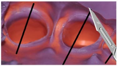
For each group, the impressions were assembled, reproducing the initial status (control group) and the new status after the gingival retraction, respectively, with a diode laser (group A) and with retraction cords (group B). The impressions were detached from the tray with a bevel knife and cut inter-dentally for each tooth in a bucco-lingual direction with a scalpel blade number #11 (Fig. 1). Each section was analyzed under 25x magnification using a stereo microscope (Wild Heerbrugg M5A, Wild Heerbrugg AG, Switzerland). The measuring process was monitored and cross-checked by an independent examiner.
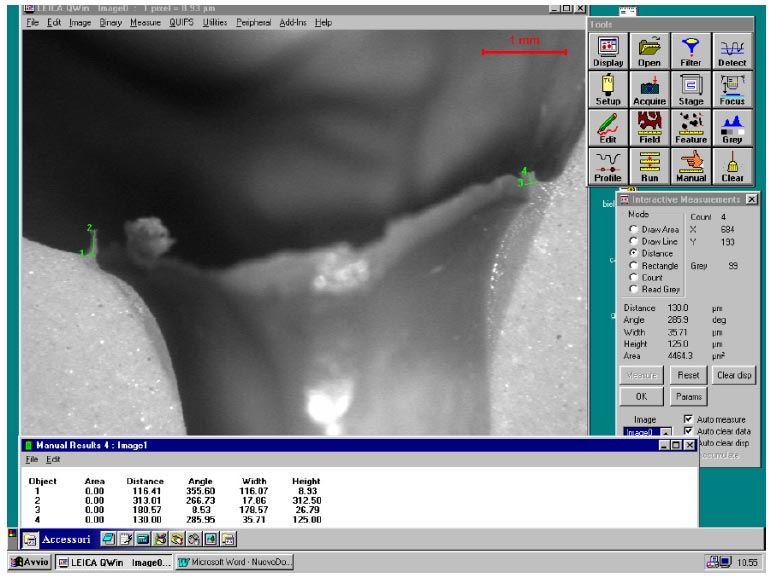
In each sample, the displacement was assessed by means of a digital image analysis system (Leica Q500IW, Leica Microsystems, Wetzlar, Germany) (Fig. 2). The amount of displacement in the vertical direction, matching the extent of penetration of the material (VD), and in the horizontal direction, corresponding to the thickness of the material penetrated (HD), was detected by measuring them in higher value points of the buccal portion. This portion was chosen from an aesthetic point of view because it represented the most visible portion of the prosthetic crown.

The impressions were cut inter-dentally for each tooth in a bucco-lingual direction with a scalpel blade number #11.
The data obtained were recorded on a computer spreadsheet (Microsoft Excel '07). To get the most reliable result, the arithmetic mean was computed considering the two buccal points as follows:
- VD: height in the vertical direction of the buccal portion (Laser, VD; Cords, VD)
- HD: width in the horizontal direction of the buccal portion (Laser, HD; Cords, HD).
Accordingly, all experimental groups have been divided into two subgroups, VD and HD.
2.4. Statistical Analysis
The difference between control and treated samples was performed by one-way Analysis of Variance (ANOVA). A non-parametric post-hoc test for multiple comparisons, Tukey’s test, or a parametric Student’s t-test were also performed when necessary. Values of p<0.05 were considered significant.
The statistical power of sample size was calculated according to Simple Interactive Statistical Analysis (α: 5%).
3. RESULTS
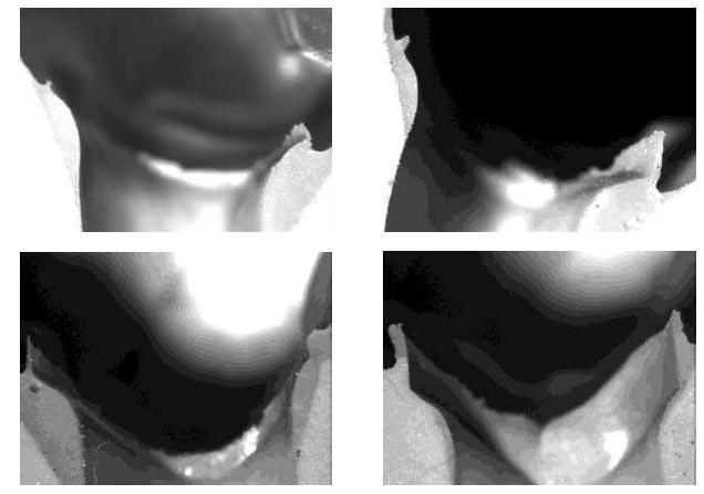
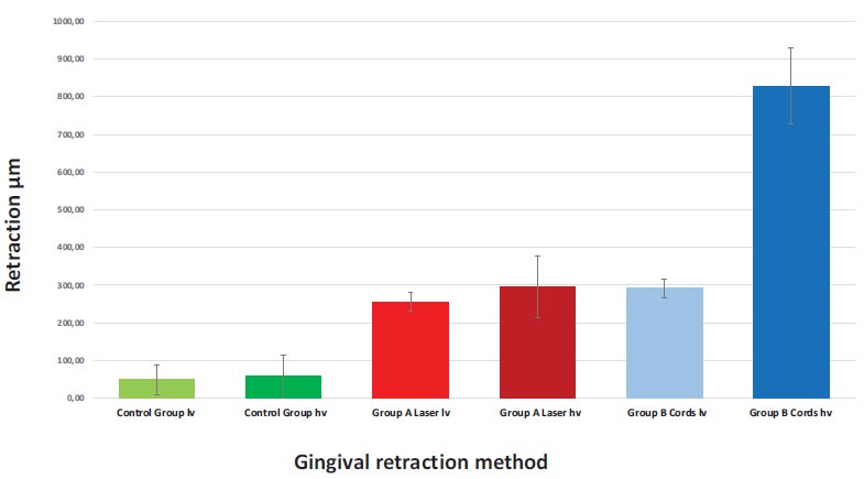
The experimental results are listed in Table 1 and represented in Figs. (3A-D and 4). Differences between the control group and treated groups (laser and cords) for each point were evaluated to obtain a comparison for each sample. The mean value was calculated from the data.

Assessment of the vertical and horizontal displacement by means of a digital image analysis system (Leica Q500IW, Leica Microsystems, Wetzlar, Germany).
| Sample | n° | Means (µm) | Standard Deviation |
|---|---|---|---|
| Control group lv | 16 | 48.93 | 39.04 |
| Control group hv | 16 | 58.25 | 55.30 |
| Group A Laser lv | 16 | 255.90* | 24.3 |
| Group A Laser hv | 16 | 294.40*§ | 82.1 |
| Group B Cords lv | 16 | 291.4* | 25.1 |
| Group B Cords hv | 16 | 828.2*§ | 101.3 |

Sections analyzed using a stereo microscope (25x). Differences between control group and treated group.
(A): control group; (B): laser treated group; (C): control group; (D): laser treated group.

Bar diagram showing the amount of retraction attained in the different experimental methods.
No statistical difference was observed between the two control groups (control VD vs. control HD). Conversely, both controls were statistically different from the treated samples (Groups A, B) (P< 0.001). Both retraction techniques produced a high amount of displacement as compared to their pre-displacement state.
The statistical tests also highlighted a statistically significant difference (P<0.05) between the means of laser VD and the cord VD. The double-cord technique provided a better vertical displacement compared to the 980 nm diode laser. Conversely, a statistically significant difference was not observed when the laser HD and cord HD were compared (P> 0.05). Indeed, the horizontal gingival retraction obtained by means of a 980 nm diode laser was similar to the retraction obtained with the double-cord technique. Consequently, only the first null hypothesis had to be rejected.
This significant result was underscored by the evidence that a group with a sample size of 16 achieved close to 95% statistical power.
4. DISCUSSION
The primary aim of the gingival retraction is to deflect or remove the inner surface of the gingival sulcus to allow accurate registration of all details of the finish line through the use of impression materials or intraoral scanners. A minimum lateral displacement of approximately 0.2 mm is required for this purpose [14].
The gingival sulcus is lined by sulcular epithelium with two basal layers of cells. Any violation of the attachment apparatus can cause the loss of connective tissue attachment and apical migration of the junctional epithelium. Gingival retraction procedures must be performed in a way that does not injure the basal cell layer or connective tissue cells in order to avoid a gingival recession related to the tissue response [4, 9, 15].
The commonly used double-cord technique for subgingival tissue displacement has certain drawbacks. These include the possibility of damaging the periodontal ligament if excessive pressure is applied during the insertion of the cords, the difficulty in removing the retraction cord without causing bleeding, and potential postoperative discomfort. Several clinical studies have demonstrated that the use of diode lasers for gingival retraction purposes causes less recession around natural teeth compared to the retraction cord [6, 9].
Comparable but not clinically significant differences in gingival recession were reported in vivo for the double-cord technique (mean = 0.26 mm) and diode laser (0.27 mm) 8 weeks after cementation [10]. On the contrary, statistical differences were found between laser and retraction cords, with the following mean values of gingival recession 4 weeks after retraction: retraction cord = 0.24 ±0.08 mm; diode = 0.13 ±0.08 mm. These results showed wider gingival width and less gingival recession for the diode laser than the retraction cord [7].
A few clinical studies comparing cord and laser have confirmed the less invasive periodontal impact of laser [13, 16]. However, Stuffken et al. did not find significant differences in the results of the double cord and laser technique [10].
A diode laser may cause minimal collateral tissue damage if used at the correct power [9]. Removing the superficial layers of the sulcular epithelium without damaging the basal cell layer and connective tissue cells may prevent shrinkage of the gingival tissue [11].
The gingival retraction has to be obtained apically as well as horizontally in order to allow the impression material to penetrate into the sulcus beyond the preparation margins or sufficiently expose the abutment surface visible to the intraoral scanner light beam. The results of the present study have shown a statistically significant difference between the means of laser vertical displacement and the cord vertical displacement, leading to the rejection of the first null hypothesis. In accordance with the present results, Gururaj R et al. reported, with respect to the depth of the gingival sulcus, that the retraction cord performed better (1.43 mm) than the diode laser (1.24 mm) [17].
On the contrary, no statistically significant differences were observed in our study when the laser group and cord group were compared for Horizontal Displacement (HD); therefore, we failed to reject the second null hypothesis. Consistent with the present results, a clinical study reported that the retraction cord produced a significantly larger lateral displacement (0.32 ±0.09 mm) than the diode laser (0.55 ±0.15 mm) [7].
In contrast to the results of our study, a clinical assessment reported that a diode laser produced greater mean lateral gingival displacement (0.48 ±0.10 mm) than the retraction cord (0.44 ±0.11 mm) [18]. Moreover, Dawani et al. reported that the diode laser produced in vivo wider lateral displacement (0.62 ±0.09 mm) than a mechanical atraumatic gingival displacement method (0.42 ±0.04 mm) [19].
The critical sulcular width has been reported to be approximately 0.2mm at the level of the finish line, which is important for good flow of the impression material beyond the finish line [20]. It has been reported that impressions with less sulcular widths have a higher incidence of tearing of impression materials and voids, and consequently a reduction in marginal accuracy [12]. Krishna et al. as well as Stuffken et al. reported that gingival troughing with the diode laser achieved greater than the minimum required sulcular width of 0.2 mm [9, 10]. Our findings, assessing both horizontal and vertical gingival displacement, confirmed the impression with the diode laser to be consistently accurate.
As a concluding remark, the present clinical study has some limitations. The measurements were taken solely at the buccal margin of the gingiva and not at the mesio- and disto-buccal gingival margins. In addition, the influence of gingival thickness, varied sulcus depth, visibility, and accessibility to the gingival retraction were not considered. Moreover, the comparative evaluation of the efficacy of the two different gingival retraction systems was performed by analyzing only traditional polyvinyl siloxane impressions. Optical impressions by means of an intraoral scanner can provide additional data. The in vitro design that limited the clinical generalizability of the results was also considered a limitation of the study.
CONCLUSION
The gingival retraction obtained by means of a 980 nm diode laser has been found to be similar to the retraction obtained with the double-cord technique. Both retraction techniques produced a highly significant amount of displacement as compared to their pre-displacement state. However, the double cord technique provided a better vertical displacement. The use of a 980 nm diode laser for gingival retraction offers benefits, such as an effective and more simplified gingival displacement for the impression materials or the intraoral scanner light beam combined with hemostasis, limited periodontal tissue invasiveness, and greater patient comfort. Further investigations are warranted to corroborate and expand upon our findings and the variables not considered in this study.
AUTHORS’ CONTRIBUTION
Luca Solimei contributed to conceptualization, methodology, investigation, data curation, resources, supervision, and project administration; Andrea Amaroli performed formal analysis, validation, data curation, and review; Anna Yu Turkina and Lavinia Signore performed validation and visualization; Stefano Benedicenti contributed to methodology and resource allocation; and Antonio Signore contributed to the writing of the original draft, conceptualization, methodology, investigation, and project administration. An English expert performed the professional English editing and proofreading.
LIST OF ABBREVIATIONS
| IOS | = Intraoral Scanner |
| PVS | = Polyvinyl Siloxane |
| ANOVA | = Analysis of Variance |
ETHICS APPROVAL AND CONSENT TO PARTICIPATE
This study received approval from the ethical committee of the University of Genoa (protocol number: 2402/2018).
HUMAN AND ANIMAL RIGHTS
All procedures performed in studies involving human participants were in accordance with the ethical standards of institutional and/or research committee, and with the 1975 Declaration of Helsinki, as revised in 2013.


