All published articles of this journal are available on ScienceDirect.

The Effect of Adding Silica Nanoparticles on the Compressive Strength and the Dimensional Accuracy of Type IV Dental Stone
Authors Info & Affiliations
Abstract
Background
Considering the need for high compressive strength and dimensional accuracy in dental casts, these properties were investigated in this study by adding silica nanoparticles to dental type IV stone (plaster).
Materials and Methods
In this in vitro study, silica nanoparticles with percentages of 0.5, 1, and 2% were added to the plaster powder. Dental plaster without silica nanoparticles was used as a control group. The prepared samples were examined in terms of compressive strength properties and dimensional accuracy (dimensional changes). A universal testing machine was used to check the compressive strength, and a stereomicroscope was used to check the dimensional accuracy.
Results
The addition of silica nanoparticles to the plaster increases the dimensional accuracy, which requires at least 1% silica nanoparticles for this increase (P<0.05). Adding the silica nanoparticles had no significant effect on the compressive strength compared to the control group (P>0.05).
Conclusion
The addition of silica nanoparticles to dental plaster Type IV at low concentrations can be considered in dentistry to increase dimensional accuracy without negative effects on compressive strength.
1. INTRODUCTION
Dental stone (plaster) is used in manufacturing dental prostheses such as full or partial dentures, fixed dentures, or a mobile orthodontic device [1]. Dental gypsum should have enough compressive strength and wear resistance, especially in the margin, to resist the carving process [2]. Type IV or 5 gypsum products are widely used in dental prostheses, both artificially and naturally [2]. Due to low fracture resistance, dimensional instability, technical sensitivity, and low wear resistance, the researchers state that gypsums cannot reconstruct the details of elastomer molds [3]. In general, the reason for the high use of gypsum products is that their characteristics can be changed by chemical or physical methods [4].
Today, the use of nanotechnology is considered an important change in fillers, which is efficient in improving the mechanical properties of dental materials [5]. Nanomaterials are materials that contain nanoparticles, at least 50% of which have an external diameter of 1 to 100 nanometers [6]. Their very small size makes their properties different compared to those materials in bulk size [7]. It should be mentioned that neutral bulk materials are catalytically activated when they are transformed into nanoparticles [8]. According to the reports, numerous treatments have been suggested to enhance the physicochemical, mechanical, and antimicrobial action of dental plasters using nanoparticles [5, 7, 9]. For the field of dentistry, antibacterial ceramics might be a good reference as they would inhibit dental problems [10, 11].
Types IV and V dental plasters are mostly used to make casts and dies [12]. Dye plaster is a copy of the tooth prepared in the mouth. For this reason, it must have certain characteristics, such as having an acceptable hardness. If it is not hard enough, it will be easily scratched by the corresponding pens during waxing. The issue eventually causes the mismatch of the wax pattern, and as a result, the final framework is disturbed [13].
The aim of this study was to investigate the compressive strength and the dimensional accuracy of dental type IV plaster by adding silica nanoparticles.
2. MATERIALS AND METHODS
In this in vitro study, the sample size for test number 1 (Dimensional changes) was 40, and for test number 2 (Compressive strength) was 80. The plaster used in this research project was Neo Stone (Type IV, Siladent, Germany).
2.1. Dimensional Changes
The sample size was calculated according to Akkus et al. [14], with an alpha of 0.05 and a power of 80%. A total of 40 samples (10 for each group) were used.
A Teflon mold was designed as a single structure with a base diameter of 38 mm and a height of 30 mm (Fig. 1). The upper surface of the mold included two lines with a triangular cross-section, the width of which was 75 micrometers. A circular metal piece with a height of 6 mm and a thickness of 4 mm was placed around this structure, and a groove was created in order to facilitate the opening and closing of the metal [15]. The distance between these 2 lines was 25 mm according to the standard (ISO48 23:1992 test method 7.7) [16].
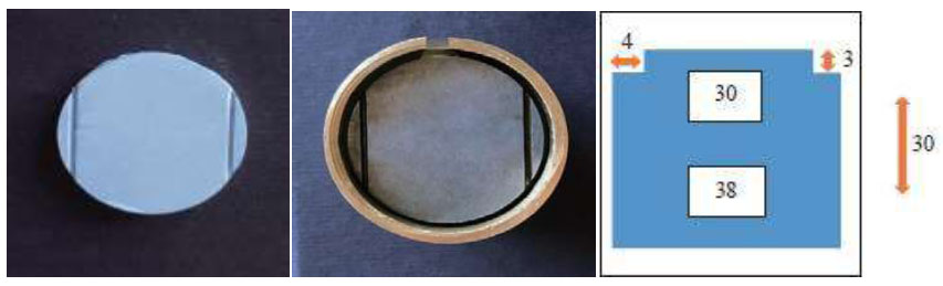
A teflon mold for dimensional change test.
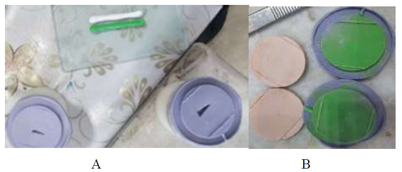
Duplicate samples of dimensional changes before (A) and after (B) silicone wash.
Also, according to Fig. (2), a vent was created for a better fit. For the control group, the plaster mixing process was performed by observing the water-to-powder ratio of 23ml to 100g at 23°C by a vacuum mixer (Multivac 4-Degussa, Germany and Renfert, Hilzingen, Germany) with a vibrator (KFP Dental, Iran).
Nanoparticles and plaster powder were weighed using a digital scale, and the volume of water was measured using a 10 ml pipette (CNWT Co. Jianxin, China). An ultrasonic mixer (sonicator, RPS-SONIC, Jiangsu, China) and a homogenizer (RoyanIran, Tehran, Iran) were used to homogenize the nanoparticles with the stone powder. Then, the ingredients were mixed with water according to the manufacturer’s instructions.
A stereo microscope (Nikon SMZ745T, Tokyo, Japan) was used to measure the dimensional changes. In all samples, the distance of two lines was measured directly, and the measurement accuracy was 0.005 mm. Magnification was set to 40 times, and low-angle light was used. The following equation was used to calculate dimensional accuracy.
AL=100 (L2-L1)
L1: Distance between two points a and b, which was measured on the Teflon model
L2: The same distance measured on the gypsum model according to (ISO 4823) [17].
2.2. Compressive Strength
The sample size was calculated according to the De Cesero study [10]. Taking an alpha of 0.05 and a power of 80%, 20 samples for each group and a total of 80 samples were used.
Improved gypsum type IV was used as a molding material, and silica nanoparticles were used as reinforcing materials.
Cylindrical samples were prepared for compressive strength test with a diameter of 3 mm and a height of 6 mm according to (ISO 6873:1992) and (ADA No. 25) [18]. These samples were prepared with the help of two Teflon molds. A layer of non-reactive separating material was added with plaster on the inner surface of the Teflon mold. Dental plaster is mechanically mixed under a vacuum mixer (Multivac 4-Degussa, Germany and Renfert, Hilzingen, Germany) with a spatula and poured into the mold under vibration (KFP Dental, Iran). Then, the materials were allowed to be set for one hour before separation from the mold. Then, they were kept in dry condition for 7 days before compressive strength tests.
The compressive strength test was measured by a universal testing machine (Hounsfield Test Equipment, Model HSK-S, Surrey, UK) with a crosshead speed of 1 mm/min. The application of pressure continued until the plaster fractured, and the compressive strength was calculated by the following formula:
б=4F/Πd^2
б = Compressive strength in MPa
F = compressive force at fracture
D = diameter of the material in millimeters [19]
3. RESULTS
3.1. Dimensional Accuracy
Table 1 shows the results for dimensional accuracy. One-way ANOVAtest showed thatthere was a significant difference between the studied groups (P<0.001). Tukey post-hoc test (Table 2) showed that the addition of silica nanoparticles in a concentration of 0.5% did not significantly change the dimensional accuracy compared to the control group (P=0.06). However, the addition of silica nanoparticles in 1 and 2 percent concentrations caused a significant change in dimensional accuracy compared to the control group (P<0.05). There was also no significant difference between the 1% and 2% groups (P=0.85). In other words, the addition of silica nanoparticles to the plaster increases the dimensional accuracy, which requires at least 1% silica nanoparticles for this increase in dimensional accuracy.
3.2. Compressive Strength
Table 3 shows the results for the compressive strength. One-way ANOVAtest showed thatthere was a significant difference between the studied groups (P=0.008). Tukey post-hoc test (Table 4) showed that the addition of silica nanoparticles in a concentration of 0.5,1 and 2% did not significantly change the dimensional accuracy compared to the control group (P=0.06).
| Groups | Sample Size | Mean±SD | P-value |
|---|---|---|---|
| Control | 10 | 0.199±0.084 | <0.001 |
| 0.5% | 10 | 0.137±0.037 | |
| 1% | 10 | 0.050±0.035 | |
| 2% | 10 | 0.069±0.038 |
Table 2.
| (I) | (J) | P-value |
|---|---|---|
| Control | 0.5% | 0.060 |
| Control | 1% | 0.0001 |
| Control | 2% | 0.0001 |
| 0.5% | 1% | 0.004 |
| 0.5% | 2% | 0.034 |
| 1% | 2% | 0.854 |
| Groups | Sample Size | Mean±SD | P-value |
|---|---|---|---|
| Control | 20 | 501.90±81.90 | 0.008 |
| 0.5% | 20 | 434.30±139.46 | |
| 1% | 20 | 419.40±119.95 | |
| 2% | 20 | 563.60±139.23 |
| (I) | (J) | P-value |
|---|---|---|
| Control | 0.5% | 0.307 |
| Control | 1% | 0.153 |
| Control | 2% | 0.388 |
| 0.5% | 1% | 0.980 |
| 0.5% | 2% | 0.007 |
| 1% | 2% | 0.002 |
4. DISCUSSION
The results of the current study showed that the addition of silica nanoparticles to dental Type IV plaster at a proper concentration can be considered in dentistry to increase the dimensional accuracy without negative effects on compressive strength.
Salah et al. [20], investigated the effect of adding silver nanoparticles on the compressive strength of type IV dental plaster (Elite stone Type IV Die stone, Zhermack, Italy). The samples were prepared according to ISO 6873 with a diameter of 20 mm and a height of 40 mm. They found that increasing the concentration of nanoparticles decreased the compressive strength of dental stone type IV in the dry state.
De Cesero et al. [10], also conducted a study on Fuji Rock gypsum and Durone gypsum and found that the compressive strength of Durone plaster did not change significantly after adding silica nanoparticles. Also, the compressive strength of Fuji Rock plaster decreased significantly after adding nanoparticles in concentrations of 1% and 5%.
Also, in the study of Salah et al. [21], which was conducted on gypsum (Ultrarock, Kalabhai Karson, Mumbai, India) and several types of nanoparticles with concentrations of 10%, the samples were tested after 24 hours. Their results showed that adding 10% of nanoparticles to all groups significantly reduced the properties of compressive strength and diagonal tensile strength compared to the control group. Also, the addition of zirconium oxide nanoparticles caused a smaller decrease in mechanical properties compared to other groups, and on the other hand, the addition of zinc oxide particles caused the greatest decrease in mechanical properties compared to other nanoparticles.
Taqa et al [22], conducted a study on two types of plaster, type IV (Zhermack, Italy) and type III (Saffa, Jordan), and the effect of adding different materials, including cured resin particles, pulverized stone, powdered plaster, and glass fiber was tested on the compressive strength of the plasters. They showed that Fiberglass significantly increased the compressive strength of plaster-based materials. Cured resin and pulverized materials can be added to dental stone and dental plaster at a concentration of 1% by weight since they increase the mechanical properties of these materials.
Akkus et al. [14], conducted a study on the effect of adding silica nanoparticles on stone type III and IV (Elite) dental plaster. The properties of compressive strength and diagonal tensile strength were investigated. Samples with a length of 6 mm and a diameter of 3 mm in a cylindrical shape were used for compressive strength tests. They concluded that the addition of silica nanoparticles significantly reduced the properties of compressive strength and diagonal tensile strength of dental stone types III and IV, and with increasing concentration of nanoparticles, the compressive strength decreased.
The results obtained for the dimensional accuracy test indicated that the dimensional accuracy in concentrations of 1 and 2% of silica nanoparticles was significantly higher than the control group (without silica nanoparticles). However, the addition of 0.5% silica nanoparticles had the same dimensional accuracy as the control group. The reason for this process may be due to the location of silica nanoparticles between the empty spaces of dental plaster particles and the elimination of voids [23].
Also, the results showed that the dimensional accuracy in concentrations of 1 and 2% is significantly higher than the 0.5% group. However, there was no significant difference in the dimensional accuracy of 1% and 2% concentrations. In fact, adding silica nanoparticles to plaster increased the dimensional accuracy, which required at least 1% of silica nanoparticles to increase the dimensional accuracy. Another reason for improving the dimensional accuracy of dental stones might be the high surface-to-volume ratio of silica nanoparticles, which reduces the surface tension and increases the wettability of the hemihydrate state. Then, the solubility increases, and as a result, the crystallization process occurs faster. Finally, the porosity of dental stone decreases, and as a result, the hardness and dimensional accuracy of this improved plaster increase [23].
Aljubori et al. [24], conducted a study on nanoparticles and their effect on the properties of dental plaster. Their results showed that adding silica nanoparticles to dental stone type IV plaster and mixing them increased the surface hardness of these materials significantly, and Linear dimensional changes were significantly reduced.
A study by Omer et al. [25], investigated the effect of adding Bergamot essential oil to dental stones and investigated its antibacterial and physical properties. In order to check the properties of compressive strength and record details, bergamot essential oil was added to plaster in three concentrations: 0%,8%, and 10%. One hour after the start of mixing, the samples were tested for compressive strength in the wet state. The results showed that Bergamot essential oil can be successfully buried inside the dental stone type 3 plaster, and it can affect the antimicrobial properties of the plaster. Furthermore, the results show that the compressive strength properties of this plaster increased significantly while the reproduction of detail ability remained intact.
5. LIMITATIONS
Due to the very small size of the nanoparticles, which gave them friction, it was very difficult to mix them with plaster powder, which may have caused errors. Of course, we have tried to reduce these errors as much as possible.
CONCLUSION
Considering the significant increase in dimensional accuracy after adding silica nanoparticles and the lack of significant effect of adding silica nanoparticles on compressive strength, the addition of nanoparticles to dental stone type IV plaster can be suggested to be used in dentistry.
AUTHORS’ CONTRIBUTION
All authors have read and agreed to the published version of the manuscript.
ABBREVIATION
| SD | = Standard Deviation |
ETHICAL STATEMENT
The present in vitro study was approved by the Ethics Committee of Tabriz University of Medical Sciences with the ethics ID of IR.TBZMED.VCR.REC.1401.233.
AVAILABILITY OF DATA AND MATERIALS
The data and supportive information are available within the article.
FUNDING
The Vice Chancellor for Tabriz University of Medical Science, Iran provided financial support.
ACKNOWLEDGEMENTS
This paper was based on the data from a proposal recorded at the Tabriz University of Medical Sciences, Iran (number 69098).

