All published articles of this journal are available on ScienceDirect.
Magnetic Resonance Imaging (MRI) findings of Cervical Spine Derangement (CSD) among Iraqi Working Dentists
Abstract
Objective:
To assess the cervical spine derangement among working dentists using MRI in order to establish a relationship between job parameters and cervical derangement changes.
Methods:
A cross-sectional study was carried out at the MRI unit of Al-Sader Medical city of Al Najaf health directorate from June 2015 to December 2016. The involved 43 working dentist volunteers of varying age and sex, who underwent an MRI of their cervical spine.
Results:
The MRI was normal in 16 (37.3%) and abnormal in 27 (62.8%) participants. The abnormality was due to cervical spine spondylitis changes.
Conclusion:
Intervertebral Discs (IVD) degeneration was the most frequent finding, followed by IVD herniation.
1. INTRODUCTION
Recently, research based on surveys suggested the prevalence of Musculoskeletal Diseases (MSDs) in dental professionals, with respect to the cervical, shoulder, and/or lumbar region [1-5] . The unsuitable posture has a greater contribution to developing such a muscular imbalance [5-9]. Work-related MSDs have shown an increasing incidence among dental professionals, and thus, proved to be one of the major occupational hazards for them [4, 11-14]. More than 86.7% of dental professionals are suffering from MSDs affecting the neck and shoulder regions. They usually complain of neck and shoulder ache [15]
The development of various diagnostic techniques like MRI has been a prime encouragement for researchers to look into such cervical changes among medical professionals. This is because MRI is very safe, non-invasive, and highly sensitive for detecting changes in the skeletal structures like Cervical Spondylotic Changes (CSC), in both symptomatic and asymptomatic patients. Thus, MRI can detect early stages of disc degeneration and has been a preferred method of choice in assessing disc integrity in clinically symptomatic individuals [3, 4, 10].
The predisposing risk factors for cervical spine changes include the nature of work, posture of the individual, age factor, gender, any medical history of trauma in the cervical region, genetic factor, smoking, and so on [14].
1.1. Anatomy of the Cervical Spine
Cervical spines constitute the upper segment of the vertebral column which extends from base of the skull to the thorax. There are seven vertebrae, namely C1- C7, with the primary function of skull support and brain and spinal cord maintenance in their relative positions [22].
1.2. MRI of Cervical Spines
MRI is a non-invasive technique that uses non-ionising electromagnetic radiation waves [26].
Age-related changes in the Intervertebral Discs (IVDs):
Decrease in the T2 signal of IVD is age-related. It predisposes to CSC in the discs such as loss in disc height, disc herniation, and annular tears as compared to normal aging discs [12].
Pathophysiology of CSC disease [25]:
1. Disc height loss causing stress on the facet and neurocentral joints,
2. Exaggerated joint motion with facet joint disease,
3. Instability of spine with arthritis,
4. Hypertrophy of joint capsule, and
5. Posterior ligament hypertrophy.
Thus, the study aims at assessing the cervical spine derangement among dental professionals using MRI in order to establish a relationship between job parameters and cervical derangement changes.
2. MATERIALS & METHODS
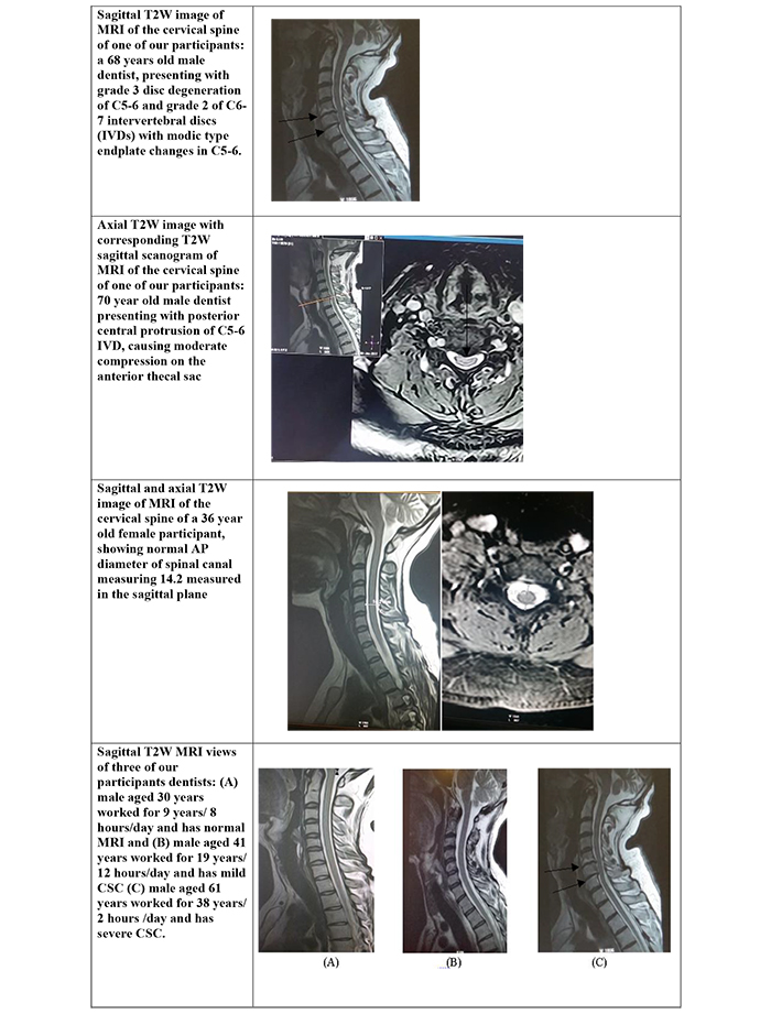
A cross-sectional study was carried out at the MRI unit of Al-Sader Medical city of Al Najaf health directorate during an 18 months period, i.e. June - 2015 to December -2016. This involved 43 working dentist volunteers of varying age and sex, who underwent an MRI of their cervical spine (Fig. 1).
All the volunteers gave their consent to participate in this study and completed a questionnaire prior to the MRI investigation. The questionnaire included details like their name, gender, weight, height, years of work, hours of daily work, work posture, previous medical history of trauma in the cervical region, previous MRI investigations, any systemic disease(s) or any problem(s) interfering with the MRI test (Fig. 1; Table 1 and Appendices A and B).
2.1. Inclusion Criteria
Any working dentist was included in this study whether they had symptoms or not. The symptoms included neck pain, upper limb radiculopathy, headache, trouble walking, etc.
2.2. Exclusion Criteria
Anyone who underwent previous surgery or significant trauma to the cervical spine or who contraindicated to MRI reports were excluded from the study.
All volunteer dentists were examined by Philips, Achieva, 1.5 tesla model 2010 MRI machine. The examination was done in the supine position with the RF coil under the neck. The protocols used were: sagittal T1W image, which is produced using a spin-echo (SE) sequence. This included a short echo time (TE < 30 ms), short repetition time (TR 300-700 ms), axial and Sagittal T2 weighting with a repetition time (TR 2000-3000 ms) and long echo time (TE 80 ms). All the results were reviewed by specialists in Diagnostic Radiology, experts in cervical changes and neuro-imaging.

| Variable | No. | % | |
|---|---|---|---|
| BMI | Normal | 8 | 18.6 |
| Overweight | 23 | 53.5 | |
| Obese | 8 | 18.6 | |
| Morbid obesity | 4 | 9.3 | |
| Loss of sphincters control due to neurological causes | Yes | 0 | 0.0 |
| No | 43 | 100 | |
| Smoking | Yes | 2 | 4.6 |
| No | 41 | 95.4 | |
| Family history of cervical spine spondylitis | Yes | 9 | 20.9 |
2.3. Study Findings
The data collected from the questionnaires stated that 13 professionals had more than 20 years of experience, followed by 9 having 16-20 years of experience, 5 having 11-15 years, 13 having 6-10 years, and 3 having <=5 years of experience. Out of this, 26 professionals worked 4-6 h per day and 5 worked more than 6 h a day. The specialty, posture of work, and other additional things have been clearly stated in Table 2. The table data stated that the maximum respondents were general dental practitioners (20 of them). of 43 respondents, 33 had twisted work posture, and around 81.4% of respondents used suntem chair type.
2.4. MRI Data Interpretation:
Disc degeneration was assessed in T2W images according to the grading system by Lloydine J. Jacobs et al. for each level alone from C2-C7 levels. Grade zero and one disc degeneration was considered to be normal while grade two and three were considered as abnormal.
Loss of disc height was assessed subjectively in comparison with higher normal levels and divided into 4 grades from 0-4 meaning 0-25%, 25-50%, 50-75%, and 75-100%.
Osteophytes were considered to be any osseous protrusion from the vertebral body outside its normal borders in anterior or posterior directions. The posterior part was divided into central or paracentral osteophytes, which were observed in T1W and T2W images in axial and sagittal views.
Disc herniation was considered as any protrusion of non-osseous disc material beyond borders of the endplate of the adjacent vertebral body, and classified as follows:
- Disc bulge when more than 50% of disc circumference was seen,
- Disc protrusion when less than 50% of disc circumference was seen with a wider base than tip,
- Disc extrusion when less than 50% of circumference was seen with a narrower base than tip.
These protrusions were classified as central, paracentral, foraminal, or extraforaminal (far lateral) according to its relationship with the spinal cord in the axial plane. Central protrusions were opposite to the middle third of the cord, paracentral was opposite to the lateral third, foraminal against neural foramen, and extraforaminal (far lateral) protrusions lateral to the cord Some of the examples can be viewed in (Fig 2).
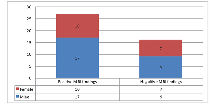
Twenty-seven (62.8%) respondents showed at least a single positive MRI change and 16 (37.3%) showed normal MRI. Positive MRI findings are mainly seen in males accounting for 17 (39.5%) participants, while females were 10 (23.2%). Negative findings were also more common in males than in females (Fig 3). All the findings were due to degenerative disc disease.
The most frequent MRI finding was disc degeneration seen in 27 respondents (62.8%), followed by disc herniation seen in 19 (44.2%). Only the degenerative type of spinal canal stenosis was seen. It was seen in 6 (14%) participants. The most frequently affected level was C5-6 (Table 3). C5-6 level was the most frequently affected level.
| Variables | - | No. | % |
|---|---|---|---|
| Specialty | General dental practitioners | 20 | 46.5 |
| - | Surgeons | 10 | 23.3 |
| - | Resident | 1 | 2.3 |
| - | Other* | 12 | 27.9 |
| Work posture | Neutral | 10 | 23.3 |
| - | Twisted | 33 | 76.7 |
| Type of chair | Suntem | 35 | 81.4 |
| - | Other** | 8 | 18.6 |
| Has a dental assistant | Yes | 17 | 39.5 |
| No | 18 | 41.8 | |
| Not always | 8 | 18.6 | |
| Instrument accessibility | Easily accessible | 31 | 72 |
| Difficultly accessible | 12 | 28 | |
| Additional works burden neck (reading, using PC or mobile phone and sewing) | Yes | 12 | 28 |
| No | 31 | 72 |


Correlation between MRI findings and demographic characteristics:
(1) Age was significantly correlated with CSC (P value= 0.003) (Table 4).
(2) Gender showed no significant correlation with CSC, although some CSCs were seen more frequently in males (8 of them showed herniation plus disc degeneration), but it did not reach values to be statistically significant (P value= 0.6).
(3) Also, CSCs were seen more frequently in participants who worked in twisted posture but it did not reach values to be statistically significant.
(4) BMI and positive family history of CSCs showed a significant correlation (Table 4).
There was also a significant correlation between CSC and increased years of work while there was no statistically significant correlation with increased hours of daily work (Tables 5 and 6).
But after adjustment of age, gender, and body mass index, there was no correlation between years of work and MRI changes, which stated that the changes could be due to the ageing process rather than dental work as shown in this study (Appendix C). There was no statistically significant correlation between symptoms and MRI changes as shown in Table 7. No significant correlation was found between CSC and work posture, type of dental chair or presence or absence of assistants and the details have been presented in Appendices D-G.
| Variables | No (n = 43) | % | Most frequently level affected |
|---|---|---|---|
| IVD degeneration | 27 | 62.8 | C5-6 |
| Disc herniation | 19 | 44.2 | C5-6 |
| Posterior osteophyte | 27 | 62.8 | C5-6 |
| Degenerative spinal canal stenosis and spinal cord compression | 6 | 18 | C5-6 |
| Ligamentum flavum hypertrophy | 14 | 32.6 | C5-6 |
| Finding | - | Age | BMI | Family history |
|---|---|---|---|---|
| loss of disc height level(s) | R* | - | 0.361 | - |
| P* | - | 0.007 | - | |
| Disc bulge | R | 0.377 | -0.323 | 0.304 |
| P | 0.005 | 0.017 | 0.025 | |
| Paracentral protrusion | R | - | - | - |
| P | - | - | - | |
| foraminal protrusion | R | 1.000 | -1.000 | -1.000 |
| P | 0.001 | 0.001 | 0.001 | |
| Ligamentum flavum hypertrophy | R | - | 0.643 | - |
| - | - | 0.013 | - |
| Working hours per day | P.value | |||||||
|---|---|---|---|---|---|---|---|---|
| < 5 | 5 - 8 | > 8 | ||||||
| No. | % | No. | % | No. | % | |||
| posterior osteophytes | Yes | 2 | 40.0 | 15 | 65.2 | 11 | 73.3 | 0.40 |
| No | 3 | 60.0 | 8 | 34.8 | 4 | 26.7 | ||
| Disc degeneration | Severe | 3 | 60.0 | 11 | 47.8 | 12 | 80.0 | 0.14 |
| Mild | 2 | 40.0 | 12 | 52.2 | 3 | 20.0 | ||
| Herniation | Yes | 4 | 80.0 | 16 | 69.6 | 13 | 86.7 | 0.47 |
| No | 1 | 20.0 | 7 | 30.4 | 2 | 13.3 | ||
| - | Years of Work | P.value | ||||||||||
|---|---|---|---|---|---|---|---|---|---|---|---|---|
| < 5 | 6 - 10 | 11 - 15 | 16 - 20 | > 20 | ||||||||
| No. | % | No. | % | No. | % | No. | % | No. | % | |||
| loss of disc height |
Yes | 1 | 33.3 | 1 | 7.7 | 0 | 0.0 | 1 | 11.1 | 5 | 38.5 | 0.18 |
| No | 2 | 66.7 | 12 | 92.3 | 5 | 100 | 8 | 88.9 | 8 | 61.5 | ||
| posterior osteophytes |
Yes | 1 | 33.3 | 5 | 38.5 | 3 | 60.0 | 7 | 77.8 | 12 | 92.3 | 0.035 |
| No | 2 | 66.7 | 8 | 61.5 | 2 | 40.0 | 2 | 22.2 | 1 | 7.7 | ||
| Disc degeneration |
Yes | 1 | 33.3 | 3 | 23.1 | 2 | 40.0 | 7 | 77.8 | 13 | 100.0 | 0.001 |
| No | 2 | 66.7 | 10 | 76.9 | 3 | 60.0 | 2 | 22.2 | 0 | 0.0 | ||
| Herniation | Yes | 2 | 66.7 | 7 | 53.8 | 4 | 80.0 | 7 | 77.8 | 13 | 100.0 | 0.093 |
| No | 1 | 33.3 | 6 | 46.2 | 1 | 20.0 | 2 | 22.2 | 0 | 0.0 | ||
| Foraminal compression |
Yes | 2 | 66.7 | 1 | 7.7 | 0 | 0.0 | 0 | 0.0 | 0 | 0.0 | 0.001 |
| No | 1 | 33.3 | 12 | 92.3 | 5 | 100.0 | 9 | 100.0 | 13 | 100.0 | ||
| Thecal Sac compression |
Yes | 2 | 66.7 | 8 | 61.5 | 4 | 80.0 | 7 | 77.8 | 13 | 100.0 | 0.19 |
| No | 1 | 33.3 | 5 | 38.5 | 1 | 20.0 | 2 | 22.2 | 0 | 0.0 | ||
| degenerative spinal canal stenosis |
Yes | 1 | 33.3 | 0 | 0.0 | 0 | 0.0 | 1 | 11.1 | 5 | 38.5 | 0.061 |
| No | 2 | 66.7 | 13 | 100.0 | 5 | 100.0 | 8 | 88.9 | 8 | 61.5 | ||
| Spinal cord compression |
Yes | 1 | 33.3 | 0 | 0.0 | 0 | 0.0 | 1 | 11.1 | 4 | 30.8 | 0.14 |
| No | 2 | 66.7 | 13 | 100.0 | 5 | 100.0 | 8 | 88.9 | 9 | 69.2 | ||
| Variables | Symptomatic (n = 28) |
Asymptomatic (n = 15) | P. value | |||
|---|---|---|---|---|---|---|
| No. | % | No. | % | |||
| Posterior osteophytes | Yes | 19 | 67.9 | 9 | 60.0 | 0.61 |
| No | 9 | 32.1 | 6 | 40.0 | ||
| Disc degeneration | Yes | 17 | 60.7 | 9 | 60.0 | 0.96 |
| No | 11 | 39.3 | 6 | 40.0 | ||
| Disc herniation | Yes | 23 | 82.1 | 10 | 66.7 | 0.25 |
| No | 5 | 17.9 | 5 | 33.3 | ||
3. DISCUSSION
This study considered is the first study using MRI to detect cervical changes among working dentists. We could not find any previous similar imaging studies which did an MRI on dental professionals to compare with. However, many clinical studies revealed that MSDs are common among dentists. They have a higher incidence of work-related MSDs than workers of other occupations [1-7] . .Neck pain and upper limb numbness are the most common symptoms and occur in more than 85% of dentists after they start working [15]
Many dentists or faciomaxillary surgeons, if not all of them, blame their routine dental work as the main cause of their neck pain and/or radiculopathy, which are relatively common symptoms among the dentists. Many of the dentists may seek multiple sick leaves, changing jobs, or even retirement due to ill health. In the latter case, MSDs are the most common causative factor accounting for 55% of all ill-health retirements [9, 18].
CONCLUSION AND FUTURE RECOMMENDATIONS
MRI of cervical spines derangement among working dentists was found to be positive (86%) and all of the changes were related to cervical spine spondylosis. The most common finding is disc degeneration followed by disc herniation. The frequency of CSC distributed by levels, i.e. from the most to the least severe levels: C5-6, C4-5, C3-4, and C6-7. There was no statistically significant correlation between symptoms and MRI findings of CSC disc degeneration with and without disc herniation, which significantly increased with an increase in the age in the case of symptomatic as well as asymptomatic participants. There was no overall statistically significant correlation between gender and CSC, whether in symptomatic or asymptomatic participants.
CSC was seen to be significantly correlated with BMI and family history of CSC. No statistically significant correlation was found between years of work and daily hours of work with CSC; however, this result may not reflect the reality, because of the small sample size in this study, which involved more younger participants that responded. No significant correlation was found between CSC and work posture, type of dental chair, or the presence or absence of assistants.
The Current Study Recommends the Following:
(1) This study is considered to have the antecedent in studying MRI of cervical spine derangement among working dentists and it is considered to be an evidence-based study.
(2) The authors encourage further studies with a larger sample size.
(3) The authors also recommend preventive strategies for working dentists by adopting neutral body postures, designing the work to allow machinery to perform/assist highly repetitive tasks, and early management of disorders.
ETHICS APPROVAL AND CONSENT TO PARTICIPATE
The research has been done according to the approval of the health authority in Najaf hospital, Iraq.
HUMAN AND ANIMAL RIGHTS
No animals were used in this research. All human research procedures followed were in accordance with the ethical standards of the committee responsible for human experimentation (institutional and national), and with the Helsinki Declaration of 1975, as revised in 2013.
CONSENT FOR PUBLICATION
Written informed consent was obtained from each participant prior to the study.
AVAILABILITY OF DATA AND MATERIALS
The authors confirm that the data supporting the findings of this study are available within the article.
FUNDING
None.
CONFLICT OF INTEREST
The author declares no conflict of interest, financial or otherwise.
ACKNOWLEDGEMENTS
We appreciate the hard work of Dr Fatimeh Shimirty and Dr Haider Al Muaala for their assistance in collecting the data and reviewing the study.
| QUESTIONERS FOR VOLNTEER'S WORKING DENTIST FOR DOING MRI CERVICAL SPINE TEST: | ||||||
| Do you have any objection to be one of the volunteer for MRI spine test? | ||||||
| Name: |
Gender: M F |
Age: |
Specialty: Dental Student: GDP: Specialist: |
|||
| Type of work: General Specialty of work: Ortho OMF Surgery, Other (Specify please) | ||||||
| Have an oriented assistant: YES NO Not always | ||||||
| Posture during work : Normal Twisted | ||||||
| Accessibility to your instruments Easy Access Difficult Access | ||||||
| How many years you are working in Dentistry: Less than ten years More than ten years | ||||||
| How many days per week you are working 1-2-3-4-6-7 day(s) | ||||||
| How many hours per day you are working (1-5) hours (6-10 )hours | ||||||
| Additional hobbies burden your neck movement Yes No if yes specify please | ||||||
| Pleas answer the following questions: | ||||||
| Question: | Yes | No | Comment | |||
|
Do you have any C/I for taking MRI ,Like
Pregnancy(female), Pacemaker or metal object in your body |
||||||
| Do you smoke | ||||||
| Do you have any family history of cervical spondylosis ? | ||||||
| Do you have any past, present medical problems | ||||||
| Do you take any medication ? | ||||||
| Have you consulted physician before for neck and /or shoulder symptoms ? | ||||||
| Other disease? | ||||||
| Symptoms: | ||||||
|
Do you have any cervical spine symptoms like:
Neck ache or stiffness (could be worse during activity) Weakness and numbness in the arm, hand and orfingers Walk trouble Imbalance Headache Grinding and popping sound/feeling in neck movement Loss of bladder or bowel control(neurological causes only) Dizziness when tilting head back Others |
||||||
| Name | Signature | Date | ||||
| Thank you for your help | ||||||
| Variable | No. | % |
|---|---|---|
| Medications use | 6 | 14 |
| Consultations for neck problems | 10 | 23.3 |
| Neck pain | 28 | 65.1 |
| Upper limb numbness | 17 | 39.5 |
| Walk troubles | 3 | 6.9 |
| Loss of balance | 3 | 6.9 |
| Headache | 17 | 39.5 |
| Popping | 9 | 20.9 |
| Loss of urine or bowel control | 0 | 0.0 |
| Tingling | 7 | 16.2 |
| Dizziness | 6 | 14 |
| low back pain | 7 | 16.2 |
| Years of Work | Working Hours on the Chair | ||
|---|---|---|---|
| Posterior osteophytes | R | 0.007 | -0.035 |
| P | 0.967 | 0.833 | |
| Herniation | R | 0.036 | 0.071 |
| P | 0.826 | 0.667 | |
| Degenerative spinal canal stenosis and spinal cord compression | R | -0.026 | -0.214 |
| P | 0.878 | 0.191 | |
| Work Posture | P | |||||
|---|---|---|---|---|---|---|
| Neutral (n = 10) | Twisted (n = 33) | |||||
| Posterior osteophytes | Yes | 6 | 60.0% | 22 | 66.7% | 0.70 |
| No | 4 | 40.0% | 11 | 33.3% | ||
| Disc degeneration | Yes | 6 | 60.0% | 20 | 60.6% | 0.97 |
| No | 4 | 40.0% | 13 | 39.4% | ||
| Herniation | Yes | 9 | 90.0% | 24 | 72.7% | 0.26 |
| No | 1 | 10.0% | 9 | 27.3% | ||
| Type of Chair | P | |||||
|---|---|---|---|---|---|---|
| Suntem (n = 36) | Other (n = 7) | |||||
| Posterior osteophytes | Yes | 23 | 63.9% | 5 | 71.4% | 0.70 |
| No | 13 | 36.1% | 2 | 28.6% | ||
| Disc degeneration | Yes | 20 | 55.6% | 6 | 85.7% | 0.14 |
| No | 16 | 44.4% | 1 | 14.3% | ||
| Herniation | Yes | 26 | 72.2% | 7 | 100.0% | 0.63 |
| No | 10 | 27.8% | 0 | 0.0% | ||
| Have An Oriented Assistant | P | ||||||||
|---|---|---|---|---|---|---|---|---|---|
| Yes (n = 16) | No (n = 18) | Not Always (n =9) | |||||||
| Posterior osteophytes | Yes | 10 | 62.5% | 14 | 77.8% | 4 | 44.4% | 0.22 | |
| No | 6 | 37.5% | 4 | 22.2% | 5 | 55.6% | |||
| Disc degeneration | Yes | 9 | 56.3% | 12 | 66.7% | 5 | 55.6% | 0.78 | |
| No | 7 | 43.8% | 6 | 33.3% | 4 | 44.4% | |||
| Herniation | Yes | 11 | 68.8% | 13 | 72.2% | 9 | 100.0% | 0.16 | |
| No | 5 | 31.3% | 5 | 27.8% | 0 | 0.0% | |||
| Smoking | P | |||||
|---|---|---|---|---|---|---|
| Yes (n =2) | No (n =41) | |||||
| Posterior osteophytes | Yes | 1 | 50.0% | 27 | 65.9% | 0.65 |
| No | 1 | 50.0% | 14 | 34.1% | ||
| Disc degeneration | Yes | 0 | 0.0% | 26 | 63.4% | 0.073 |
| No | 2 | 100.0% | 15 | 36.6% | ||
| Herniation | Yes | 2 | 100.0% | 31 | 75.6% | 0.43 |
| No | 0 | 0.0% | 10 | 24.4% | ||
| No | 0 | 0.0% | 9 | 22.0% | ||


