All published articles of this journal are available on ScienceDirect.
Replacement of Congenitally Missing Bilateral Maxillary Canines with Dental Implants: A Rare Case Report and Mini Review
Abstract
Introduction:
Maxillary canines play strategic roles in maintaining the normal function and visibility of teeth. However, rare cases of bilateral permanent congenitally missing maxillary canines were occurring. Replacing these missing canines with dental implant prostheses is an optimal treatment plan for aesthetics and functions.
Case Report:
In this case report, we described the replacement of bilateral maxillary canines with osseointegrated implants to support all-ceramic prostheses for a young female patient. This treatment avoids the conventional preparation of adjacent teeth as a part of prosthetic reconstructions. In addition to that, all-ceramic crowns are biocompatible with oral cavity structures.
Conclusion:
The stable short-term result has been achieved with the replacement of the bilateral congenitally missed canines with an implant and cemented all-ceramic crowns, with 3-years cumulative success rate.
1. INTRODUCTION
Tooth hypodontia or agenesis is one of the most common human tooth anomalies characterized by the developmental absence of one or more teeth. It is non-syndromic and can occur in association with a recognized genetic syndrome or as a solitary anomaly [1]. The absence of eruption of one or more permanent teeth in the oral cavity without any systemic disorder is the most common phenotype of tooth hypodontia [1, 2].
A retained canine is a tooth that has reached its normal time for eruption (11-13 years old for the maxillary arch), but its full development (formed teeth) remains locked inside a maxilla or a mandible, thereby maintaining its intact pericoronary sac [3].
Maxillary canines are the second most common ectopic teeth in the dental arch after the third molar, thereby affecting approximately 2- 4% of the all human population worldwide [4]. For example, it is a rare finding in Europe and Caucasian populations as mentioned by the previous case reports those recorded that the prevalence of maxillary permanent canine agenesis varies between 0.07%, 0.13%, and 0.91% [3-7], in Germany, Italy, UK, Hungary, and Netherlands, respectively.
Agenesis of maxillary canines is a relatively common occurrence in Asian races as in China [2, 8, 9]. Besides, it registered in selective cities of Saudi Arabia such as in Jazan, Qassim, Riyadh, and Al-Jouf [10-14] and Recorded 2.2%, 6.3%, 4.45%, and 4.9%, respectively. Also, in other Arab countries, the hypodontia of canine is higher than other countries worldwide and registered as 3.55% in Yemen, 6.2% - 7.8% in Qatar, while in Libya and Jordan are 1.5% and 2.6%, respectively [14-18].
Some researches or case reports found agenesis of canine as in the maxillary arch, unilaterally (either right or left), and among female patients [11, 13, 19, 20] or male patients [21]. Also, others found that canine hypodontia is bilateral, in the maxilla and among females [14, 17], or males [22].
Replacing a congenitally missing tooth in the maxillary region with implants is often preferred to conventional tooth-supported restorations such as bridges or an adhesive restor- ation because this technique of replacement avoids the preparation of an intact adjacent tooth substance and requires a relatively short treatment time [20]. Osseointegrated implants were first introduced in the mid-1960s, and the original concept of “ad modum” was based on the placement of four to six standard 3.75 mm titanium implants in an edentulous maxillary arch [2]. This technology has shown great potential. Further- more, titanium implants have broadened their indications and have become a reliable and widely acceptable treatment modality in all fields of dentistry [23].
Canines are important teeth within the dentition and implicated in dental aesthetic, facial appearance, arch development, masticatory efficiency, and functional occlusion [24, 25]. This case report described the replacement of bilateral congenitally missed canines in the maxillary arch of a 30-year-old female patient with a 3-years follow-up period.
2. CASE REPORT
A 30-year-old female Yemeni patient visited a private clinic and complained of up normal size and shape of bilateral maxillary canines and pain whenever she chewed hard foods in the same area. She was medically fit, normal extraoral findings, without symptoms or pain in temporomandibular joints and muscles involved in mastication. Intraoral examination revealed bilateral retained deciduous canines in the maxillary arch, an evident small space between the left lateral and premolar maxillary teeth, and a crown on tooth #45 (Fig. 1A-B). The patient exhibited a skeletal class I and Angle Class I malocclusion on both sides. No family history of missing permanent teeth or personal history of permanent canine extraction. Radiographic examination showed the bilateral retained crowns of deciduous maxillary canines with the complete absence of the permanent once and mandibular third molars (Fig. 1C-E). At the end of this visit, the teeth were scaled and polished, and then alginate dust-free impression was obtained for both arches. From the poured impression, diagnostic casts were prepared and mounted in the maximum intercuspation occlusion on an articulator.
In the next appointment, different treatment plans were discussed with the patient. He agreed on conservative treatment with dental implants. Local anesthesia was administered, and her bilateral deciduous maxillary canines were extracted using regular forceps (Fig. 2A). Care was provided to preserve the tissue of the interdental papilla. An implant depth gauge was used to ensure that the buccal bone plates remained intact. With an osteotomy, a 2 mm pilot drill was placed in the selected area with a 12 mm implant. Drilling was performed under copious amounts of saline irrigation. A 3.4 mm × 12 mm long conical threaded implant fixture (B&B, Italy) was placed in the extracted sites (Fig. 2B). A periapical radiograph was taken to check the position of the implants concerning the extraction sites (Fig. 2C). The primary implant stability of 35 Nm torque was confirmed. The flap was sutured in place by using the vertical mattress technique.

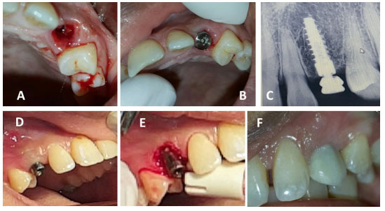
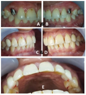
The patient was recalled after 3 weeks for the second stage of the surgery. During this visit, the level of free gingiva with the adjacent teeth were evaluated (Fig. 2D). Gingivectomy was performed bilaterally to be in harmony with the free gingiva of the adjacent teeth (Fig. 2E). Composite contouring was established on the maxillary anterior tooth for aesthetic reasons (Fig. 2F). The cover screws were removed, and gingiva formers were placed for 3 weeks. Provisional crowns (Success SD, Promedica, Neumunster, Germany) were made and cemented with temporary cement (Temp-Bond NE, Italy). We ensured that the new customized temporary abutments and their corresponding provisional restorations registered the cervical gingival emergence of the extracted teeth (Fig. 3A-E).
Three weeks later, the provisional restorations were removed, and impression copings were screwed onto the top of the implant. A light-polymerizing resin material was injected around the impression copings to prevent the soft tissues from collapsing onto the impression copings. An open-tray tech- nique was used for the final implant level impression of the maxillary arch by using a plastic stock tray with additional silicone (Zermakh, Italy). A 2M2 3D-Master shade was selected using a digital VITA System 3D-Master shade guide (VITA Easyshade Compact, Vita, Germany) in accordance with the manufacturer’s instructions. A maxillary master cast was poured with an improved dental stone and mounted manually in maximum intercuspation with the mandibular cast by using a Di-Lok tray (Di-Equi Dental Products, Wappingers Falls, NY).
The final cast was scanned using an S600 ARTI scanner (ICE Zirkon, Zirkonzahn-ZA9246A, Italy) to transfer the prepared bilateral maxillary canines directly to the software. The finished design was directly milled using Zirkonzahn’s Screw-Tec system, and zirconia cores were obtained. The inner surface of the milled cores was placed under an infrared lamp to dry for 40 min and then sintered overnight at 1600 °C in a Zirkonofen 700 sintering furnace. Zirkonzahn’s ceramic layers were fired onto the core to obtain the final shape, contour, and then glazed.
All-ceramic crowns were tried in the patient’s mouth. Interocclusal adjustment, canine guidance, protrusive and lateral movements were checked. The glazed all-ceramic crowns were cemented with Rely X, TM, Unicem AppliCap Resin Cement (3M ESPE, Germany) following the manufac- turer’s instructions (Fig. 4A-C).
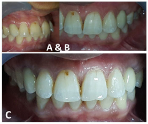
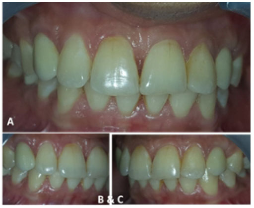
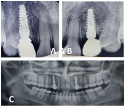
The patient was scheduled for follow up after 6, 12, 24, and 36 months. The implant-supported restorations showed good soft tissue appearances with the absence of inflammation and gingival recession. The interdental papillae looked normal and enhanced the optimal aesthetic result obtained using the definitive all-ceramic crowns (Fig. 5A-C and Fig. 6A-C). The patient had given standard oral hygiene instructions during her recall appointments.
3. DISCUSSION
Agenesis or hypodontia of permanent canines is an uncommon condition in human teeth. One of the characteristics of hypodontia is that it can occur as either isolated hypodontia or syndromic hypodontia. Isolated hypodontia refers to cases without any syndrome, affects several teeth, and appears as either a sporadic condition or an inherited condition within a family pedigree [7, 24]. In this report, the case was related to isolate hypodontia because the patient’s family members did not suffer from this condition.
The surgical placement of a single implant to replace the root of a missing maxillary canine does not usually pose any problems, and this is correct in most cases, but occasionally not possible because of unique patient anatomy such as diastema or distally curved roots of adjacent teeth. Access is easy when a large amount of bone is available, and the space between the first premolar and the lateral incisor is adequate. However, in most cases, surgeons should carefully design to Insert implants palatably and exactly at the midpoint between the premolar and the lateral incisor to leave a sufficient amount of gingival tissue in place, thereby providing a natural appearance to artificial teeth [25].
The prevalence of retained canines is 2 to 3 times greater in females than in males. Ericson and Kurol [26] reported a 1.17% prevalence in females and 0.51% in males. Other studies indicated that the prevalence of maxillary permanent canine agenesis varies between 0.07% and 0.13% [5].
In 2017, Al-Motareb et al. conducted a study in Sana’a City, Yemen, and reported a 3.55% canine impaction among a Yemeni population. Canine impactions in the maxillary arch were more prevalent in females 2.1% than in males 1.06% and more unilaterally 2.35% than bilateral 0.83%. The most prevalent was ectopic eruption 61.1%, followed by the retention of deciduous canine 51.5% and inadequate space 28.2% [14]. Another study performed among groups of Asian (Qatari population) has revealed that the prevalence rates of canine hypodontia ranged between 6.2% and 7.8%, which are within the normal range of the majority of other populations. Hypodontia is more significantly prevalent in the maxilla than in the mandibles, in females than in males, and in unilateral cases than in bilateral cases [15, 16]. These findings were recorded in our case except that describing the bilateral impactions of the maxillary canines. The bilateral impactions were possibly related to environmental or genetic factors.
The objectives in some cases were to keep the primary canine as long as possible with periapical x-rays monitoring and follow up the patient [27]. In the present case, the patient came for evaluating the existing maxillary deciduous canines. Periapical x-rays showed retained primary bilateral maxillary canines with resorbed roots (Fig. 1C & D). Further investigation was done by a panoramic x-ray which shows bilateral congenitally missing maxillary permanent canines (Fig. 1E), this is agreed with a case reported by Ramalingam et al. [17]), for a young female patient at Libya. Also, our findings during follow-up periods were totally in parallel with the finding of the one-year post-periodic evaluation of the implant abutment and the all-ceramic zirconia crown [17, 28].
The current case finding is agreed with Al-Motareb et al. and Ramalingam et al. [14, 17], Those mentioned that canine hypodontia was found more frequently in the maxilla and females patients than in the mandible and females subjects. Also, it is not agreed with researches recorded that canine agenesis is more unilaterally [14, 17].
In the literature, two abutments are recommended from each side to replace a missing canine, if conventionally a fixed prosthesis is selected to replace missing canines. This means that we need to prepare eight abutments in the maxillary arch to replace the missing two canines. However, this treatment is characterized by a complex fixed partial denture design [29]. But in our case, the treatment modality avoids the need for conventional preparation of teeth as part of a prosthetic reconstruction or prolonged orthodontic treatment aimed at bringing the impacted canine to the dental arch. Combining the implantation with bone augmentation preserved the alveolar bone and shortened the treatment period [30]. Conversely, the replacement of missing teeth with implants in the current case was clinically significant in terms of the preservation of adjacent teeth and the achievement of maximum aesthetics and functions, in addition, the biocompatibility due to the using of all ceramic materials as supra structures for the dental implant [31].
The final aesthetic goals included the closure of the spaces between the maxillary anteriors with composite restoration and the re-contouring of teeth to improve the patient’s aesthetic and self-esteem.
CONCLUSION
The proper treatment plan and the placements of the implants in the proper position are essential and important factors to achieve successful treatment results. Our case demonstrated an extraction of retained deciduous canine’s teeth replaced by immediate osseointegrated implants. This case presentation aimed to evaluate the success of bilateral maxillary canine. Stable short-term results were achieved by implants and cemented all-ceramic crowns on the bilateral maxillary canines. A 3-year prolonged follow-up of this case was conducted precisely and still we are following-up the case.
ETHICS APPROVAL AND CONSENT TO PARTICIPATE
The study was approved by the ethical committee of "Al Etkan Dental Center, Hodaidah, Republic of Yemen", Code No. 1A/3/2/2016.
HUMAN AND ANIMAL RIGHTS
No animals were used in this research. All research procedures on humans were followed in accordance with the ethical standards of the committee responsible for human experimentation (institutional and national), and with the Helsinki Declaration of 1975, as revised in 2013. (http://ethics.iit.edu/ecodes/node/3931).
CONSENT FOR PUBLICATION
The patient signed a consent form to be involved in this study.
AVAILABILITY OF DATA AND MATERIALS
All data supporting this article are available with Dr. Mohammed M. Al Moaleem.
FUNDING
None.
CONFLICT OF INTEREST
The authors declare no conflict of interest, financial or otherwise.
ACKNOWLEDGEMENTS
Not applicable.


