All published articles of this journal are available on ScienceDirect.
Influence of the Arabic-Coffee on the Overall Color of Glazed or Polished Porcelain Veneers – In vitro Study
Abstract
Background:
Sometimes, porcelain restorations experience some degree of colour change in oral environment, which could be related to the quality of diet and surface roughness of these restorations.
Objective:
The aim of this in-vitro study was to evaluate the influence of Arabic-Coffee on the overall color of glazed or polished porcelain veneers fabricated from four different porcelain materials and two different thicknesses.
Materials and Methods:
A total of 160 porcelain disc of tested specimens were fabricated to a standardized thickness of 1.00 mm and 0.6mm using the following materials: Feldspathic porcelain, Zircon, E-max Press, and E-max CAD; (80 discs for each thickness and 20 specimens of each material used). Veneer specimens from each material were randomly divided into two subgroups (n = 10): 10 specimens were kept as glazed, were the other 10 tested specimens were adjusted with diamond burs, and then polished with Ivoclar Vivadent ceramic polishing kits using the recommended protocol for polishing provided by the company. A color of all tested specimens was measured using Vita Easy Shade Spectrophotometer. Then, all specimens were immersed in Arabic coffee (Al Mosafer Coffee, Saudi Arabia) and theromcycled for 1 week, and the colors of all tested specimens were then recorded again.
Results:
It was shown that there is a significant difference in the average color changes before and after immersing in Arabic-Coffee for all materials and thicknesses used in the current study. In-addition, significant differences in color changes were noticed between glazed and polished specimens. Moreover, colour change caused by the coffee was not significantly related to the thickness of the specimens used.
Conclusion:
Color stability of porcelain materials could be affected by surface treatment whether glazing or polishing. All aesthetic restorations should be deglazed whenever any adjustments have been done to maintain the color match and stability in an oral environment. Also, Arabic-Coffee is considered as a staining drink to a limited extend where patient should be assured to maintain their oral health to maintain the colour stability of their restorations.
1. INTRODUCTION
Teeth appearance has become a growing concern to the majority of patients. And it may have an effect on patients' psychology (i.e. confidence and self-esteem) [1-6]. One option for aesthetic restoration is ceramic veneers which can be used in several aesthetic treatments to improve tooth shape, color, contour, size, and malalignment [7, 8]. In a survey, 91% of dental practitioners, identified veneers as an ethical choice to treat aesthetic problems. Based on their strength, longevity, texture, conservative nature, good wear resistance, biocompatibility, aesthetics, porcelain veneers have been considered as an ultimate option for conservative aesthetic treatment modalities [9, 10].
Several materials are being used to fabricate ceramic veneers including: feldspathic porcelain and glass-based ceramics [11]. Feldspathic porcelain has been widely used to fabricate porcelain veneers, and it has continued to be the material of choice in some aesthetic cases. Moreover, since the introduction of lithium di-silicate E-max system, it becomes one of the best materials to fabricate porcelain veneers. Moreover, Zircon based ceramic is considered the material of choice in some cases to fabricate porcelain veneers, especially when we have deeply discolored teeth.
It is important to achieve an acceptable color match between the natural teeth and the aesthetic restorations; however, it is also essential to maintain such color match in oral environment. Several studies have assessed the color stability in relation to the surface texture of different restorative materials [12]. Ceramic surfaces adjustment can remove the glazed layer, which reveals the pores of the ceramic material, creating a rough surface and may lead to discoloration of the restoration that requires re-glazing or polishing after grinding [13-19] Many studies have proven that polishing of grinded porcelain provides as smooth surface as glazed one which may even be esthetically better. Moreover, some studies supported the use of polishing as an alternative to glazing [13]. On the other hand, some studies demonstrated that porcelain staining is correlated with diet, immersion time along with type of surface treatments [12, 20]
Moreover, it is not clearly stated yet if that the porcelain thickness is considered a significant factor in surface discoloration of ceramic veneers. However, majority of studies reported the importance of ceramic thickness in masking or showing the discoloration of underlying resin cement rather than the discoloration of the ceramic material itself [9, 12, 13].
In a study was done by Gupta et al. to evaluate color stability of a porcelain materials after exposure to commonly consumed beverages i.e. tea, coffee, and Coca-Cola, it has been concluded that porcelain veneers is affected by dietary habits [20]. Furthermore, coffee is considered to be the most beverage cause discoloration to porcelain veneer [21, 22].
In fact, Coffee is known as one of the most popular beverages worldwide. Saudi population consumes a special type of coffee so-called: Arabic-Coffee. It contains some additives including: Saffron, Ginger and Cardamom. And it has been suggested that it might be a staining factor to aesthetic restorations [23, 24].
The value of average Colour change (ΔE), is clinically important, and challenging in different levels of colour measrments. It has been shown that the borderline ΔE* which is perceptible to all people in a color test is 2.5 [25]. A scale of perceptible color difference has also been proposed with a ΔE* < 1 regarded as not appreciable to the human eye and a ΔE* > 2 appreciable by non-skilled persons and therefore of clinical significance [26]. Moreover, it has been found that3.3 units of color difference have been considered unacceptable by 50% of observers. Similarly, 50% of observers had rejected the color difference of 2.72 ΔE units between the samples [27]. Additionally, an in vivo study has shown that the average ΔE* between teeth assessed to be a complete color match intra-orally is 3.28 While the average ΔE* of 6.8 units has been assessed to present the clinically color mismatch [28]. However, a recent in vivo study has presented the clinically acceptable threshold to be ΔE* 4.2 units [29-33]. Therefore, ΔE* perceptible and clinically acceptable thresholds should be borne in mind when assessing restorations spectrophotometrically. This study aims to evaluate the influence of Arabic-Coffee on the overall color of glazed or polished porcelain veneers fabricated from four different porcelain materials and two different thicknesses.
2. MATERIALS AND METHODS
2.1. Study Design
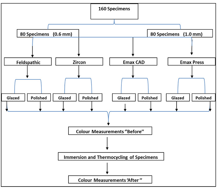
A total of 160 ceramic disc specimens of 10 mm diameter were prepared for this study. Ceramic discs were produced to a standardized thickness of 1.00 mm (80 samples) and 0.6 mm (80 samples) using the following materials (20 samples of each material): Feldspathic, Zircon, E-max press, and E-max CAD (Table 1). Veneer Specimens from each material were divided into two subgroups (n = 10) according to the surface management: polished or glazed.
| Material | Type (brand name) |
Manufacture | Shades |
|---|---|---|---|
| Feldspathic | Enamel Vita VM 13 | (Vita, Zahnfabrik, Bad Sackigen, Germany) | Shade: B1 |
| Zircon | Ceramill Zolid PS | (Aman Girrbach, Germany) | Shade: B light |
| Emax press | IPS Ema Press HT | (Ivoclar Vivadent, Liechtenstein) | Shade: B1 |
| Emax CAD | IPS Emax CAD HT | (Ivoclar Vivadent, Liechtenstein) | Shade: B1 |
Color was measured with the samples placed on gray background using vita Easy shade spectrophotometer. Then all specimens were immersed in Arabic coffee for four weeks, and the colors of all specimens were measured again. A flow chart demonstrated the study design and specimens distribution Fig. (1).

2.2. Specimens Fabrication
For the first group, specimens of shade B1 enamel feldspathic ceramic (Vita, Germany) were fabricated by a single operator using two Teflon molds with a diameter of 10 mm and a depth of 1.1 mm and 0.7 mm respectively. These shades were selected as such light shades are the most used shades in porcelain veneers fabrication. Both the porcelain and modeling liquid were mixed, packed and dried and then placed onto platinum foil and fired according to the manufacturer’s instruction. Specimens were then glazed according to the company instructions.
For the second group, presintered zirconia blocks attached to the milling machine (Amann Girrbach, Germany) to produce 20 specimens of 10 mm diameter × 0.6 mm and 20 specimens of 10 mm diameter x 1.00 mm thickness), and then samples were glazed according to the manufacturer’s recommendations.
For the third group, 40 specimens were made, designed from IPS Emax CAD, and milled into the desired dimensions using the CAM milling machine (Amann Girrbach, Germany) and then were crystallized and glazed according to the manufacturer instructions.
For the last group, test specimens of IPS Emax Press were fabricated from casting wax (Ivoclar Vivadent) using same Teflon mould. Then these discs were invested and heated to melt the wax away, and then IPS E-max ceramic were pressed and then glazed according to the manufacturer’s recommendations.
Both surfaces of the specimens (Feldspathic and E-max Press) were finished using abrasive papers to give a finished thickness of 1.0 mm +/- 0.025 mm and 0.6 +/-0.025 (measured with digital calipers and rejected if outside given range). Colour measurements for all tested specimens were then recorded by single operator and considered as a baseline colour readings.
2.3. Materials' Surface Treatments
Tested specimens of each thickness and material were divided into 2 subgroups: First subgroup specimens were glazed according to the manufacturer’s instructions for each material, where the other subgroup specimens were adjusted with diamond burs then polished with ceramic polishing kit (Ivoclar Vivadent) using the recommended protocol for polishing provided by the company.
2.4. Color Measurements
Color measurements were made using an ‘Easy shade’ Vita probe spectrophotometer (Vita Easy shade, Vita, Germany). Spectrophotometers measure CIE-LAB values giving a numerical representation of a 3D measure of color. These measurements have been previously used in studies assessing shades of both porcelain and teeth. Readings of L*, a* and b* were performed three times against the same (gray) background and the mean value used. Means of color data with the standard deviations of tooth surfaces were calculated.
2.5. Immersion of Tested Specimens in the Arabic-Coffee
All the tested specimens were immersed in an Arabic-Coffee (Almosafer Coffee, Saudi Arabia) for four weeks. During this immersion period, an ageing process was conducted using a thermocycling machine where 10 cycles were accomplished every day; first in 5 °C cold water and then in 55 °C hot water (Ivoclar Vivadent). The all the tested specimens were then dipped in distilled water following removal from the Arabic-Coffee drink solution, and moved up and down for 10 times. The tested specimens were then wiped dry with tissue paper, and then placed in viewing port for color measurement. Colors were measured again by the same operator, same settings and same gray background.
2.6. Data Analysis
SPSS 22.0 software (Chicago, USA) and excel Microsoft 10 were used to insert the data. Data were analyzed to determine any differences in the colour of the tested specimens after immersion in coffee, and difference in colour change between glazed and polished groups, and difference in colour change in between 0.6 mm and 1.00 mm specimens. To determine these differences, one-way analysis of variance (ANOVA) and T-test analytical tests were used at P-value of 0.05 [34, 35]. Then comparing colour change values to the perceptible threshold 2.8 and clinically acceptable threshold 4.2 [31].
The ΔE* values were calculated for the different materials and thicknesses using the following equation:
ΔE* = [(L1*- L2*)2 + (a1٭ -a2*)2+ (b1٭ -b2*)2]1/2
3. RESULTS
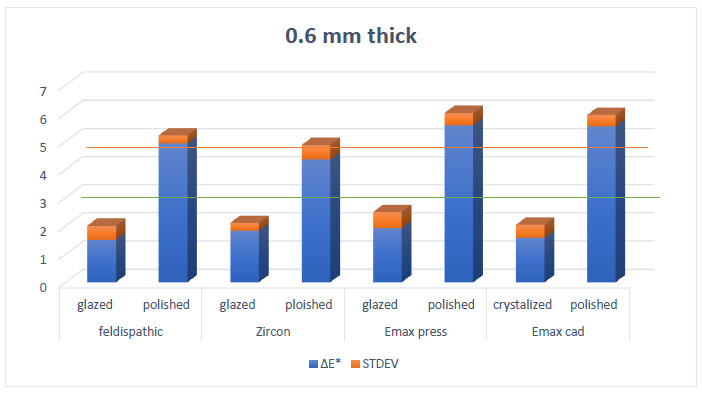
For 0.6 mm thick samples (Table 2 and Graph 1); the mean ΔE* of the Feldspathic porcelain samples was (1.5) for the glazed group and (4.92) for the polished group. Where Mean ΔE* for Zircon samples was (1.82) for the glazed group and (4.35) for the polished group. Mean ΔE* for E-max Press was (1.92) for the glazed group and (5.57) for the polished group. Finally Mean ΔE* of the Emax Press was (1.58) for the glazed group and (5.53) for the polished group.
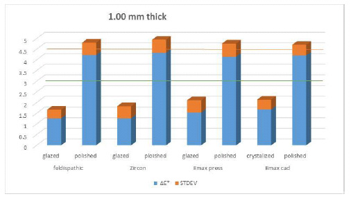
For 1.0 mm thickness (Table 3 and Graph 2); the mean ΔE* of the Feldspathic porcelain samples was 1.26 for the glazed group and 4.23 for the polished group. Where ΔE* of the Zircon samples was 1.261 for the glazed group and 4.34 for the polished group. Mean ΔE* of the Emax Press was 1.54 for the glazed group and 4.15 for the polished group. Finally, Mean ΔE*of the Emax CAD was 1.68 for the glazed group and 4.21 for the polished group.
| Materials Used | Feldispathic | Zircon | Emax Press | Emax CAD | ||||
|---|---|---|---|---|---|---|---|---|
| - | - | - | - | - | - | - | - | - |
| Surface | Glazed | Polished | Glazed | Polished | Glazed | Polished | Glazed | Polished |
| - | - | - | - | - | - | - | - | - |
| Mean ΔE* | 1.5 | 4.92 | 1.82 | 4.35 | 1.92 | 5.57 | 1.58 | 5.53 |
| - | - | - | - | - | - | - | - | - |
| STDEV | 0.50 | 0.29 | 0.28 | 0.52 | 0.57 | 0.44 | 0.46 | 0.41 |
| P-value | P ≤ 0.01 | P ≤ 0.01 | P ≤ 0.01 | P≤ 0.01 | ||||
Based on analysis tests; significant differences in color change were noticed before and after immersing with coffee for all materials and thicknesses used (p < 0.01). Significant differences in color changes were noticed between glazed and polished specimens (p < 0.01). No significant differences in color change were noticed when using different thickness for all materials used p ≥ 0.05).
| Materials Used | Feldspathic | Zircon | Emax Press | Emax CAD | ||||
|---|---|---|---|---|---|---|---|---|
| - | - | - | - | - | - | - | - | - |
| Surface | Glazed | Polished | Glazed | Polished | Glazed | Polished | Glazed | Polished |
| - | - | - | - | - | - | - | - | - |
| Mean ΔE* | 1.26 | 4.23 | 1.261 | 4.34 | 1.54 | 4.15 | 1.68 | 4.21 |
| - | - | - | - | - | - | - | - | - |
| STDEV | 0.41 | 0.58 | 0.57 | 0.61 | 0.57 | 0.62 | 0.46 | 0.50 |
| P -value | P ≤ 0.01 | P ≤ 0.01 | P ≤ 0.01 | P ≤ 0.01 | ||||


4. DISCUSSION
Nowadays patients demand for aesthetic restorations with long-term color stability to enhance their teeth appearance [1-6]. These considerations have led to the use of all-ceramic materials. Which still can experience some degrees of colour changes related to some consumed foods or beverages?
Arabic-Coffee is considered one of these staining drinks as it has special additive ingredients [23, 24]. This current study was conducted to evaluate the effect Arabic coffee on the overall all color of glazed and polished porcelain specimens fabricated from different porcelain material and two different thicknesses. Statistically significant differences were noticed for all tested specimens after immersion in this type of Coffee, which assures the fact that this Arabic Coffee is considered as a staining factor for porcelain restorations and patients, in their turns, supposed to be instructed to clean their teeth well after consumption of such coffee in an attempt to maintain the colour stability of their restorations.
In considerable number of cases, dentists tend to do some adjustments of their aesthetic restorations, and some of them tend to or prefer to do only polishing while neglecting to do the glazing step. Based on that, we attempted in this current study to compare the color changes of polished and glazed porcelain specimens. Noticed significant differences in color changes between glazed and polished specimens were noticed for all ceramic materials used, and for both thicknesses. This assures the importance of glazing any ceramic restoration after doing any type of adjustments, even when doing a proper polishing protocol.
Several studies have demonstrated the relationship between the color changes and ceramic thickness. Bulent Uludag et al. conducted a study to evaluate the effect of ceramic thickness on the color stability, and concluded that as the ceramic thickness increased, significant decreases in ΔE* values were recorded [36, 37]. However, in this study, no significant difference in color change was noticed when using different thicknesses of all porcelain materials used. This might be explained by different study settings used in different studies.
Furthermore, in most specimens, the colour changes of glazed materials were less than the perceptibility threshold (2.8). This confirms the fact that porcelain materials experience limited and imperceptible colour change to the human eye despite the statistically significant difference caused by Arabic-Coffee. While the colour changes of the polished specimens were above the perceptibility threshold and even above the acceptability threshold (4.2) in most specimens which demonstrate that such colour changes are clinically unacceptable to the human eye [38]. This, again, assures the importance of glazing all aesthetic restorations before cementing them into the patient’s mouth.
Clinicians have to put in his consideration the possible color changes that might result after any intra-oral adjustment followed by polishing procedures to the ceramic surface. In-addition, it is usually preferable to reglaze the all ceramic restorations after any surface treatments and before final cementation.
CONCLUSION
Different porcelain materials used have shown a difference in terms of color change after immersing in Arabic-Coffee. The difference between glazed and polished porcelain specimens were obvious for all materials and thicknesses used. The average color change was not significant with different thicknesses. Color change of polished specimens where remarkably higher than those of glazed specimens and was clinically unacceptable, which suggest glazing of all aesthetic restorations after any adjustments might be done and before cementation to maintain the color match in oral environment.
ETHICS APPROVAL AND CONSENT TO PARTICIPATE
Not applicable
HUMAN AND ANIMAL RIGHTS
No animals/humans were used for studies that are the basis of this research.
CONSENT FOR PUBLICATION
Not applicable.
AVAILABILITY OF DATA AND MATERIALS:
All data supporting this article are available with Dr Mohammed M. Al Moaleem on reasonable request.
FUNDING
None.
CONFLICT OF INTEREST
The authors declare no conflict of interest, financial or otherwise.
ACKNOWLEDGEMENTS
Declared none.


