All published articles of this journal are available on ScienceDirect.
Lipoma of the Palate: An Uncommon Finding
Abstract
Background:
Lipoma is a benign neoplasm originated from adipose cells circumscribed by connective tissue. This neoplasm represents about 1% to 4.4% of all oral benign tumors and it is rarely located in the palate area.
Objective:
This case reports the occurrence of an oral lipoma in the hard palate of a 57-year-old woman and discusses its etiology and treatment.
Case Report:
The treatment consisted in the total resection of the lesion and laser therapy. The patient is being followed up for forty three months with no signs of recurrence.
Conclusion:
Lipoma in hard palate is a rare entity that may be associated with endocrine factors and local inflammation.
INTRODUCTION
Lipoma is a benign mesenchymal tumor constituted by encapsulated mature adipose tissue with variably sized adipocytes [1]. Clinically, it is characterized as a painless well delimited nodular mass with yellow surfaces located mainly in the back, neck, chest wall, face, and femur [2].
When at the oral cavity, lipomas are majorly located in this order: tongue; floor of mouth; buccal vestibule; lip; palate; gingiva and retromolar region [3].
The case of a lipoma located in the hard palate is presented, highlighting its clinical and imaginological aspects, therapeutics and follow up.
CASE REPORT
A 57-year-old woman attended the Mato Grosso Cancer Hospital referred by a general dentist to evaluate and treat a lesion on hard palate impeding her to use the upper denture. She reported having Diabetes Mellitus type 2, in use of metformin hydrochloride 850 mg and denied any drug allergy.
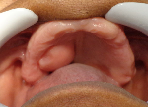
The clinical assessment revealed a normal colored volumetric tissue increase with fibrous consistence and smooth surface of approximately 30 mm of diameter located on the right side of the hard palate with one year of evolution (Fig. 1).

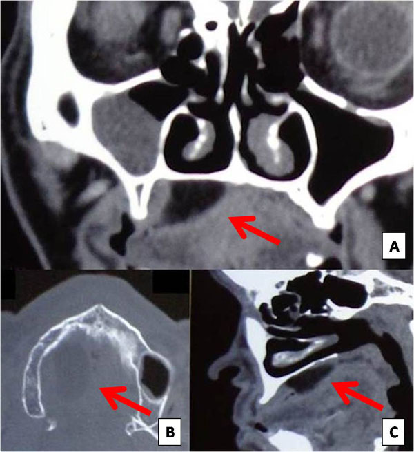
The computed tomography, in axial cut, showed a hypodense area in the right side of the hard palate, corroborating to the diagnostic hypothesis (Fig. 2).

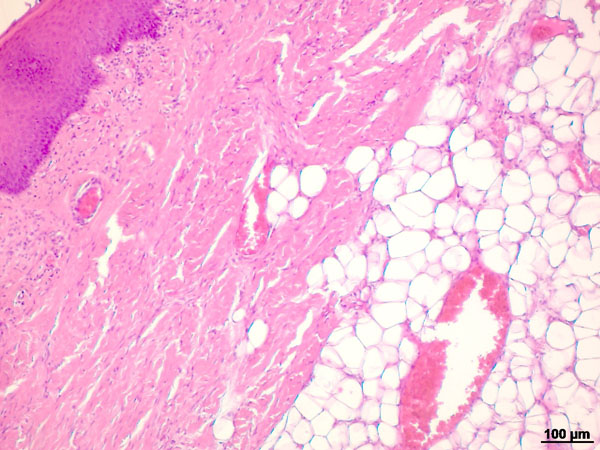
Excisional biopsy of lesion was performed. Histological examination of the excised specimen revealed a mucosal fragment covered by parakeratinized stratified squamous epithelium and skin basement membrane formed by fibrous connective tissue (Fig. 3). In the submucosa, proliferation of mature adipose cells was noted, leading to the diagnosis of lipoma.

After biopsy, low level laser therapy was performed aiming to improve wound healing. The protocol used was punctual application around the operated area, as follows: four points with red laser (wavelength of 660 nm ± 10 nm) and four points with infra-red laser (wavelength of 808 nm ± 10 nm), 40 seconds per point, totalizing the fluence of 4J/cm2 with a proper device (XT Therapy – DMC, São Paulo, Brazil). This protocol was executed in 14 sessions (3 times/week).
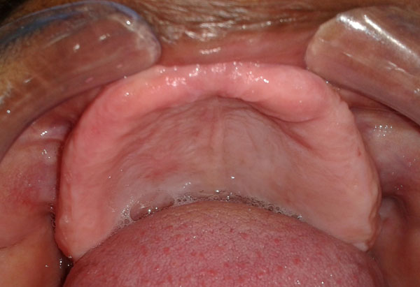
The patient was followed up for forty three months without recurrence (Fig. 4).

DISCUSSION
Lipoma is a benign tumor that arises of mature adipose cells in the mesenchymal tissue and because of their abundance it can occur in several soft tissues as retroperitoneum, and the omentum. However, in the head and neck region it is a lesion extremely rare reported in hypopharynx, larynx, oral cavity, nasopharynx, and the retropharyngeal space. As there is little adipose tissue in the hard palate, it is not often its emergence at that site [3, 4] as occurred in this case.
In the presented case the lesion had a pink coloring aspect. However it is common for lipomas to exhibit yellow color, characteristic of adipose tissue and visible through the thin overlying epithelium [3]. Clinically, differential diagnosis of lipomas include dermoid cysts, ranulae, thyroglossal duct cysts, pleomorphic adenomas, ectopic thyroid tissues, mucoepidermoid carcinomas, angiolipomas, fibrolipomas and malignant lymphomas [5].
Patient was 57 years old. Typical lipoma occurs in patients most often between the ages of 40 and 60 years and it is uncommon for pediatric patients [1, 2].
The ultrasound and magnetic resonance imaging can be used to differentiate infiltrating lipoma from well-limited lipoma that shows hypodense homogenous mass [4]. In the presented case, radiograph and CT were necessary to establish the lesion limits, guiding the complete surgical resection of the lesion.
After the resection of the lesion, the wound site was treated with laser therapy to improve wound healing and post-operative comfort. The application of low level laser is known to promote tissue regeneration, reduce inflammation and relieve pain in a healing area [6]. The recurrence of lipoma is unlikely when there is the resection of entire encapsulated lesions but its follow up is necessary because of the possibility of transformation into a malign lesion [2]. In the present case, after for forty three months of follow up there is no sign of recurrence of the lesion.
CONFLICT OF INTEREST
The authors confirm that this article content has no conflict of interest.
ACKNOWLEDGEMENTS
Declared none.


