All published articles of this journal are available on ScienceDirect.
Green Tea (Camellia Sinensis): Chemistry and Oral Health
Abstract
Green tea is a widely consumed beverage worldwide. Numerous studies have suggested about the beneficial effects of green tea on oral conditions such as dental caries, periodontal diseases and halitosis. However, to date there have not been many review articles published that focus on beneficial effects of green tea on oral disease. The aim of this publication is to summarize the research conducted on the effects of green tea on oral cavity. Green tea might help reduce the bacterial activity in the oral cavity that in turn, can reduce the aforementioned oral afflictions. Furthermore, the antioxidant effect of the tea may reduce the chances of oral cancer. However, more clinical data is required to ascertain the possible benefits of green tea consumption on oral health.
INTRODUCTION
Before the development of pharmaceutical industry, herbal products were used for curing various conditions. The concept of using natural and herbal products is very old. Humans have used these products for curing various diseases and become popular accordingly. In terms of oral care, most commonly used herbal product is “Miswak” that become popular in several communities as an effective, cheap and efficient way of maintaining oral hygiene [1-4]. Similarly, beneficial effects of using various types of tea leaves have been a common topic of discussion in general public and professionals. According to Chinese legend, tea has a known history of over 4000 years ago which was discovered accidentally by an emperor, while according to other sources the Chinese have been drinking tea since 3000 B.C. and over millions of acres are devoted to its cultivation. Currently, it is being used in almost every country around the world and is now been grown in India, Sri Lanka, Iran, Java and Japan [5].
Green tea (botanical name of tea plant is Camellia Sinensis) belonging to the family Theaceae (Table 1) has been explored in the recent years for its beneficial effects on oral health. It can be available as a shrub or evergreen tree. Its leaves may vary from exstipulate, lanceolate to obovate up to 30 cm long, 2–5 cm broad, pubescent, sometimes becoming glabrous, serrate, acute or acuminate [6].
According to an estimate, daily consumption of tea is more than 3 billion cups; almost 75 percent consumption by China [7]. The standard method defined in Chinese culture is illustrated Fig. (1). Tea leaves are available in a variety of forms depending on processing techniques illustrated in Fig. (2) [8]. Dissimilarity between green tea and black tea is due to processing methods adopted during manufacturing. As a consequence of fermentation of black tea, chemical substance known as catechins (i.e. polyphenols with molecular weight less than 450Da) are oxidized and condensed while these are present in green tea. A third type of tea, known as oolong, contains mixture of monomeric and oligomeric catechins and is in semi fermented form [9].
Distinctive composition of green is listed in Table 2 [10, 11]. It is reported that one third part of bioactive compounds in green tea is contributed by polyphenols that is the most interesting constituent [12]. The main type of polyphenols are called catechins (flavan-3-ols) also known as tannins serve as astringency constituent. Dr. Michiyo Tsujimura extracted the catechins from tea leaves in 1929 and studied their functions (Fig. 3) for the first time during his work at The Institute of Physical and Chemical Research, Japan.
Most important kind of catechins include epigallocatechin-3-gallate (EGCG; 59%), epigallocatechin (EGC; 19%), epicatechin-3-gallate (ECG; 13.6%) and epicatechin (EC; 6.4%) including EGCG and EGC are reported for their presence in green tea. Both the types along with ECG exhibit antimicrobial activity [13]. ECG reviles the possibility of protection against urinary tract infections (UTI) as it is reported to be excreted by kidney while it is also extracted in bile along with EGCG. Large variations can be observed in the chemical structures of catechins due to dissimilar composition (Fig. 4). Green tea also serves as a great source of anti-oxidants.
Numerous therapeutic effects of green tea are reported after human clinica trials [13, 14] . It contributes to improved oral health and decreases the chances of cardiovascular diseases, cancer and acts as an anti-hypertensive, anti-bacterial, anti-viral agent. It also exhibits protection against ultraviolet radiations, helps in weight loss, causes increase in bone mineral density and provides neuro-protective power. Other than the mentioned advantages of green tea, it is popularly utilized for the treatment of stomach discomfort, indigestion, vomiting, diarrhea and flatulence. It also enhances brain functioning and helps in maintaining blood glucose balance and body weight [15]. Due to numerous health benefits, there has been a significant increase in the popularity and consumption of green tea among various cultures and populations.
Classification of green tea plant.
| Kingdom | Plantae |
|---|---|
| Subkingdom | Tracheobionta |
| Super-division | Spermatophyte |
| Division | Magnoliophyta |
| Class | Magnoliopsida |
| Sub-class | Dillenidea |
| Order | Theales |
| Family | Theaceae |
| Genus | Camellia L. |
| Species | Camellia Sinensis |
GREEN TEA EFFECT ON ORAL TISSUES
Green tea which is essential source for providing polyphenol antioxidants has the ability to protect against various oral diseases such as dental caries, gingivitis, periodontitis, halitosis and oral malignancy (protection and regression). In addition it can prevent from oral oxidative stress, inflammation resulting due to cigarette smoke and reduces dentin erosion and abrasion. The therapeutic roles of green tea related to oral health are given.
a) Role as Antioxidants
Polyphenols have shown anti-oxidative activity by neutralization of free radicals in the body. They are also known to reduce or prevent their detrimental effects and restrain ROS generation to inhibit the lysosomal secretions. The hydrogen releasing property of catechin and epicatechin results in scavenging effects [16]. Polyphenols have following processes that reduce oxidation level (Fig. 5).
b) Therapeutic Effects on Periodontal and Gingival Health
The gingival crevice is a physiological zone surrounded by gingiva and tooth margins. A colony of microorganisms resides in this space and most common of that are anaerobes. In pathological conditions, these spaces extend and periodontal pockets filled with serum exudates and large colonies of polymorphs. Additionally, oxidative stress supports the development of diseased conditions. The microbes commonly present in such conditions are Prevotella spp and Porphyromonas gingivalis (black pigmented anaerobes) [18].
Periodontal health is inversely related to consumption of green tea, an epidemiological study proves that people have better periodontal health if they drink green tea very often for example during meals or at breaks from work [19]. Green tea plays supportive role in the maintenance of periodontal health, as suggested by an in vitro study catechins (e.g. EGCG) restrict the development and colonization of harmful bacteria such as Porphyromonas gingivalis, Prevotella intermedia, and Prevotella nigrescens [20]. These bacteria cause severe harm to periodontal tissues for example Porphyromonas gingivalis develop adhesion to buccal mucosa and cause destruction [21]. Recently, Nadeem et al. [22] studied the influence of green tea consumption versus black tea on periodontal health of 240 dental students and noticed that students who were consuming green tea had good periodontal health status with minimal plaque accumulation in comparison with consumers of black tea. Literature exhibited that better periodontal health status of regular green tea using individuals is mainly due to catechins, as these are steric structures of 3-galloyl radial, ECG, EGCG and gallocatechin gallate (GCG) (major polyphenols) and responsible for restrain release of toxic end metabolites from Porphyromonas gingivalis [23].

Standard method to make green tea in chinese history.
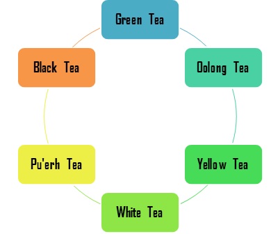
Different kinds of tea according to their processing.
c) Dental Caries
Dental carries is a pathological condition resulting due to demineralization of tooth structure because of bacterial infections and/or deficiency of nutrients. Oral microbes are responsible for carries development, out of which mainly streptococcus mutants are most active [24]. The oral antimicrobial peptides are known to reduce the bacterial activity [25]. Green tea leaves are known to be rich in fluoride and other components such as polyphenols (catechins) that play a supportive role in resisting dental carried as reported in many studies [23, 26-28]. The beneficial role of fluoride is well known as it inhibits the bacterial growth and helps re-mineralization of dental tissues [29, 30]. Cariogenic bacteria release glucans with the help of glucosyltransferase (GTase) which are branched and facilitate adherence of microbes to tooth surface [24]. GC and EGC are the catechins that restrict the growth of 10 types of carries causing bacteria. Otake et al. [31] showed that total amount of catechins, mainly EGCG and its epimer gallocatechin, EC and ECG present in one cup of green tea prevent streptococcus mutants from adhering to tooth surfaces. The catechins were extracted from green tea with concentration of 100mg/L and their effects on bacterial adhesion to salivary coated hydroxyapatites were observed. Other studies also exhibit that consumption of tea constrains the release of salivary enzyme, amylase as observed by Kashket and Paolino [32]. Later it was supported by Zhang and Kashket [33] proving that tea, either black or green restrains the enzymatic activity of Streptococcus mutans’ amylase on tooth structure preventing deminerlization.
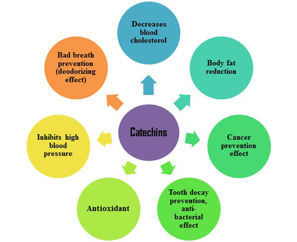
Functions of catechins in human body.
d) Bad Breath (halitosis)
The main reagents responsible for halitosis (bad breath) are volatile sulfide compounds such as hydrogen sulfide (H2S), dimethylsulfide [(CH3)2S] and methyl mercaptan (CH3SH). These reagents degrade in the oral cavity by proteolytic reactions primarily by anaerobic gram negative bacteria consuming numerous sulfur-containing substrates including saliva, food debris, epithelial cells and blood [34]. Green tea is well known for antibacterial properties against anaerobic microorganisms. Green tea can abolish bad breath by suppressing anaerobic bacteria and eliminating the production of volatile sulfur compounds [35]. Deodorant action of ingredients decreases in the following order: EGCG > EGC > ECG > EC. The deodorizing action of EGCG is based on a chemical reaction of EGCG and MSH and introducing methylsulfinyl/methylthio group into the B ring of EGCG. During this reaction, a methylthio group is supplemented in orthoquinone form of the catechin produced by oxidation, hence eliminating the halitosis [36].
Chemical composition of green tea plant.
| Protein | Peptides and enzymes |
| Carbohydrates | Fructose, sucrose, cellulose, pectin and glucose |
| Vitamins | Vitamins (B, C, and E) |
| Xanthic bases | Caffeine, Theophylline |
| Pigments | Chlorophyll, Carotenoids |
| Minerals and trace elements | Calcium, Magnesium, Sodium, Chromium, Manganese, Copper, Cobalt, Nickel, Iron, Molybdenum, Selenium, Phosphorus, Potassium, Fluoride, Zinc, Aluminum |
e) Cigarette Smoke Induced Inflammation
Smoking affects the homeostasis and injurious to oral health [37, 38]. The effects of smoking range from simple mucosal erythema to premalignant conditions and oral cancer. Smoking intensifies oral malignancies in response to oral inflammatory diseases due to compromised status of salivary antioxidants [39]. Diminished activity of numerous salivary enzymes is observed during smoking hence compromising the protection against oxidative damage [17]. In addition, smoke coming from tobacco comprised of reactive oxygen species (ROS) including superoxides, hydroxyl radical/hydrogen peroxide. The toxicity is further added due to the formation of nitric oxide (NO). Superoxide reacts chemically with NO to form peroxynitrite (ONOO). The inflammatory transcription factor pathway (NFkB) is triggered by ONOO due to activation of IkB kinase (IKK). The cascade of changes results in the intensification of expressions, iNOS activity and chronic inflammation in tissues even by exposing to a very minute dose of smoke [40].
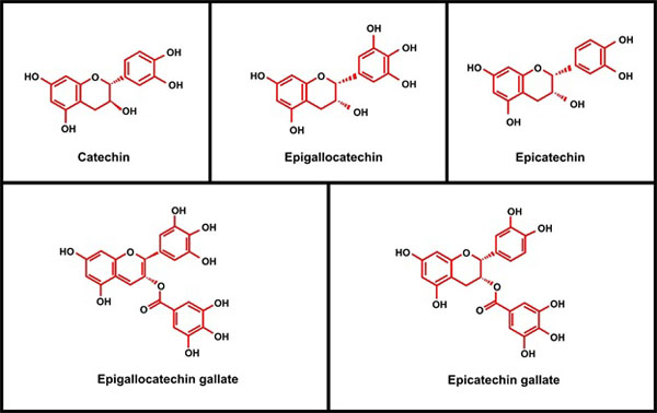
Chemical structures of catechin and its family.
Green tea can be beneficial for cigarette smokers. Catechins present in green tea are capable of scavenging superoxide oxide, NO, and ONOO [41, 42]. In addition, EGCG has the ability to suppress activation of NF-kB that leads to inhibition of phosphorylation. It can further trigger the collapse of the inhibitory sections IkB-a in human pulp cells. The IkBa is also accountable for overturning nuclear transfer of NF-kB operating subunits (p65 and p50) and stimulation of pro-inflammatory genes [43, 44]. EGCG resulted in decreased NF-kB expression and reduction of proteins facilitated including matrix metalloproteinase9 (MMP-9). The MMP-9 is implicated in the breakdown of extracellular matrix, nterleukin-8 (IL-8) and iNOS present in bronchial epithelium [14]. There are more than 4700 oxygen/nitrogen species and reactive chemical compounds in the cigarette smoke. Nicotine is the main chemical compound released from cigarettes that affects gingival and periodontal health [45]. Nicotine stimulates apoptosis across ROS generations of human gingival fibroblasts (HuGF) [45]. The metabolites of nicotine such as tobacco-specific nitrosamines (TSNAs) are considered major carcinogens.
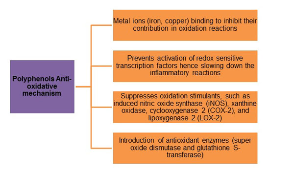
Representing steps of oxidative reduction mechanism by polyphenols.
Weitberg et al. [46] treated cultured human pulmonary cells through EGCG prior to introducing to TSNA 4-(Nmethyl- N-n-trosamino)-1-(3-pyridyl)-1-butanone (NNK). EGCG exhibited the ability to protect against the initiation of genetic injury to DNA of cultivated human pulmonary cells [46]. Another smoke compound (acrolein) is a risk factor for developing periodontal diseases and toxic to HuGF. It inhibits normal functioning of gingival fibroblasts due to interruption with cells attachment and proliferation [47]. In advance stages, acrolein can prompt inflammatory conditions and cancer onset. Green tea extract and EGCG can extinguish acrolein and other unsaturated aldehydes, hence reducing acrolein toxicity [48]. Green tea consumption can diminish oxidative and inflammatory tissue injuries in the oral cavity caused by cigarette smoking.
CONCLUSION
There are numerous beneficial effects of green tea on oral health. Green tea has valuable effects on oral conditions such as dental caries, periodontal disease and halitosis. Research suggests that green tea helps to reduce the bacterial activity in the oral cavity that in turn, can reduce the aforementioned oral afflictions. Furthermore, the antioxidant effect of the tea may reduce the chances of oral cancer in tobacco users. However, more clinical data is require to ascertain benefits of using green tea for oral tissues and prevention of oral diseases
CONFLICT OF INTEREST
The authors confirm that this article content has no conflict of interest.
ACKNOWLEDGEMENTS
Declared none.


